



Juglanin
Based on 7 publication(s) in Google Scholar
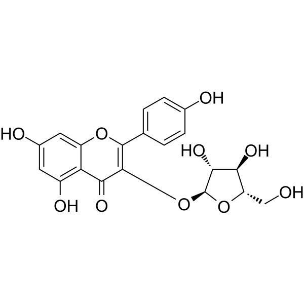
Juglanin, a occurring flavonoid that can be isolated from crude Polygonum aviculare, is a JNK acticator, with anti-inflammatory, anti-oxidant and anti-tumor activities. Juglanin can induce apoptosis and autophagy on human breast cancer cells.
Nur für Forschungszwecke. Wir verkaufen nicht an Patienten.
- Reinheit : 99.76%
- CAS. Nr.: 5041-67-8
- Formel: C20H18O10
- Molecular Weight:418.35
-
Speicherung:
4°C, sealed storage, away from moisture
* In solvent : -80°C, 6 months; -20°C, 1 month (sealed storage, away from moisture)

To place orders, for customer services and technical support, please contact: MedChemExpress USA
Tel: 609-228-6898 E-mail: [email protected] [email protected]


-
 Biologische Aktivität
-
Chemical Information
-
Lösungsmittel & Löslichkeit
- Protokoll
- Reinheit & Dokumentation
- Verweise
-
 Help & FAQs


-
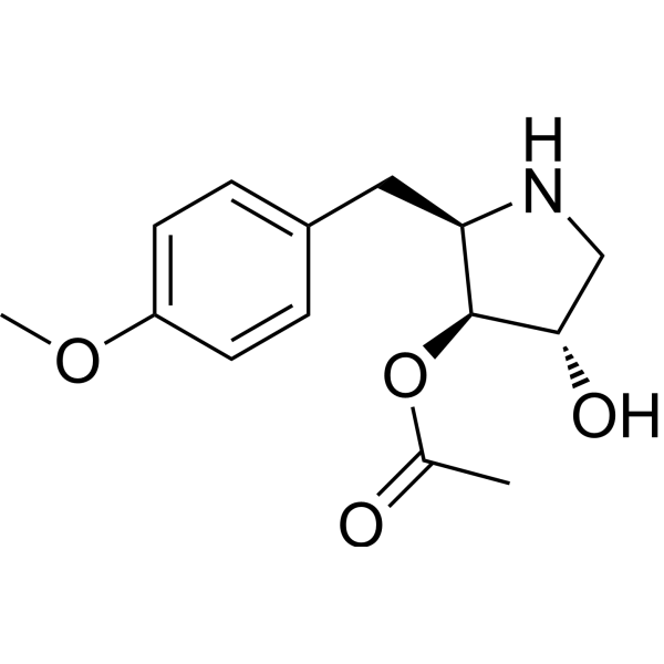
HY-18982
AnisomycinProtein Synthesis Inhibitor -
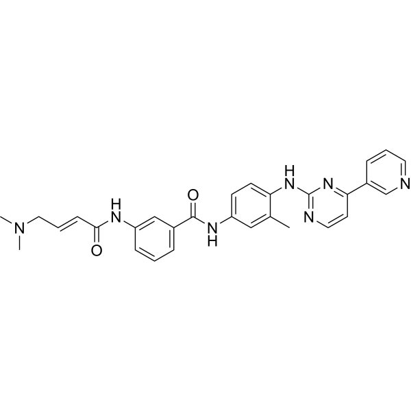
HY-13319
JNK-IN-8JNK Inhibitor -
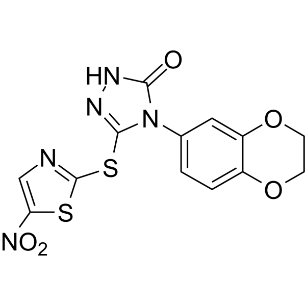
HY-10366
BI-78D3JNK Inhibitor -
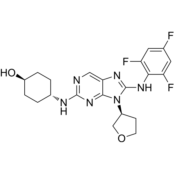
HY-15495
Tanzisertib -
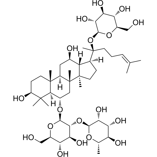
HY-N0044
Ginsenoside ReEndogenous Metabolite -
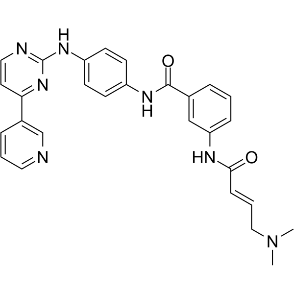
HY-15617
JNK-IN-7JNK Inhibitor -
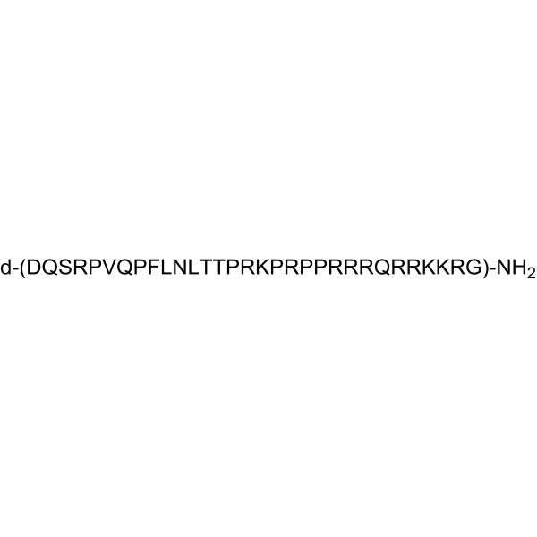
HY-P0069
D-JNKI-1 -
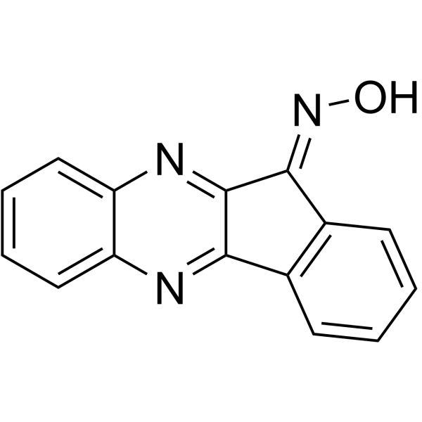
HY-100233
IQ-1S free acidJNK Inhibitor -
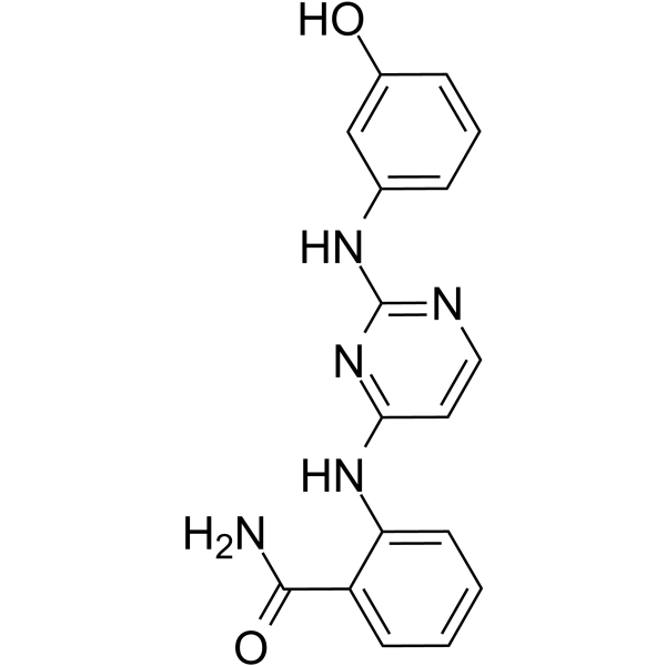
HY-15737
DB07268JNK Inhibitor -
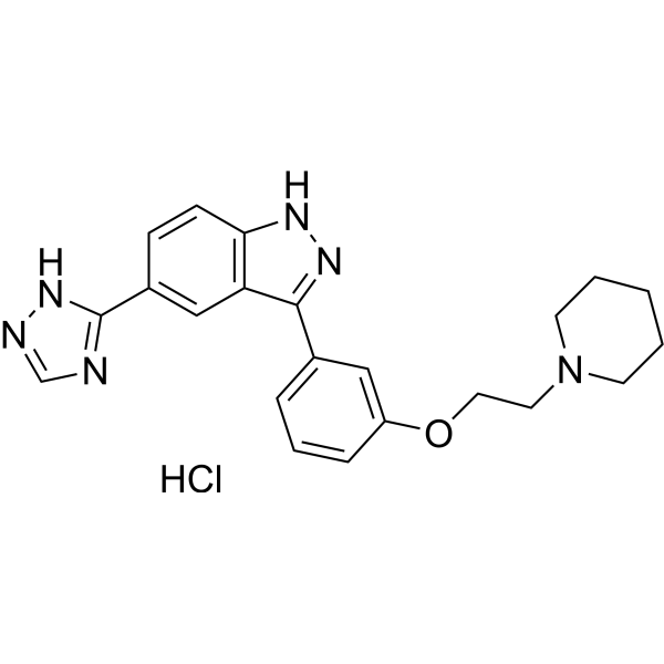
HY-13022
CC-401 hydrochlorideJNK Inhibitor -
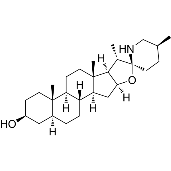
HY-N2149
TomatidineAutophagy Inducer -
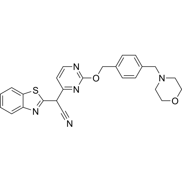
HY-14761
BentamapimodJNK Inhibitor
Publications Citing Use of MedChemExpress (MCE) Juglanin
More- Nat Commun. 2025 Jun 2;16(1):5101. [Abstract]
- Acta Pharm Sin B. 2021 Jan;11(1):143-155. [Abstract]
- Cell Mol Neurobiol. 2022 Nov;42(8):2643-2653. [Abstract]
- Int Immunopharmacol. 2020 Dec;89(Pt B):107048. [Abstract]
- Pestic Biochem Physiol. 2025 Dec:215:106655. [Abstract]
- Autoimmunity. 2023 Dec;56(1):2200908. [Abstract]
- Biomed Pharmacother. 2020 Jul:127:110119. [Abstract]


Biologische Aktivität
Beschreibung
IC50 & Target
[1]|
JNK |
Cellular Effect
|
Cell Line

|
Type | Value | Description | References |
|---|---|---|---|---|
| C3H 10T1/2 | IC50 |
>100 μM
Compound: 8
|
Cytotoxicity against mouse C3H10T1/2 cells after 24 hrs by FMCA method
Cytotoxicity against mouse C3H10T1/2 cells after 24 hrs by FMCA method
|
[PMID: 21190854] |
| DU-145 | IC50 |
2.4 μM
Compound: 8
|
Cytotoxicity against human DU145 cells after 24 hrs by FMCA method
Cytotoxicity against human DU145 cells after 24 hrs by FMCA method
|
[PMID: 21190854] |
| HaCaT | IC50 |
2 μM
Compound: 8
|
Inhibition of human HH/Gli1-mediated transcriptional activity in tetracycline-treated human HaCaT cells treated after 12 hrs of tetracycline addition by luciferase reporter gene assay
Inhibition of human HH/Gli1-mediated transcriptional activity in tetracycline-treated human HaCaT cells treated after 12 hrs of tetracycline addition by luciferase reporter gene assay
|
[PMID: 21190854] |
| PANC-1 | IC50 |
1.8 μM
Compound: 8
|
Cytotoxicity against human PANC1 cells after 24 hrs by FMCA method
Cytotoxicity against human PANC1 cells after 24 hrs by FMCA method
|
[PMID: 21190854] |
In Vitro
Juglanin (0-40 μM, 24-48 h) inhibits the proliferation of breast cancer cell in a dose- and time-dependent
manner and presents less cytotoxic against the normal cells[1].
Juglanin (0-10 μM, 24 h) induces the G2/M phase arrest and Caspase-dependent apoptosis in MCF-7/SKBR3 cells[1].
Juglanin (10 μM, 24 h) promotes ROS generation and enhances JNK activation in MCF-7/SKBR3 cells[1].
Juglanin (40-160 μM, 24 h) inhibits inflammation and attenuates neuroinflammation in Lipopolysaccharides (LPS, HY-D1056)-treated AST cells[2].
Juglanin (40 μM, 24 h) inhibits Sting expression in TGF-β1-incubated MRC-5 cells[3].
MedChemExpress (MCE) has not independently confirmed the accuracy of these methods. They are for reference only.
-
Cell Line:MCR-5 cell
-
Concentration:0-40 μM
-
Incubation Time:0-72 h
-
Result:Reduced the cell viability of MCR-5 after 24 h exposure at 160 μM.
Reduced the cell viability of MCR-5 after 72 h exposure at 40 μM.
-
Cell Line:MCF-7, SKBR3
-
Concentration:0-10 μM
-
Incubation Time:24 h
-
Result:Increased the expressions of phosphorylated Chk2, Chk2, phosphorylated Cdc25C, phosphorylated Cdc2, p27, cyclin D, Bad, Bax and decreased the levels of Cdc25C, Cdc2, Bcl-2.
Activated caspases and led to Casapse-9, Casapse-8 and Caspase-3 cleavage.
Increased the level of JNK phosphorylation.
-
Cell Line:Lipopolysaccharides (LPS, HY-D1056)-treated AST cells
-
Concentration:40-160 μM
-
Incubation Time:24 h
-
Result:Decreased the expression of TLR4, MyD88, CD14, p-IKKα, p-IκBα, p-NF-κB, GFAP, BDNF, Aβ and p-Tau induced by LPS (HY-D1056).
Increased the level of SYP, PSD-25 and SNAP-25.
In Vivo
Juglanin (10-30 mg/kg, i.p., 5 times weekly) ameliorates neuroinflammation-related memory impairment and neurodegeneration through impeding TLR4/NF-κB in C57BL6 male mice[2].
Juglanin (80 mg/kg, i.g., daily, 21 days) protects against bleomycin (BLM, HY-108345)-induced lung injury by inhibiting fibrosis in C57BL/6 male mice[3].
MedChemExpress (MCE) has not independently confirmed the accuracy of these methods. They are for reference only.
-
Animal Model:Tumor-transplanted mouse model [1]
-
Dosage:10-20 mg/kg
-
Administration:Intraperitoneal injection (i.p.), daily, 7 days
-
Result:Increased levels of cleaved Caspase-9 and Caspase-3, LC3BI, LC3BII and phosphorylated JNK.
Increased dead cells and apoptosis proportion in juglanin-treated tumor tissues.
Increase in mean areas that stained positively cleaved Caspase-3 in juglanin-treated tumor tissues.
-
Animal Model:C57BL6 male mice[2]
-
Dosage:10-30 mg/kg
-
Administration:Intraperitoneal injection (i.p.), 5 times weekly
-
Result:Increased number of platform crossings and expanded time in target quadrant.
Decreased the expression level of α-synuclein, pro-inflammatory cytokines (IL-1β, TNF-α, IL-18 and COX-2), TLR-4, MyD88, CD14, Aβ, phosphorylation of Tau, Ibal and neurotrophic factor (BDNF).
Reversed LPS-triggered effect on synaptic markers, up-regulating the expression of SYP, PSD-95 and SNAP-25.
Restored TH levels in the hippocampus of mice in a dose-dependent manner.
Decreased p-IKKα, p-IκBα and p-NF-κB levels combinated with LPS.
Decreased the GFAP-positive cells.
-
Animal Model:C57BL/6 male mice[3]
-
Dosage:80 mg/kg
-
Administration:i.g., daily, 1-21 days
-
Result:Improved the survial rate of the BLM (HY-108345)-treated mice.
Decreased the expression of BALF, CXCL1, IL-6, TNF-α, MPO activity, TGF-β1, and its down-streaming proteins (fibronectin/MMP-9/α-SMA/collagen I) Reduced the number of total cells, neutrophils and macrophages in BALF.
- Detection of Apoptosis
- TUNEL staining for apoptotic DNA fragmentation
- Annexin V plus membrane-impermeant dye apoptosis staining
- Autophagy
- Apoptosis Solutions
- Research Protocol for Inflammation-related Diseases
- Lysosome and acidic-vesicle live-cell staining
- Macroautophagy Solutions
- Breast Cancer Modeling


Chemical Information
-
CAS. Nr. 5041-67-8
-
Appearance Solid
-
Molecular Weight 418.35
-
Formel C20H18O10
-
Color White to yellow
-
SMILES
O=C1C(O[C@@H]2O[C@@H](CO)[C@H](O)[C@H]2O)=C(C3=CC=C(O)C=C3)OC4=CC(O)=CC(O)=C41
-
Structure Classification
-
Initial Source
-
Versand
Room temperature in continental US; may vary elsewhere.
-
Speicherung
4°C, sealed storage, away from moisture
* In solvent : -80°C, 6 months; -20°C, 1 month (sealed storage, away from moisture)


Publications (7)
-
Journal Impact Factor
-
Most Recent
-
Nat Commun
Myocardial mitochondrial antiviral signaling protein promotes heart Ischemia-reperfusion injury via RIG-I signaling in mice. [Abstract]2025 Jun 2;16(1):5101. PMID: 40456736 -
Acta Pharm Sin B
Chrysin serves as a novel inhibitor of DGK α/FAK interaction to suppress the malignancy of esophageal squamous cell carcinoma (ESCC). [Abstract]2021 Jan;11(1):143-155. PMID: 33532186 -
Cell Mol Neurobiol
2022 Nov;42(8):2643-2653. PMID: 34227028 -
Int Immunopharmacol
Juglanin suppresses oscillatory shear stress-induced endothelial dysfunction: An implication in atherosclerosis. [Abstract]2020 Dec;89(Pt B):107048. PMID: 33049495 -
Pestic Biochem Physiol
Mechanistic insights into chlorogenic acid and caffeic acid as novel juvenile hormone antagonists. [Abstract]2025 Dec:215:106655. PMID: 41162045 -
Autoimmunity
Sema3A inactivates the ERK/JNK signalling pathways to alleviate inflammation and oxidative stress in lipopolysaccharide-stimulated rat endothelial cells and lung tissues. [Abstract]2023 Dec;56(1):2200908. PMID: 37128697 -
Biomed Pharmacother
Juglanin alleviates bleomycin-induced lung injury by suppressing inflammation and fibrosis via targeting sting signaling. [Abstract]2020 Jul:127:110119. PMID: 32276127


Lösungsmittel & Löslichkeit
In Vitro:
DMSO : 100 mg/mL (239.03 mM; Need ultrasonic; Hygroscopic DMSO has a significant impact on the solubility of product, please use newly opened DMSO)
Please refer to the solubility information to select the appropriate solvent. Once prepared, please aliquot and store the solution to prevent product inactivation from repeated freeze-thaw cycles.
Storage method and period of stock solution: -80°C, 6 months; -20°C, 1 month (sealed storage, away from moisture). When stored at -80°C, please use it within 6 months. When stored at -20°C, please use it within 1 month.
Please refer to the solubility information to select the appropriate solvent. Once prepared, please aliquot and store the solution to prevent product inactivation from repeated freeze-thaw cycles.
Storage method and period of stock solution: -80°C, 6 months; -20°C, 1 month (sealed storage, away from moisture). When stored at -80°C, please use it within 6 months. When stored at -20°C, please use it within 1 month.
Konzentration (Stammlösung) × Volumen (Stammlösung) = Konzentration (Ziellösung) × Volumen (Ziellösung)
In Vivo:
Select the appropriate dissolution method based on your experimental animal and administration route.
- For the following dissolution methods, please ensure to first prepare a clear stock solution using an In Vitro approach and then sequentially add co-solvents:
- To ensure reliable experimental results, the clarified stock solution can be appropriately stored based on storage conditions. As for the working solution for In Vivo experiments, it is recommended to prepare freshly and use it on the same day.
- The percentages shown for the solvents indicate their volumetric ratio in the final prepared solution. If precipitation or phase separation occurs during preparation, heat and/or sonication can be used to aid dissolution.
Add each solvent one by one: 10% DMSO 40% PEG300 5% Tween-80 45% Saline
Solubility: ≥ 2.5 mg/mL (5.98 mM); Clear solution
This protocol yields a clear solution of ≥ 2.5 mg/mL (saturation unknown).
Taking 1 mL working solution as an example, add 100 μL DMSO stock solution (25.0 mg/mL) to 400 μL PEG300, and mix evenly; then add 50 μL Tween-80 and mix evenly; then add 450 μL Saline to adjust the volume to 1 mL.
Preparation of Saline: Dissolve 0.9 g sodium chloride in ddH₂O and dilute to 100 mL to obtain a clear Saline solution.
Add each solvent one by one: 10% DMSO 90% (20% SBE-β-CD in Saline)
Solubility: ≥ 2.5 mg/mL (5.98 mM); Clear solution
This protocol yields a clear solution of ≥ 2.5 mg/mL (saturation unknown).
Taking 1 mL working solution as an example, add 100 μL DMSO stock solution (25.0 mg/mL) to 900 μL 20% SBE-β-CD in Saline, and mix evenly.
Preparation of 20% SBE-β-CD in Saline (4°C, storage for one week): 2 g SBE-β-CD powder is dissolved in 10 mL Saline, completely dissolve until clear.
In Vivo Dissolution Calculator
Please enter the basic information of animal experiments:
-
-
-
-
Recommended: Prepare an additional quantity of animals to account for potential losses during experiments.
Please enter your animal formula composition:
-
%DMSO +
Recommended: Keep the proportion of DMSO in working solution below 2% if your animal is weak.
-
%+
-
+%Tween-80 + +
-
%Saline +
The co-solvents required include: DMSO, . All of co-solvents are available by MedChemExpress (MCE). , Tween 80. All of co-solvents are available by MedChemExpress (MCE).
Working solution concentration: 0.22 mg/mL
Method for preparing stock solution: mg drug dissolved in μL DMSO. Stock solution concentration: mg/mL. * In solvent : -80°C, 6 months; -20°C, 1 month (sealed storage, away from moisture)
1. Take μL DMSO stock solution;
2. Add μL .
μL , mix evenly;
3. Then add μL Tween 80, mix evenly;
4. Then add μL
Please ensure that the stock solution in the first step is dissolved to a clear state, and add co-solvents in sequence. You can use ultrasonic heating (ultrasonic cleaner, recommended frequency 20-40 kHz), vortexing, etc. to assist dissolution.


Reinheit & Dokumentation
-
Data Sheet (284 KB)
-
SDS (393 KB)
- English - EN (393 KB)
- Français - FR (393 KB)
- Deutsch - DE (393 KB)
- Norwegian - NO (393 KB)
- Español - ES (393 KB)
- Swedish - SV (393 KB)
- Italian - IT (393 KB)
- Korean - KR (393 KB)
- Portuguese - PT (393 KB)
-
Handling Instructions (2659 KB)
Verweise
[1]. Sun ZL, et al. Juglanin induces apoptosis and autophagy in human breast cancer progression via ROS/JNK promotion. Biomed Pharmacother. 2017 Jan;85:303-312. [Content Brief]
[2]. Zhang FX, et al. Juglanin ameliorates LPS-induced neuroinflammation in animal models of Parkinson's disease and cell culture via inactivating TLR4/NF-κB pathway. Biomed Pharmacother. 2018 Jan;97:1011-1019. [Content Brief]
[3]. Sun SC, et al. Juglanin alleviates bleomycin-induced lung injury by suppressing inflammation and fibrosis via targeting sting signaling. Biomed Pharmacother. 2020 Jul;127:110119. [Content Brief]


Complete Stock Solution Preparation Table
Please refer to the solubility information to select the appropriate solvent. Once prepared, please aliquot and store the solution to prevent product inactivation from repeated freeze-thaw cycles.
Storage method and period of stock solution: -80°C, 6 months; -20°C, 1 month (sealed storage, away from moisture). When stored at -80°C, please use it within 6 months. When stored at -20°C, please use it within 1 month.
| Optional Solvent | Concentration Solvent Mass | 1 mg | 5 mg | 10 mg | 25 mg |
|---|---|---|---|---|---|
| DMSO | 1 mM | 2.3903 mL | 11.9517 mL | 23.9034 mL | 59.7586 mL |
| 5 mM | 0.4781 mL | 2.3903 mL | 4.7807 mL | 11.9517 mL | |
| 10 mM | 0.2390 mL | 1.1952 mL | 2.3903 mL | 5.9759 mL | |
| 15 mM | 0.1594 mL | 0.7968 mL | 1.5936 mL | 3.9839 mL | |
| 20 mM | 0.1195 mL | 0.5976 mL | 1.1952 mL | 2.9879 mL | |
| 25 mM | 0.0956 mL | 0.4781 mL | 0.9561 mL | 2.3903 mL | |
| 30 mM | 0.0797 mL | 0.3984 mL | 0.7968 mL | 1.9920 mL | |
| 40 mM | 0.0598 mL | 0.2988 mL | 0.5976 mL | 1.4940 mL | |
| 50 mM | 0.0478 mL | 0.2390 mL | 0.4781 mL | 1.1952 mL | |
| 60 mM | 0.0398 mL | 0.1992 mL | 0.3984 mL | 0.9960 mL | |
| 80 mM | 0.0299 mL | 0.1494 mL | 0.2988 mL | 0.7470 mL | |
| 100 mM | 0.0239 mL | 0.1195 mL | 0.2390 mL | 0.5976 mL |


Juglanin Related Classifications
HY-N3442 Related Classifications
 Powered by Bioz