



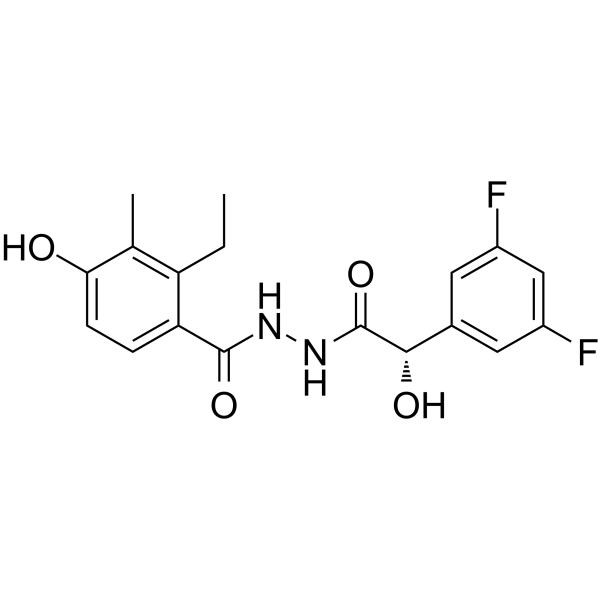
EMD638683 (S-Form)

Based on 1 Customer Validation
EMD638683 (S-Form) (Compound 1a), the S-enantiomer of EMD638683 (HY-15193), is a SGK1 inhibitor with an IC50 value > 300 nM. EMD638683 is an orally active SGK1 inhibitor with an IC50 of 3 μM. EMD638683 exhibits strong inhibition against SGK1, moderate inhibition against SGK2 and SGK3, and shows excellent selectivity for other AGC kinase family members. EMD638683 has antihypertensive activity by inhibiting SGK1, and independently of the blood pressure-lowering effect, it effectively prevents heart inflammation and fibrosis caused by hypertension by inhibiting the cardiac NLRP3 inflammation body/ IL-1β axis. EMD638683 promotes apoptosis of colon cancer cells and sensitizes radiotherapy. EMD638683 (S-Form) can be used in research related to hypertension, hypertensive heart damage, and colon cancer.
For research use only. We do not sell to patients.
- Purity : 99.72%
- CAS No.: 1184940-46-2
- Formula: C18H18F2N2O4
- Molecular Weight:364.34
-
Storage:
4°C, sealed storage, away from moisture
* In solvent : -80°C, 6 months; -20°C, 1 month (sealed storage, away from moisture)

To place orders, for customer services and technical support, please contact: MedChemExpress USA
Tel: 609-228-6898 E-mail: [email protected] [email protected]


-
 Biological Activity
-
Chemical Information
-
Solvent & Solubility
- Protocols
- Purity & Documentation
- References
-
 Help & FAQs


-
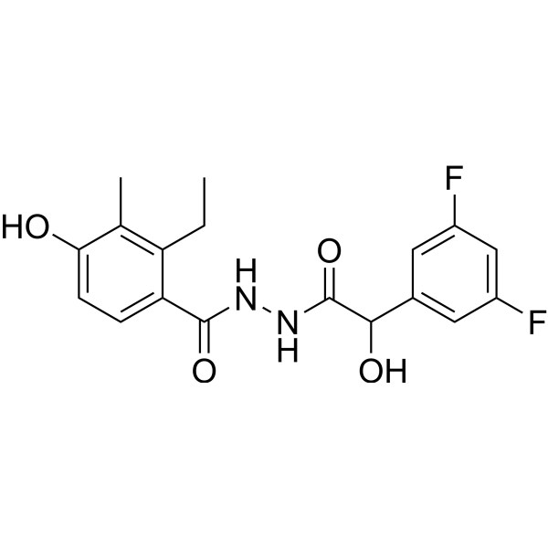
HY-15193
EMD638683SGK1 Inhibitor -
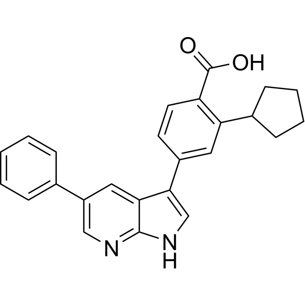
HY-15192
GSK 650394SGK Inhibitor -
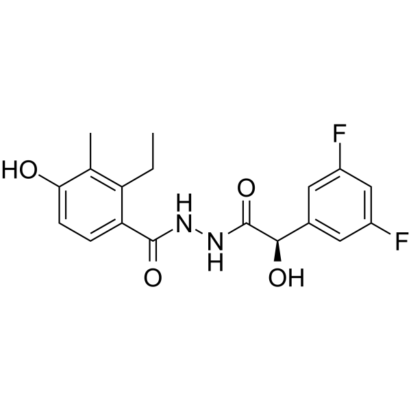
HY-15193A
EMD638683 (R-Form)R-form -

HY-15193R
EMD638683 (Standard)Standard of EMD638683 -
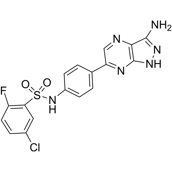
HY-18607
SGK1-IN-1SGK-1 Inhibitor -

HY-15192R
GSK 650394 (Standard)SGK Inhibitor -
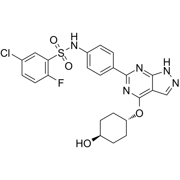
HY-142687
SGK1-IN-4SGK1 Inhibitor -
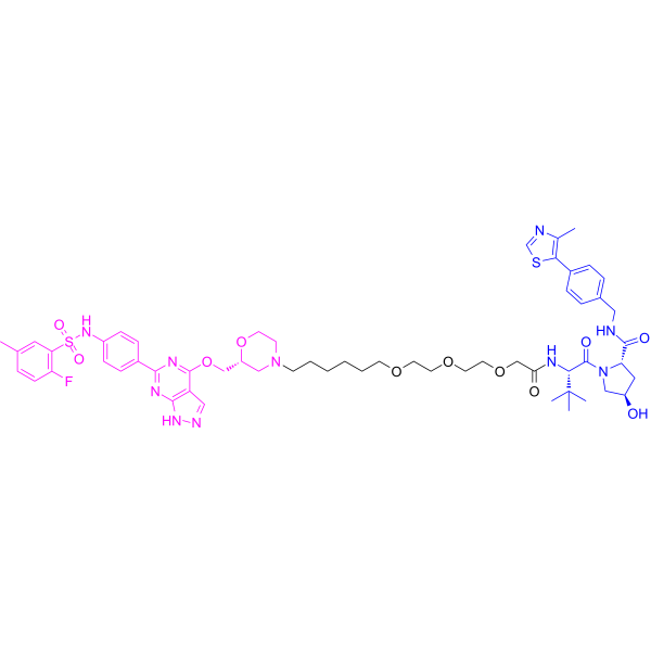
HY-125878
PROTAC SGK3 degrader-1PROTAC SGK3 Degrader -
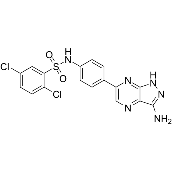
HY-135893
SGK1-IN-2SGK1 Inhibitor -
HY-117357
SI-113SGK1 Inhibitor -

HY-128221
SGK1-IN-5SGK1 Inhibitor


Biological Activity
Description
IC50 & Target
[4]|
SGK1 > 300 nM (IC50) |
In Vitro
EMD638683 (50 μM; 9-25 h) inhibits NLRP3 inflammasome activation and IL-1β secretion in mouse bone marrow-derived macrophages induced by angiotensin II (Ang II) by targeting NLRP3 expression[2].
EMD638683 (50 μM; 25-49 h) inhibits angiotensin II-induced transformation of primary mouse cardiac fibroblasts into myofibroblasts by reducing IL-1β secretion from macrophages[2].
EMD638683 (50 μM; 96 h) enhances radiation-induced apoptotic death of Caco-2 colon cancer cells[3].
MedChemExpress (MCE) has not independently confirmed the accuracy of these methods. They are for reference only. Further protocols information, click here.
-
Cell Line:Caco-2 cells
-
Concentration:50 μM
-
Incubation Time:24 h pre-incubation, 72 h post-radiation incubation
-
Result:Enhanced radiation-induced apoptotic cell death in Caco-2 colon carcinoma cells, including augmented cell shrinkage, mitochondrial depolarization, caspase 3 activation, phosphatidylserine exposure, and late apoptosis.
In Vivo
EMD638683 (600 mg/kg, administered via mixed feed for 4 weeks) prevents hypertension in wild-type mice treated with 10% fructose in isotonic saline for 4 weeks[1].
MedChemExpress (MCE) has not independently confirmed the accuracy of these methods. They are for reference only.
-
Animal Model:SV129 original background backcrossed 4 generations to C57BL/6J sgk1+/+ wild type (mixed gender, 4 months old, hyperinsulinemic salt-loaded)[1]
-
Dosage:600 mg/kg
-
Administration:p.o.; continuous in chow; 4 days
-
Result:Normalized systolic blood pressure from 111 mmHg to 87 mmHg.
Significantly increased urinary flow rate.
Significantly decreased body weight.
Caused no significant changes to fluid intake, food intake, or urinary Na+ and K+ excretion.
Increased blood pressure to 106 mmHg after returning to placebo food.
-
Animal Model:SV129 original background backcrossed 4 generations to C57BL/6J sgk1+/+ wild type (mixed gender, 7 to 10 months old, fructose/salt-induced hypertension)[1]
-
Dosage:600 mg/kg
-
Administration:p.o.; continuous in chow; 4 weeks
-
Result:Prevented the fructose/salt-induced rise in systolic blood pressure.
Resulted in significantly lower systolic blood pressure compared to placebo-treated mice.


Chemical Information
-
CAS No. 1184940-46-2
-
Appearance Solid
-
Molecular Weight 364.34
-
Formula C18H18F2N2O4
-
Color Off-white to pink
-
SMILES
CC1=C(CC)C(C(NNC([C@@H](O)C2=CC(F)=CC(F)=C2)=O)=O)=CC=C1O
-
Shipping
Room temperature in continental US; may vary elsewhere.
-
Storage
4°C, sealed storage, away from moisture
* In solvent : -80°C, 6 months; -20°C, 1 month (sealed storage, away from moisture)


Solvent & Solubility
In Vitro:
DMSO : ≥ 100 mg/mL (274.47 mM; Hygroscopic DMSO has a significant impact on the solubility of product, please use newly opened DMSO)
* "≥" means soluble, but saturation unknown.
Please refer to the solubility information to select the appropriate solvent. Once prepared, please aliquot and store the solution to prevent product inactivation from repeated freeze-thaw cycles.
Storage method and period of stock solution: -80°C, 6 months; -20°C, 1 month (sealed storage, away from moisture). When stored at -80°C, please use it within 6 months. When stored at -20°C, please use it within 1 month.
Please refer to the solubility information to select the appropriate solvent. Once prepared, please aliquot and store the solution to prevent product inactivation from repeated freeze-thaw cycles.
Storage method and period of stock solution: -80°C, 6 months; -20°C, 1 month (sealed storage, away from moisture). When stored at -80°C, please use it within 6 months. When stored at -20°C, please use it within 1 month.
Concentration (start) × Volume (start) = Concentration (final) × Volume (final)
In Vivo:
Select the appropriate dissolution method based on your experimental animal and administration route.
- For the following dissolution methods, please ensure to first prepare a clear stock solution using an In Vitro approach and then sequentially add co-solvents:
- To ensure reliable experimental results, the clarified stock solution can be appropriately stored based on storage conditions. As for the working solution for In Vivo experiments, it is recommended to prepare freshly and use it on the same day.
- The percentages shown for the solvents indicate their volumetric ratio in the final prepared solution. If precipitation or phase separation occurs during preparation, heat and/or sonication can be used to aid dissolution.
Add each solvent one by one: 10% DMSO 40% PEG300 5% Tween-80 45% Saline
Solubility: ≥ 2.75 mg/mL (7.55 mM); Clear solution
This protocol yields a clear solution of ≥ 2.75 mg/mL (saturation unknown).
Taking 1 mL working solution as an example, add 100 μL DMSO stock solution (27.5 mg/mL) to 400 μL PEG300, and mix evenly; then add 50 μL Tween-80 and mix evenly; then add 450 μL Saline to adjust the volume to 1 mL.
Preparation of Saline: Dissolve 0.9 g sodium chloride in ddH₂O and dilute to 100 mL to obtain a clear Saline solution.
Add each solvent one by one: 10% DMSO 90% (20% SBE-β-CD in Saline)
Solubility: ≥ 2.75 mg/mL (7.55 mM); Clear solution
This protocol yields a clear solution of ≥ 2.75 mg/mL (saturation unknown).
Taking 1 mL working solution as an example, add 100 μL DMSO stock solution (27.5 mg/mL) to 900 μL 20% SBE-β-CD in Saline, and mix evenly.
Preparation of 20% SBE-β-CD in Saline (4°C, storage for one week): 2 g SBE-β-CD powder is dissolved in 10 mL Saline, completely dissolve until clear.
In Vivo Dissolution Calculator
Please enter the basic information of animal experiments:
-
-
-
-
Recommended: Prepare an additional quantity of animals to account for potential losses during experiments.
Please enter your animal formula composition:
-
%DMSO +
Recommended: Keep the proportion of DMSO in working solution below 2% if your animal is weak.
-
%+
-
+%Tween-80 + +
-
%Saline +
The co-solvents required include: DMSO, . All of co-solvents are available by MedChemExpress (MCE). , Tween 80. All of co-solvents are available by MedChemExpress (MCE).
Working solution concentration: 0.22 mg/mL
Method for preparing stock solution: mg drug dissolved in μL DMSO. Stock solution concentration: mg/mL. * In solvent : -80°C, 6 months; -20°C, 1 month (sealed storage, away from moisture)
1. Take μL DMSO stock solution;
2. Add μL .
μL , mix evenly;
3. Then add μL Tween 80, mix evenly;
4. Then add μL
Please ensure that the stock solution in the first step is dissolved to a clear state, and add co-solvents in sequence. You can use ultrasonic heating (ultrasonic cleaner, recommended frequency 20-40 kHz), vortexing, etc. to assist dissolution.

Protocols
-
Fibrosis/Collagen Morphometry
Fibrosis and collagen morphometry is based on the quantitative visualization of fibrillar collagen deposition in tissue sections using histochemical stains such as Sirius Red (Picrosirius Red) or Masson's trichrome, followed by image-based or polarization-enhanced analysis to estimate collagen proportional area as a surrogate of extracellular matrix accumulation during fibrotic remodeling. Sirius Red combined with polarized light microscopy enhances detection of collagen fibers due to birefringence properties, enabling more specific visualization of collagen type I and III fibrils compared to conventional bright-field histology, while whole-section or region-restricted digital morphometry reduces field-selection bias in fibrosis assessment. Alternative quantitative approaches include second harmonic generation (SHG) and two-photon excited fluorescence microscopy, which enable label-free detection of fibrillar collagen and have been validated against histological staining and biochemica
-
Collagen: Sirius Red Staining
Sirius Red or picrosirius red staining is a histochemical method for visualizing collagen-rich extracellular matrix in tissue sections, and collagen fibers are detected as red-stained structures under bright-field microscopy with enhanced birefringence under polarized light. Picrosirius red is useful for assessing total collagen organization, distribution, and fibrosis burden, but polarized color should not be interpreted as a definitive collagen type I versus type III readout because color is affected by fiber orientation, thickness, and packing.
-
Apoptosis
Apoptosis, also called programmed cell death, is generally characterized by distinct morphological characteristics.
-
TUNEL staining for apoptotic DNA fragmentation
TUNEL staining detects DNA strand breaks by using terminal deoxynucleotidyl transferase to add labeled nucleotides to exposed 3′-OH DNA termini, generating either microscopic staining in fixed cells or tissue sections, or fluorescence/cytometric signal in cell suspensions. TUNEL positivity reflects DNA fragmentation but should not be interpreted alone as definitive apoptosis, because TUNEL can also label necrotic, autolytic, mechanically damaged, or DNA-repair-associated DNA breaks.
-
Cell Cytotoxicity Assay
Cytotoxicity assays are usually based on the assessment of cell membrane damage, which can also be indirectly detected by measuring cell viability. Detection methods include MTT assay, CKK-8 assay, LDH assay and ATP assay, etc.
-
Annexin V plus membrane-impermeant dye apoptosis staining
Annexin V-based apoptosis assays rely on the detection of phosphatidylserine (PS) externalization from the inner leaflet of the plasma membrane to the outer leaflet, an early biochemical hallmark of apoptosis. Fluorescently labeled Annexin V binds PS in a calcium-dependent manner, enabling identification of early apoptotic cells by flow cytometry or fluorescence microscopy. When combined with a membrane-impermeant DNA-binding dye (e. g. , propidium iodide), this approach allows discrimination between viable (Annexin V−/dye−), early apoptotic (Annexin V+/dye−), and late apoptotic or necrotic (Annexin V+/dye+) cell populations by assessing membrane integrity and PS exposure.
-
Connective Tissue: Masson's Trichrome/Collagen Trichrome Staining
Masson’s Trichrome (collagen/trichrome staining) is a histological technique that differentially stains tissue compartments using sequential acidic dyes to distinguish collagen from muscle and cytoplasmic components based on dye affinity and tissue permeability differences, enabling visualization of fibrosis and connective tissue architecture in histological sections. The classical formulation typically uses Weigert's iron hematoxylin for nuclear staining, Biebrich scarlet-acid fuchsin for cytoplasm and muscle, and aniline blue (or light green variants) for collagen, producing a characteristic blue/green collagen signal contrasted against red cytoplasm and dark nuclei. The staining principle relies on selective displacement of smaller dye molecules by larger anionic dyes in collagen-rich regions under controlled acidified conditions, which enhances collagen-specific dye retention. This property makes the method widely used for fibrosis assessment in organs such as heart, liver, lung, a
-
Apoptosis Solutions
Apoptosis is a regulated, generally non-lytic cell-death pathway that removes unwanted, damaged, infected, or abnormal cells through coordinated morphological changes, caspase activation, DNA fragmentation, and membrane remodeling. The intrinsic apoptosis pathway is controlled mainly by mitochondrial outer membrane permeabilization, BCL-2 family proteins, cytochrome c release, apoptosome formation, caspase-9 activation, and downstream executioner caspase-3/7 activation. The extrinsic apoptosis pathway is initiated by death receptors such as Fas, TNFR, and TRAIL receptors, which recruit adaptor proteins and activate caspase-8 before engaging executioner caspases or mitochondrial amplification through BID cleavage. Apoptosis is linked to many phenotypes, including cancer cell killing, tissue homeostasis, immune regulation, neurodegeneration, infection response, and treatment-induced cytotoxicity; unresolved questions include how apoptosis interacts with necroptosis, pyroptosis, ferroptos
-
Research Protocol for Cardiovascular Diseases
Cardiovascular disease can be modeled as maladaptive cardiac remodeling, where ischemic injury or pressure overload activates inflammatory signaling, fibroblast activation, extracellular-matrix deposition, cardiomyocyte hypertrophy, vascular remodeling, and progressive ventricular dysfunction. The TGF-β/SMAD axis is a central profibrotic pathway after myocardial injury and pressure overload, while innate immune and cytokine pathways regulate leukocyte recruitment, scar formation, and adverse remodeling. Key unresolved questions include which inflammatory signals are reparative versus harmful, when fibrosis is protective versus maladaptive, and whether pathway inhibition improves function without weakening necessary infarct healing or compensatory remodeling.
-
Research Protocol for Inflammation-related Diseases
The NLRP3 inflammasome is a cytosolic innate immune signaling platform that integrates priming signals and danger-signal activation to promote caspase-1 activation, maturation of IL-1β and IL-18, and gasdermin D-mediated pyroptotic cell death. The core experimental logic is to determine whether inflammatory disease phenotypes are driven by increased NLRP3 expression, ASC-containing inflammasome assembly, caspase-1 cleavage, GSDMD cleavage, and extracellular release of IL-1β/IL-18 rather than by nonspecific cell injury alone. The pathway is strongly linked to inflammation-related disease phenotypes because monosodium urate crystals activate NALP3/NLRP3 inflammasome signaling in gout-like crystal inflammation, cholesterol crystals activate NLRP3 inflammasomes in atherogenesis models, and DSS-induced intestinal inflammation has been reported to involve NLRP3 inflammasome activity. However, experimental colitis studies also show context-dependent protective effects of NLRP3 inflammasome co
-
Pyroptosis Solutions
Pyroptosis is a lytic inflammatory cell-death pathway executed by gasdermin pores, most classically through inflammasome-mediated activation of caspase-1, cleavage of gasdermin D, membrane pore formation, LDH release, and secretion of IL-1β and IL-18. The canonical pathway is commonly modeled by priming cells with an inflammatory signal such as LPS to induce pro-IL-1β and inflammasome components, followed by an activation signal such as ATP or nigericin to activate NLRP3, ASC speck formation, caspase-1 cleavage, GSDMD cleavage, cytokine release, and pyroptotic membrane rupture. The non-canonical pathway is triggered when cytosolic LPS activates mouse caspase-11 or human caspase-4/5, leading to GSDMD cleavage and pyroptosis, and this can secondarily activate NLRP3-dependent IL-1β release. Pyroptosis is linked to inflammatory injury, infection, cancer, liver disease, ocular disease, placental inflammation, and other disease phenotypes, but unresolved questions include which gasdermin fam


Purity & Documentation
-
Data Sheet (277 KB)
-
SDS (252 KB)
- English - EN (252 KB)
- Français - FR (252 KB)
- Deutsch - DE (252 KB)
- Norwegian - NO (252 KB)
- Español - ES (252 KB)
- Swedish - SV (252 KB)
- Italian - IT (252 KB)
- Korean - KR (252 KB)
- Portuguese - PT (252 KB)
-
Handling Instructions (2659 KB)
References
[1]. Ackermann TF, et al. EMD638683, a novel SGK inhibitor with antihypertensive potency. Cell Physiol Biochem. 2011;28(1):137-146. [Content Brief]
[2]. Towhid ST, et al. Inhibition of colonic tumor growth by the selective SGK inhibitor EMD638683. Cell Physiol Biochem. 2013;32(4):838-848. [Content Brief]
[3]. Gan W, et al. The SGK1 inhibitor EMD638683, prevents Angiotensin II-induced cardiac inflammation and fibrosis by blocking NLRP3 inflammasome activation. Biochim Biophys Acta Mol Basis Dis. 2018;1864(1):1-10. [Content Brief]


Complete Stock Solution Preparation Table
Please refer to the solubility information to select the appropriate solvent. Once prepared, please aliquot and store the solution to prevent product inactivation from repeated freeze-thaw cycles.
Storage method and period of stock solution: -80°C, 6 months; -20°C, 1 month (sealed storage, away from moisture). When stored at -80°C, please use it within 6 months. When stored at -20°C, please use it within 1 month.
| Optional Solvent | Concentration Solvent Mass | 1 mg | 5 mg | 10 mg | 25 mg |
|---|---|---|---|---|---|
| DMSO | 1 mM | 2.7447 mL | 13.7234 mL | 27.4469 mL | 68.6172 mL |
| 5 mM | 0.5489 mL | 2.7447 mL | 5.4894 mL | 13.7234 mL | |
| 10 mM | 0.2745 mL | 1.3723 mL | 2.7447 mL | 6.8617 mL | |
| 15 mM | 0.1830 mL | 0.9149 mL | 1.8298 mL | 4.5745 mL | |
| 20 mM | 0.1372 mL | 0.6862 mL | 1.3723 mL | 3.4309 mL | |
| 25 mM | 0.1098 mL | 0.5489 mL | 1.0979 mL | 2.7447 mL | |
| 30 mM | 0.0915 mL | 0.4574 mL | 0.9149 mL | 2.2872 mL | |
| 40 mM | 0.0686 mL | 0.3431 mL | 0.6862 mL | 1.7154 mL | |
| 50 mM | 0.0549 mL | 0.2745 mL | 0.5489 mL | 1.3723 mL | |
| 60 mM | 0.0457 mL | 0.2287 mL | 0.4574 mL | 1.1436 mL | |
| 80 mM | 0.0343 mL | 0.1715 mL | 0.3431 mL | 0.8577 mL | |
| 100 mM | 0.0274 mL | 0.1372 mL | 0.2745 mL | 0.6862 mL |


EMD638683 (S-Form) Related Classifications
HY-15193B Related Classifications
 Powered by Bioz