



Procainamide
Based on 7 publication(s) in Google Scholar
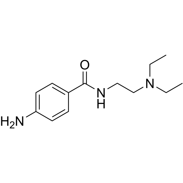
Procainamide (Procaine amide) is a specific and potent inhibitor of DNA methyltransferase 1 (DNMT1), which reactivates the expression of tumor suppressor factors by demethylating tumor suppressor genes. Procainamide induces vacuolization in various cell types and reduces cell proliferation and migration. Procainamide relaxes airway smooth muscle by activating potassium channels. Procainamide can be used in cancer and arrhythmia research.
For research use only. We do not sell to patients.
- Purity : 99.77%
- CAS No.: 51-06-9
- Formula: C13H21N3O
- Molecular Weight:235.33
-
Storage:
4°C, protect from light
* In solvent : -80°C, 6 months; -20°C, 1 month (protect from light)

To place orders, for customer services and technical support, please contact: MedChemExpress USA
Tel: 609-228-6898 E-mail: [email protected] [email protected]


-
 Biological Activity
-
Chemical Information
-
Solvent & Solubility
- Protocols
- Purity & Documentation
- References
-
 Help & FAQs


-
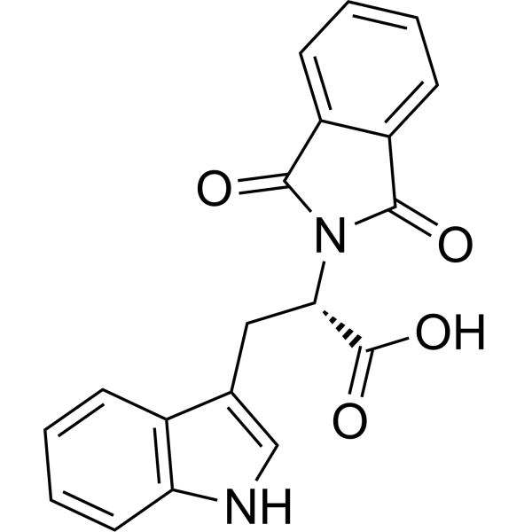
HY-13642
RG108DNMT Inhibitor -
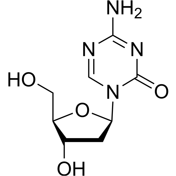
HY-A0004
DecitabineDNMT Inhibitor -
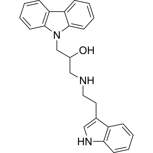
HY-12746
DC-05DNA Methyltransferase Inhibitor -
HY-15229
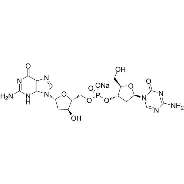
Guadecitabine sodiumDNMT Inhibitor -
HY-12747
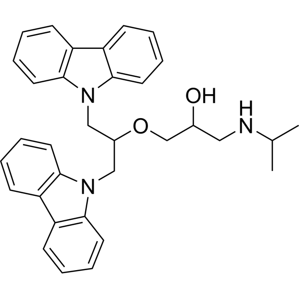
DC_517DNA Methyltransferase Inhibitor -
HY-10586
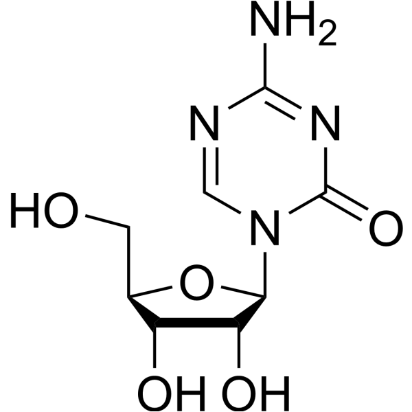
5-AzacytidineDNMT Inhibitor -
HY-135146
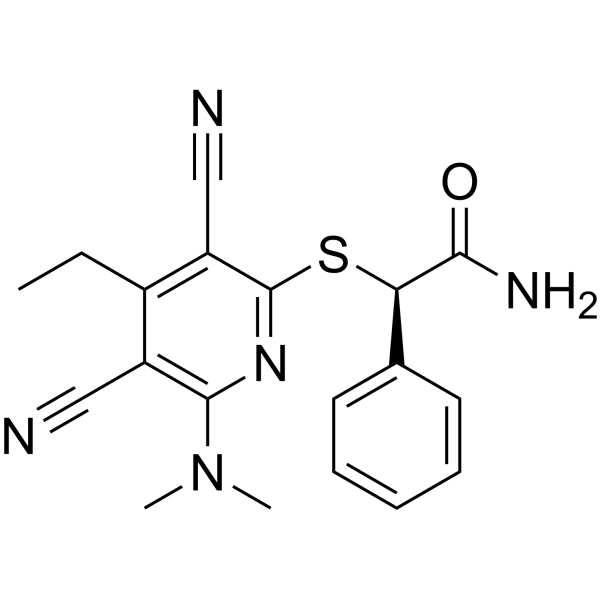
GSK-3484862DNMT1 Inhibitor -
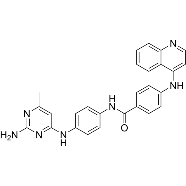
HY-13962
SGI-1027Apoptosis Inducer -
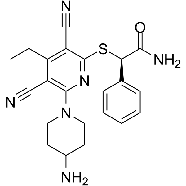
HY-139664A
(R)-GSK-3685032DNMT1 Inhibitor -
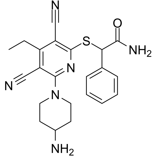
HY-139664
GSK-3685032DNMT1 Inhibitor -

HY-B2194
γ-OryzanolDNA Methyltransferase Inhibitor -
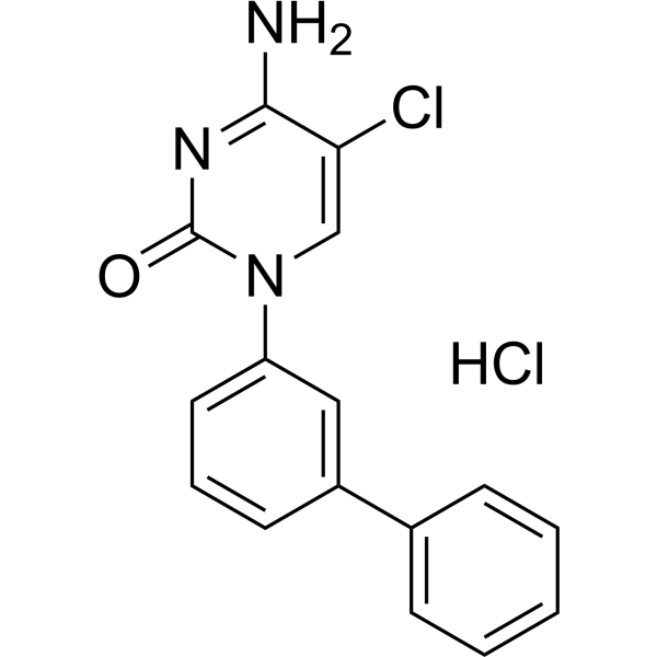
HY-111558A
Bobcat339 hydrochlorideTET Inhibitor
Publications Citing Use of MedChemExpress (MCE) Procainamide
More- Adv Sci (Weinh). 2025 Jan;12(4):e2410360. [Abstract]
- Clin Chem. 2019 Dec;65(12):1522-1531. [Abstract]
- Free Radic Biol Med. 2024 Sep:222:288-303. [Abstract]
- Environ Pollut. 2026 Feb 1:390:127432. [Abstract]
- Environ Pollut. 2023 Sep 1:332:121931. [Abstract]
- Drug Metab Dispos. 2026 Jun;54(6):100324. [Abstract]
- Patent. US20180263995A1.
All DNA Methyltransferase Isoforms
More

Biological Activity
Description
IC50 & Target
DNMT1[1]
Cellular Effect
|
Cell Line

|
Type | Value | Description | References |
|---|---|---|---|---|
| CHO | IC50 |
389.5 μM
Compound: procainamide
|
Inhibition of Cav1.2 current measured using QPatch automatic path clamp system in CHO cells expressing Cav1.2, beta-2 and alpha-2/delta-1 subunits
Inhibition of Cav1.2 current measured using QPatch automatic path clamp system in CHO cells expressing Cav1.2, beta-2 and alpha-2/delta-1 subunits
|
[PMID: 23812503] |
| HEK293 | IC50 |
12 μM
Compound: Procainamide
|
TP_TRANSPORTER: inhibition of TEA uptake (in the absence of bicarbonate) (TEA: 20 uM) in OCT1-expressing HEK293 cells
TP_TRANSPORTER: inhibition of TEA uptake (in the absence of bicarbonate) (TEA: 20 uM) in OCT1-expressing HEK293 cells
|
[PMID: 12438515] |
| HEK293 | IC50 |
215 μM
Compound: Procainamide
|
TP_TRANSPORTER: inhibition of TEA uptake (in the absence of bicarbonate) (TEA: 20 uM) in OCT2-expressing HEK293 cells
TP_TRANSPORTER: inhibition of TEA uptake (in the absence of bicarbonate) (TEA: 20 uM) in OCT2-expressing HEK293 cells
|
[PMID: 12438515] |
| HEK293 | IC50 |
7 μM
Compound: Procainamide
|
TP_TRANSPORTER: inhibition of TEA uptake (in the presence of bicarbonate) (TEA: 20 uM) in OCT1-expressing HEK293 cells
TP_TRANSPORTER: inhibition of TEA uptake (in the presence of bicarbonate) (TEA: 20 uM) in OCT1-expressing HEK293 cells
|
[PMID: 12438515] |
| HEK293 | IC50 |
90 μM
Compound: Procainamide
|
TP_TRANSPORTER: inhibition of TEA uptake (in the presence of bicarbonate) (TEA: 20 uM) in OCT2-expressing HEK293 cells
TP_TRANSPORTER: inhibition of TEA uptake (in the presence of bicarbonate) (TEA: 20 uM) in OCT2-expressing HEK293 cells
|
[PMID: 12438515] |
| HRPE | IC50 |
1 mM
Compound: Procainamide
|
TP_TRANSPORTER: inhibition of TEA uptake in Octn1-HRPE cells
TP_TRANSPORTER: inhibition of TEA uptake in Octn1-HRPE cells
|
[PMID: 10825452] |
| Oocyte | IC50 |
445 μM
Compound: Procainamide
|
TP_TRANSPORTER: inhibition of TEA uptake (TEA: 10 uM) in Xenopus laevis oocytes
TP_TRANSPORTER: inhibition of TEA uptake (TEA: 10 uM) in Xenopus laevis oocytes
|
[PMID: 11502595] |
| Oocyte | IC50 |
167 μM
Compound: Procainamide
|
TP_TRANSPORTER: inhibition pramipexole uptake in rOCT2-injected oocytes
TP_TRANSPORTER: inhibition pramipexole uptake in rOCT2-injected oocytes
|
[PMID: 15640376] |
| Oocyte | IC50 |
7.7 μM
Compound: Procainamide
|
TP_TRANSPORTER: inhibition pramipexole uptake in rOCT1-injected oocytes
TP_TRANSPORTER: inhibition pramipexole uptake in rOCT1-injected oocytes
|
[PMID: 15640376] |
| Sf9 | IC50 |
>300 μM
Compound: 8
|
Inhibition of His6-tagged human recombinant DNMT3A/DNMT3L expressed in insect Sf9 cells assessed as reduction in DNA methyltransferase activity using 5'-biotinylated 45-bp unmethylated or hemimethylated oligonucleotide substrates and [3H]-AdoMet by liquid
Inhibition of His6-tagged human recombinant DNMT3A/DNMT3L expressed in insect Sf9 cells assessed as reduction in DNA methyltransferase activity using 5'-biotinylated 45-bp unmethylated or hemimethylated oligonucleotide substrates and [3H]-AdoMet by liquid
|
[PMID: 25406944] |
| Sf9 | IC50 |
>500 μM
Compound: 8
|
Inhibition of His6-tagged human recombinant DNMT1 expressed in insect Sf9 cells assessed as reduction in DNA methyltransferase activity using 5'-biotinylated 45-bp unmethylated or hemimethylated oligonucleotide substrates and [3H]-AdoMet by liquid scintil
Inhibition of His6-tagged human recombinant DNMT1 expressed in insect Sf9 cells assessed as reduction in DNA methyltransferase activity using 5'-biotinylated 45-bp unmethylated or hemimethylated oligonucleotide substrates and [3H]-AdoMet by liquid scintil
|
[PMID: 25406944] |
In Vitro
Procainamide has a Ki of 7.2 μM for DNMT1 inhibition on hemimethylated oligonucleotide substrates and a Ki of 4600 μM for DNMT1 inhibition on unmethylated oligonucleotide substrates[1]. Procainamide (0.5 mM; 96 h) causes loss of CpG methylation in HCT116 cells and affects centromeric repeat sequences and single-copy genes[1]. Procainamide (0-10 mM) concentration-dependently relaxes bovine tracheal contractions induced by methacholine (0.3 μM) but does not significantly relax contractions induced by potassium ions (40 mM)[4]. Procainamide (250 μM-2.5 mM; 0-48 h) does not induce cellular vacuolization at a dose of 250 μM, but induces cellular vacuolization in many cells at a dose of 2.5 mM[5]. Procainamide (10 μM; 5 d) demethylates ER, p16, and RAR genes in MDA-231, T24, and MCF-7 cell lines[6].
MedChemExpress (MCE) has not independently confirmed the accuracy of these methods. They are for reference only.
In Vivo
MedChemExpress (MCE) has not independently confirmed the accuracy of these methods. They are for reference only.
- RNA extraction experimental
- BrdU Incorporation Assay
- Cell migration
- Somatic Cell Culture
- CFSE Dye Dilution Proliferation Assay
- Dye-dilution cell tracking and proliferation staining
- CCK-8/WST-8 Cell Proliferation Assay
- Protocol for Cell Cycle
- Cell Counting-Based Growth Curve Assay
- Colony Formation (Clonogenic) Assay
- Cell Viability Determination by MTT Colorimetric Assay
- EdU Incorporation Assay (Click Chemistry-Based DNA Synthesis Measurement)
- Ki-67 Immunostaining Proliferation Assay
- PCNA Immunodetection Proliferation Assay
- Cardiac voltage-sensitive optical mapping
- Protocol for Cell Counting and Cell Density Analysis
- MTT Cell Proliferation Assay
Clinical Trial
| NCT Number | Sponsor | Condition | Start Date |
Phase
|
|---|---|---|---|---|
| NCT01329991 | 2011-05 | PHASE1 |


Chemical Information
-
CAS No. 51-06-9
-
Appearance <47°C Solid,>47°C Liquid
-
Molecular Weight 235.33
-
Formula C13H21N3O
-
Color White to light yellow
-
SMILES
O=C(NCCN(CC)CC)C1=CC=C(N)C=C1
-
Synonyms
Procaine amide; SP 100
-
Shipping
Room temperature in continental US; may vary elsewhere.
-
Storage
4°C, protect from light
* In solvent : -80°C, 6 months; -20°C, 1 month (protect from light)


Publications (7)
-
Journal Impact Factor
-
Most Recent
-
Adv Sci (Weinh)
Targeted SPP1 Inhibition of Tumor-Associated Myeloid Cells Effectively Decreases Tumor Sizes. [Abstract]2025 Jan;12(4):e2410360. PMID: 39639496 -
Clin Chem
Discovering Cross-Reactivity in Urine Drug Screening Immunoassays through Large-Scale Analysis of Electronic Health Records. [Abstract]2019 Dec;65(12):1522-1531. PMID: 31578215 -
Free Radic Biol Med
Cannabidiol mitigates radiation-induced intestine ferroptosis via facilitating the heterodimerization of RUNX3 with CBFβ thereby promoting transactivation of GPX4. [Abstract]2024 Sep:222:288-303. PMID: 38830513 -
Environ Pollut
Investigating the role and mechanism of methionine in different types of skeletal fluorosis based on Siglec-15 methylation. [Abstract]2026 Feb 1:390:127432. PMID: 41349947 -
Environ Pollut
Screening of differentially methylated genes in skeletal fluorosis of rats with different types and involvement of aberrant methylation of Cthrc1. [Abstract]2023 Sep 1:332:121931. PMID: 37268221 -
Drug Metab Dispos
An in vitro approach to predict idiosyncratic drug-induced agranulocytosis using myeloperoxidase-derived reactive metabolites. [Abstract]2026 Jun;54(6):100324. PMID: 42248123 -


Solvent & Solubility
In Vitro:
DMSO : 50 mg/mL (212.47 mM; Need ultrasonic; Hygroscopic DMSO has a significant impact on the solubility of product, please use newly opened DMSO)
Please refer to the solubility information to select the appropriate solvent. Once prepared, please aliquot and store the solution to prevent product inactivation from repeated freeze-thaw cycles.
Storage method and period of stock solution: -80°C, 6 months; -20°C, 1 month (protect from light). When stored at -80°C, please use it within 6 months. When stored at -20°C, please use it within 1 month.
Please refer to the solubility information to select the appropriate solvent. Once prepared, please aliquot and store the solution to prevent product inactivation from repeated freeze-thaw cycles.
Storage method and period of stock solution: -80°C, 6 months; -20°C, 1 month (protect from light). When stored at -80°C, please use it within 6 months. When stored at -20°C, please use it within 1 month.
Concentration (start) × Volume (start) = Concentration (final) × Volume (final)
In Vivo:
Select the appropriate dissolution method based on your experimental animal and administration route.
- For the following dissolution methods, please ensure to first prepare a clear stock solution using an In Vitro approach and then sequentially add co-solvents:
- To ensure reliable experimental results, the clarified stock solution can be appropriately stored based on storage conditions. As for the working solution for In Vivo experiments, it is recommended to prepare freshly and use it on the same day.
- The percentages shown for the solvents indicate their volumetric ratio in the final prepared solution. If precipitation or phase separation occurs during preparation, heat and/or sonication can be used to aid dissolution.
Add each solvent one by one: 10% DMSO 40% PEG300 5% Tween-80 45% Saline
Solubility: ≥ 1.25 mg/mL (5.31 mM); Clear solution
This protocol yields a clear solution of ≥ 1.25 mg/mL (saturation unknown).
Taking 1 mL working solution as an example, add 100 μL DMSO stock solution (12.5 mg/mL) to 400 μL PEG300, and mix evenly; then add 50 μL Tween-80 and mix evenly; then add 450 μL Saline to adjust the volume to 1 mL.
Preparation of Saline: Dissolve 0.9 g sodium chloride in ddH₂O and dilute to 100 mL to obtain a clear Saline solution.
Add each solvent one by one: 10% DMSO 90% (20% SBE-β-CD in Saline)
Solubility: ≥ 1.25 mg/mL (5.31 mM); Clear solution
This protocol yields a clear solution of ≥ 1.25 mg/mL (saturation unknown).
Taking 1 mL working solution as an example, add 100 μL DMSO stock solution (12.5 mg/mL) to 900 μL 20% SBE-β-CD in Saline, and mix evenly.
Preparation of 20% SBE-β-CD in Saline (4°C, storage for one week): 2 g SBE-β-CD powder is dissolved in 10 mL Saline, completely dissolve until clear.
In Vivo Dissolution Calculator
Please enter the basic information of animal experiments:
-
-
-
-
Recommended: Prepare an additional quantity of animals to account for potential losses during experiments.
Please enter your animal formula composition:
-
%DMSO +
Recommended: Keep the proportion of DMSO in working solution below 2% if your animal is weak.
-
%+
-
+%Tween-80 + +
-
%Saline +
The co-solvents required include: DMSO, . All of co-solvents are available by MedChemExpress (MCE). , Tween 80. All of co-solvents are available by MedChemExpress (MCE).
Working solution concentration: 0.22 mg/mL
Method for preparing stock solution: mg drug dissolved in μL DMSO. Stock solution concentration: mg/mL. * In solvent : -80°C, 6 months; -20°C, 1 month (protect from light)
1. Take μL DMSO stock solution;
2. Add μL .
μL , mix evenly;
3. Then add μL Tween 80, mix evenly;
4. Then add μL
Please ensure that the stock solution in the first step is dissolved to a clear state, and add co-solvents in sequence. You can use ultrasonic heating (ultrasonic cleaner, recommended frequency 20-40 kHz), vortexing, etc. to assist dissolution.


Purity & Documentation
-
Data Sheet (273 KB)
-
SDS (481 KB)
- English - EN (481 KB)
- Français - FR (481 KB)
- Deutsch - DE (481 KB)
- Norwegian - NO (481 KB)
- Español - ES (481 KB)
- Swedish - SV (481 KB)
- Italian - IT (481 KB)
- Korean - KR (481 KB)
- Portuguese - PT (481 KB)
-
Handling Instructions (2659 KB)
References
[1]. Lee BH, et al. Procainamide is a specific inhibitor of DNA methyltransferase 1. J Biol Chem. 2005;280(49):40749-40756. [Content Brief]
[3]. Gardner I, et al. A comparison of the covalent binding of clozapine, procainamide, and vesnarinone to human neutrophils in vitro and rat tissues in vitro and in vivo[J]. Chemical research in toxicology, 2005, 18(9): 1384-1394. [Content Brief]
[4]. Nakahara T, et al. Involvement of K+ channel in procainamide-induced relaxation of bovine tracheal smooth muscle[J]. European journal of pharmacology, 2000, 402(1-2): 143-149. [Content Brief]
[5]. Morissette G, et al. Massive cell vacuolization induced by organic amines such as procainamide[J]. Journal of Pharmacology and Experimental Therapeutics, 2004, 310(1): 395-406. [Content Brief]
[6]. Segura-Pacheco B, et al. Reactivation of tumor suppressor genes by the cardiovascular drugs hydralazine and procainamide and their potential use in cancer therapy[J]. Clinical Cancer Research, 2003, 9(5): 1596-1603. [Content Brief]


Complete Stock Solution Preparation Table
Please refer to the solubility information to select the appropriate solvent. Once prepared, please aliquot and store the solution to prevent product inactivation from repeated freeze-thaw cycles.
Storage method and period of stock solution: -80°C, 6 months; -20°C, 1 month (protect from light). When stored at -80°C, please use it within 6 months. When stored at -20°C, please use it within 1 month.
| Optional Solvent | Concentration Solvent Mass | 1 mg | 5 mg | 10 mg | 25 mg |
|---|---|---|---|---|---|
| DMSO | 1 mM | 4.2494 mL | 21.2468 mL | 42.4935 mL | 106.2338 mL |
| 5 mM | 0.8499 mL | 4.2494 mL | 8.4987 mL | 21.2468 mL | |
| 10 mM | 0.4249 mL | 2.1247 mL | 4.2494 mL | 10.6234 mL | |
| 15 mM | 0.2833 mL | 1.4165 mL | 2.8329 mL | 7.0823 mL | |
| 20 mM | 0.2125 mL | 1.0623 mL | 2.1247 mL | 5.3117 mL | |
| 25 mM | 0.1700 mL | 0.8499 mL | 1.6997 mL | 4.2494 mL | |
| 30 mM | 0.1416 mL | 0.7082 mL | 1.4165 mL | 3.5411 mL | |
| 40 mM | 0.1062 mL | 0.5312 mL | 1.0623 mL | 2.6558 mL | |
| 50 mM | 0.0850 mL | 0.4249 mL | 0.8499 mL | 2.1247 mL | |
| 60 mM | 0.0708 mL | 0.3541 mL | 0.7082 mL | 1.7706 mL | |
| 80 mM | 0.0531 mL | 0.2656 mL | 0.5312 mL | 1.3279 mL | |
| 100 mM | 0.0425 mL | 0.2125 mL | 0.4249 mL | 1.0623 mL |


Procainamide Related Classifications
HY-A0084A Related Classifications
 Powered by Bioz