



Teclistamab
Based on 1 publication(s) in Google Scholar
Teclistamab is a human bispecific antibody to BCMA and CD3 that recognizes BCMA on target cells and CD3 on T cells and induces T cell-mediated cytotoxicity leading to T cell activation and subsequent target cell lysis. Teclistamab can be used in studies of diseases related to multiple myeloma (MM).
For research use only. We do not sell to patients.
- Purity : 99.87%
- CAS No.: 2119595-80-9
- Molecular Weight:143.64 kDa
-
Storage:
Please store the product under the recommended conditions in the Certificate of Analysis.


To place orders, for customer services and technical support, please contact: MedChemExpress USA
Tel: 609-228-6898 E-mail: [email protected] [email protected]


-
 Biological Activity
-
Chemical Information
- Protocols
- Purity & Documentation
- References
-
 Help & FAQs


-

HY-P99394
TalquetamabAnti-GPRC5D/CD3 Antibody -

HY-P99909
ElranatamabAnti-CD3E/TNFRSF17 Antibody -

HY-P99024
GlofitamabT-Cell-Engaging Bispecific Antibody -

HY-P99033
MosunetuzumabCD20/CD3 Inhibitor -

HY-P99931
EpcoritamabAnti-CD3/CD20 Antibody -

HY-P9963
BlinatumomabAntineoplastic Agent -

HY-P99802
PasotuxizumabAnti-PSMA/CD3 mAb -
HY-P99601
CevostamabAnti-FcRH5/CD3 TDB -
HY-P99575
Tarlatamabanti-DLL3/CD3 BiTE antibody -

HY-P99592
SolitomabAnti-CD3/EpCAM Antibody -
HY-P9954
CatumaxomabAntibody Against CD3/EpCAM -

HY-P9989
LinvoseltamabBCMA/CD3 Antibody
Publications Citing Use of MedChemExpress (MCE) Teclistamab
More

Biological Activity
Description
Isotype
Human IgG4 lambda
Recommend Isotype Controls
Species Reactivity
Human
IC50 & Target
CD3 & BCMA
In Vitro
Teclistamab (0.01-1000 nM, 48 h) induces T cell-mediated cytotoxicity with the EC50 values of 0.15 nM in H929 cells, 0.06 nM in MM.1R cell lines, and 0.45 nM in RPMI 8226 cell lines, respectively[1].
Teclistamab (0.01-1000 nM, 48 h) can bind CD138 + human BM monocytes in a dose-dependent manner and induces cytotoxicity of human BM monocytes[1].
MedChemExpress (MCE) has not independently confirmed the accuracy of these methods. They are for reference only.
In Vivo
MedChemExpress (MCE) has not independently confirmed the accuracy of these methods. They are for reference only.
-
Animal Model:Female NSG mice infected with H929 cell lines[1]
-
Dosage:0.1 µg (0.005 mg/kg), 0.5 µg (0.025 mg/kg), or 1 µg (0.05 mg/kg)
-
Administration:i.v., on days 0, 3, 5, 7, and 10
-
Result:Completely inhibited tumor growth at doses of 0.5 and 1 μg, while no inhibition of tumor growth was observed at a dose of 0.1 μg.
Clinical Trial
| NCT Number | Sponsor | Condition | Start Date |
Phase

|
|---|---|---|---|---|
| NCT01329991 | Plexxikon| | 2011-05 | PHASE1 |
Gene ID
Accession
Conjugated
Unconjugated
Reconsititution
The product can be reconstituted/diluted with sterile PBS or saline.
Format
-
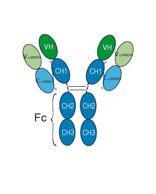
Human IgG4 lambda
Application
ELISA, FACS, Functional assay
Verified Bioactivity
-
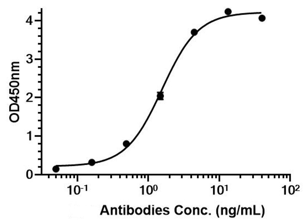 Immobilized Human CD3 epsilon/CD3e Protein (ECD, His Tag) can bind Teclistamab. The EC50 for this effect is 1.56 ng/mL.
 -
 Immobilized Human BCMA Protein (ECD, His Tag) can bind Teclistamab. The EC50 for this effect is 0.18 ng/mL.
 -
 Flow Cytometry analysis of K562 cells labelling BCMA (red) with Teclistamab (HY-P99392). Cells were fixed with 4% paraformaldehyde and permeabilised with 90% methanol. Then cells were stained with the primary antibody at 1/200 dilution for an hour at 4℃. Goat Anti-Human IgG H&L (AF488) (HY-P83776) was used as the secondary antibody at 1/1,000 dilution for 30 minutes at 4℃. Human IgG4 lambda (HY-P99987, blue) was used as the isotype control, cells without incubation with primary antibody were used as the unlabeled control (black).


Chemical Information
-
CAS No. 2119595-80-9
-
Appearance Liquid
-
Molecular Weight 143.64 kDa
-
Color Colorless to light yellow
-
SMILES
[Teclistamab]
-
Synonyms
JNJ-7957; JNJ-64007957; Tecvayli
-
Shipping
Shipping with dry ice.
-
Formulation
Please refer to the lot-specific COA for specific buffer information.
-
Storage
Please store the product under the recommended conditions in the Certificate of Analysis.


Publications (1)
-
Journal Impact Factor
-
Most Recent


Purity & Documentation
-
Data Sheet (259 KB)
-
SDS (252 KB)
- English - EN (252 KB)
- Français - FR (252 KB)
- Deutsch - DE (252 KB)
- Norwegian - NO (252 KB)
- Español - ES (252 KB)
- Swedish - SV (252 KB)
- Italian - IT (252 KB)
- Korean - KR (252 KB)
- Portuguese - PT (252 KB)
-
Inhibitory Antibodies User Guide (603 KB)
References

Calculators
Concentration (start) × Volume (start) = Concentration (final) × Volume (final)


Teclistamab Related Classifications
HY-P99392 Related Classifications
 Powered by Bioz