



Zorifertinib
Based on 7 publication(s) in Google Scholar
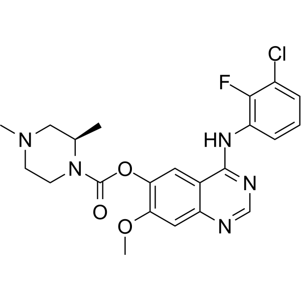
Zorifertinib (AZD3759) is a potent, orally active, BBB-penetrant, EGFR inhibitor. At Km ATP concentrations, the IC50s are 0.3, 0.2, and 0.2 nM for EGFRwt, EGFRL858R, and EGFRexon 19Del, respectively. Zorifertinib induces cancer cell apoptosis. Zorifertinib has antitumor activity, and can be used for NSCLC, HCC etc. research.
For research use only. We do not sell to patients.
- Purity : 99.37%
- CAS No.: 1626387-80-1
- Formula: C22H23ClFN5O3
- Molecular Weight:459.90
-
Storage:Powder -20°C, 3 years , 4°C, 2 years ; In solvent -80°C, 2 years , -20°C, 1 year

To place orders, for customer services and technical support, please contact: MedChemExpress USA
Tel: 609-228-6898 E-mail: [email protected] [email protected]


-
 Biological Activity
-
Chemical Information
-
Solvent & Solubility
- Protocols
- Purity & Documentation
- References
-
 Help & FAQs


-
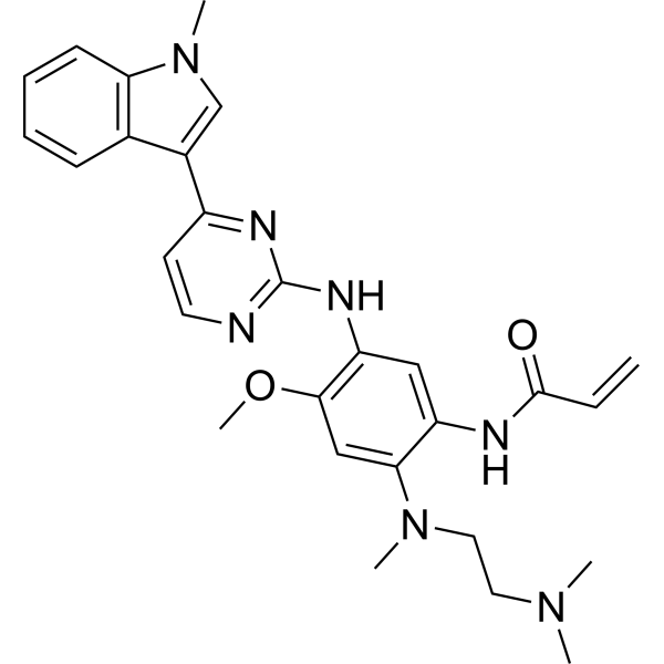
HY-15772
OsimertinibMutant-Selective EGFR Inhibitor -
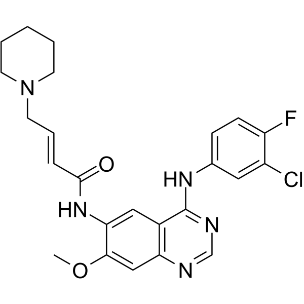
HY-13272
DacomitinibErbB Inhibitor -
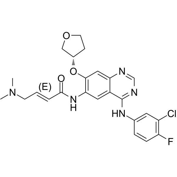
HY-10261
AfatinibEGFR/HER2 Inhibitor -
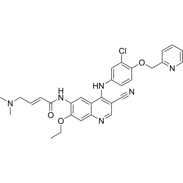
HY-32721
NeratinibHER2/EGFR Inhibitor -
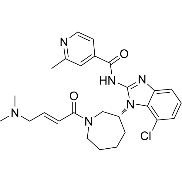
HY-12872
NazartinibEGFR Inhibitor -
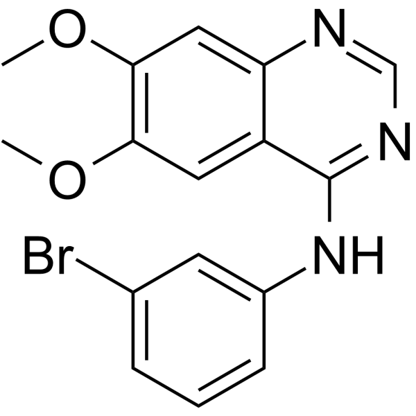
HY-14346
PD153035EGFR Inhibitor -
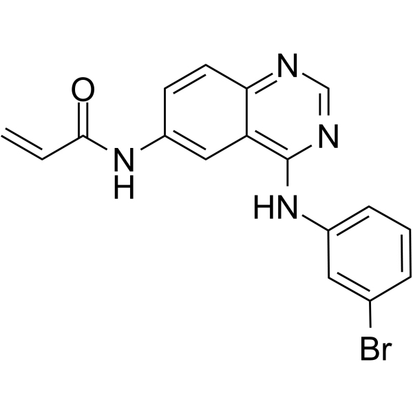
HY-13896
PD168393EGFR Tyrosine Kinase Inhibitor -
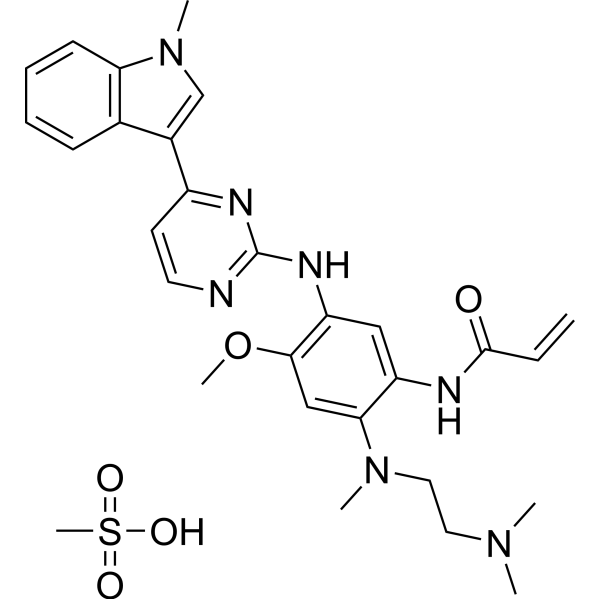
HY-15772A
Osimertinib mesylateMutant-Selective EGFR Inhibitor -
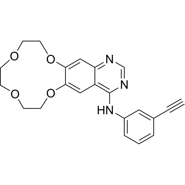
HY-15164A
IcotinibEGFR Inhibitor -
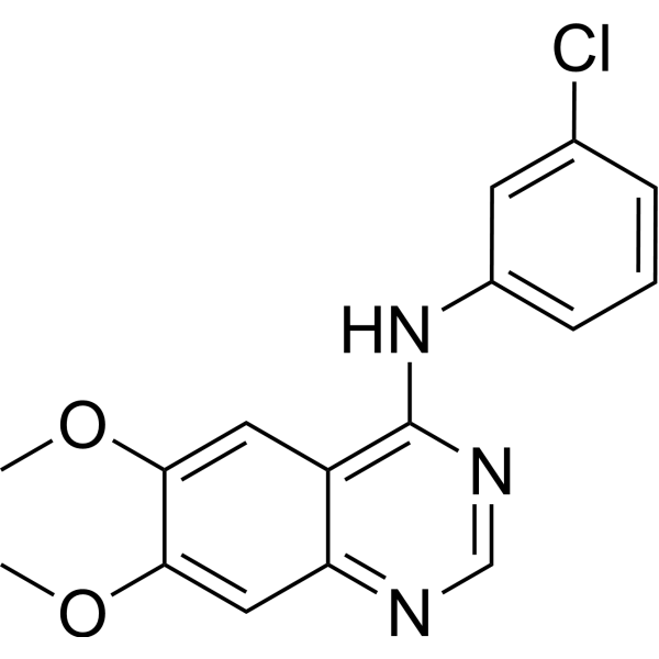
HY-13524
AG-1478EGFR Inhibitor -
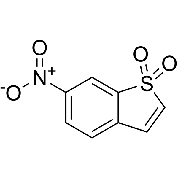
HY-13818
StatticSTAT3 Inhibitor -

HY-15772R
Osimertinib (Standard)Mutant-Selective EGFR Inhibitor
Publications Citing Use of MedChemExpress (MCE) Zorifertinib
More- Nat Commun. 2025 Dec 17;16(1):11181. [Abstract]
- Sci Adv. Sci Adv. 2024 Jun 21;10(25):eadk2299. d [Abstract]
- RSC Adv. 2022 Jul 21;12(32):20991-21003. [Abstract]
- J Neurochem. 2024 Mar;168(3):205-223. [Abstract]
- J Pharm Biomed Anal. 2022 Mar 20:211:114626. [Abstract]
- bioRxiv. 2023 Jun 9:2023.06.07.544128. [Abstract]
- Patent. US20220177473A1.
All EGFR Isoforms
More

Biological Activity
Description
IC50 & Target
[1]|
EGFR 0.3 nM (IC50) |
EGFRL858R 0.2 nM (IC50) |
EGFRExon 19 deletion 0.2 nM (IC50) |
Cellular Effect
|
Cell Line

|
Type | Value | Description | References |
|---|---|---|---|---|
| A-431 | IC50 |
3.5 μM
Compound: AZD3759
|
Antiproliferative activity against human A431 cells after 72 hrs by MTT assay
Antiproliferative activity against human A431 cells after 72 hrs by MTT assay
|
[PMID: 30195240] |
| A549 | IC50 |
28.67 μM
Compound: 2; AZD3759
|
Cytotoxicity against human A549 cells after 72 hrs by MTT assay
Cytotoxicity against human A549 cells after 72 hrs by MTT assay
|
[PMID: 30655941] |
| BEAS-2B | IC50 |
18.66 μM
Compound: AZD3759
|
Cytotoxicity against human BEAS2B cells after 72 hrs by MTT assay
Cytotoxicity against human BEAS2B cells after 72 hrs by MTT assay
|
[PMID: 30195240] |
| HCC827 | IC50 |
0.03 μM
Compound: AZD3759
|
Antiproliferative activity against human HCC827 cells after 72 hrs by MTT assay
Antiproliferative activity against human HCC827 cells after 72 hrs by MTT assay
|
[PMID: 30195240] |
| NCI-H1975 | IC50 |
24.16 μM
Compound: AZD3759
|
Antiproliferative activity against human NCI-H1975 cells after 72 hrs by MTT assay
Antiproliferative activity against human NCI-H1975 cells after 72 hrs by MTT assay
|
[PMID: 30195240] |
| NCI-H3255 | IC50 |
0.09 μM
Compound: AZD3759
|
Antiproliferative activity against human H3255 cells after 72 hrs by MTT assay
Antiproliferative activity against human H3255 cells after 72 hrs by MTT assay
|
[PMID: 30195240] |
| NCI-H3255 | IC50 |
7 nM
Compound: 1m, AZD3759
|
Antiproliferative against human H3255 cells expressing EGFR L858R mutant after 72 hrs by MTS assay
Antiproliferative against human H3255 cells expressing EGFR L858R mutant after 72 hrs by MTS assay
|
[PMID: 26313252] |
| NCI-H838 | IC50 |
21556 nM
Compound: 1m, AZD3759
|
Antiproliferative against human NCI-H838 cells expressing EGFR wild-type after 72 hrs by MTS assay
Antiproliferative against human NCI-H838 cells expressing EGFR wild-type after 72 hrs by MTS assay
|
[PMID: 26313252] |
| PC-9 | IC50 |
0.05 μM
Compound: AZD3759
|
Antiproliferative activity against human PC9 cells after 72 hrs by MTT assay
Antiproliferative activity against human PC9 cells after 72 hrs by MTT assay
|
[PMID: 30195240] |
| PC-9 | IC50 |
7.4 nM
Compound: 1m, AZD3759
|
Inhibition of EGFR exon19 deletion mutant phosphorylation in human PC9 cells
Inhibition of EGFR exon19 deletion mutant phosphorylation in human PC9 cells
|
[PMID: 26313252] |
| PC-9 | IC50 |
7.7 nM
Compound: 1m, AZD3759
|
Antiproliferative against human PC9 cells expressing EGFR exon19 deletion mutant after 72 hrs by MTS assay
Antiproliferative against human PC9 cells expressing EGFR exon19 deletion mutant after 72 hrs by MTS assay
|
[PMID: 26313252] |
In Vitro
At 2 mM of ATP concentrations, the IC50s are 102, 7.6, and 2.4 nM for EGFRwt, EGFRL858R, and EGFRexon 19Del, respectively. Zorifertinib (AZD3759) also inhibits pEGFR in H838wt, H3255L858R, and PC-9exon 19Del with IC50 of 64.5, 7.2, and 7.4 nM, respectively. In cellular phosphorylation studies, Zorifertinib also demonstrates 9-fold inhibition selectivity in EGFR-activating mutant cell lines over EGFR wild-type cell lines (H838 cell line)[1].
MedChemExpress (MCE) has not independently confirmed the accuracy of these methods. They are for reference only. Further protocols information, click here.
In Vivo
MedChemExpress (MCE) has not independently confirmed the accuracy of these methods. They are for reference only.
Clinical Trial
| NCT Number | Sponsor | Condition | Start Date |
Phase
|
|---|---|---|---|---|
| NCT01329991 | Plexxikon| | 2011-05 | PHASE1 |


Chemical Information
-
CAS No. 1626387-80-1
-
Appearance Solid
-
Molecular Weight 459.90
-
Formula C22H23ClFN5O3
-
Color White to off-white
-
SMILES
ClC1=CC=CC(NC2=NC=NC3=CC(OC)=C(OC(N4[C@H](C)CN(C)CC4)=O)C=C23)=C1F
-
Synonyms
AZD3759
-
Shipping
Room temperature in continental US; may vary elsewhere.
-
Storage
Powder -20°C 3 years 4°C 2 years In solvent -80°C 2 years -20°C 1 year


Publications (7)
-
Journal Impact Factor
-
Most Recent
-
Nat Commun
Supporting cells orchestrate noise-induced hearing loss via a Gasdermin D-dependent signaling loop with hair cells. [Abstract]2025 Dec 17;16(1):11181. PMID: 41407982 -
Sci Adv
In silico transcriptome screens identify epidermal growth factor receptor inhibitors as therapeutics for noise-induced hearing loss. [Abstract]Sci Adv. 2024 Jun 21;10(25):eadk2299. d PMID: 38896614 -
RSC Adv
Profiling of in vivo, in vitro and reactive zorifertinib metabolites using liquid chromatography ion trap mass spectrometry. [Abstract]2022 Jul 21;12(32):20991-21003. PMID: 35919181 -
J Neurochem
Molecular prognostic of nine parthanatos death-related genes in glioma, particularly in COL8A1 identification. [Abstract]2024 Mar;168(3):205-223. PMID: 38225203 -
J Pharm Biomed Anal
2022 Mar 20:211:114626. PMID: 35123331 -
bioRxiv
In Silico Transcriptome-based Screens Identify Epidermal Growth Factor Receptor Inhibitors as Therapeutics for Noise-induced Hearing Loss. [Abstract]2023 Jun 9:2023.06.07.544128. PMID: 37333346 -


Solvent & Solubility
In Vitro:
DMSO : ≥ 50 mg/mL (108.72 mM; Hygroscopic DMSO has a significant impact on the solubility of product, please use newly opened DMSO)
* "≥" means soluble, but saturation unknown.
Please refer to the solubility information to select the appropriate solvent. Once prepared, please aliquot and store the solution to prevent product inactivation from repeated freeze-thaw cycles.
Storage method and period of stock solution: -80°C, 2 years; -20°C, 1 year. When stored at -80°C, please use it within 2 years. When stored at -20°C, please use it within 1 year.
Please refer to the solubility information to select the appropriate solvent. Once prepared, please aliquot and store the solution to prevent product inactivation from repeated freeze-thaw cycles.
Storage method and period of stock solution: -80°C, 2 years; -20°C, 1 year. When stored at -80°C, please use it within 2 years. When stored at -20°C, please use it within 1 year.
Concentration (start) × Volume (start) = Concentration (final) × Volume (final)
In Vivo:
Select the appropriate dissolution method based on your experimental animal and administration route.
- For the following dissolution methods, please ensure to first prepare a clear stock solution using an In Vitro approach and then sequentially add co-solvents:
- To ensure reliable experimental results, the clarified stock solution can be appropriately stored based on storage conditions. As for the working solution for In Vivo experiments, it is recommended to prepare freshly and use it on the same day.
- The percentages shown for the solvents indicate their volumetric ratio in the final prepared solution. If precipitation or phase separation occurs during preparation, heat and/or sonication can be used to aid dissolution.
Add each solvent one by one: 10% DMSO 40% PEG300 5% Tween-80 45% Saline
Solubility: ≥ 2.5 mg/mL (5.44 mM); Clear solution
This protocol yields a clear solution of ≥ 2.5 mg/mL (saturation unknown).
Taking 1 mL working solution as an example, add 100 μL DMSO stock solution (25.0 mg/mL) to 400 μL PEG300, and mix evenly; then add 50 μL Tween-80 and mix evenly; then add 450 μL Saline to adjust the volume to 1 mL.
Preparation of Saline: Dissolve 0.9 g sodium chloride in ddH₂O and dilute to 100 mL to obtain a clear Saline solution.
Add each solvent one by one: 10% DMSO 90% (20% SBE-β-CD in Saline)
Solubility: 2.5 mg/mL (5.44 mM); Suspended solution; Need ultrasonic
This protocol yields a suspended solution of 2.5 mg/mL. Suspended solution can be used for oral and intraperitoneal injection.
Taking 1 mL working solution as an example, add 100 μL DMSO stock solution (25.0 mg/mL) to 900 μL 20% SBE-β-CD in Saline, and mix evenly.
Preparation of 20% SBE-β-CD in Saline (4°C, storage for one week): 2 g SBE-β-CD powder is dissolved in 10 mL Saline, completely dissolve until clear.
In Vivo Dissolution Calculator
Please enter the basic information of animal experiments:
-
-
-
-
Recommended: Prepare an additional quantity of animals to account for potential losses during experiments.
Please enter your animal formula composition:
-
%DMSO +
Recommended: Keep the proportion of DMSO in working solution below 2% if your animal is weak.
-
%+
-
+%Tween-80 + +
-
%Saline +
The co-solvents required include: DMSO, . All of co-solvents are available by MedChemExpress (MCE). , Tween 80. All of co-solvents are available by MedChemExpress (MCE).
Working solution concentration: 0.22 mg/mL
Method for preparing stock solution: mg drug dissolved in μL DMSO. Stock solution concentration: mg/mL.
1. Take μL DMSO stock solution;
2. Add μL .
μL , mix evenly;
3. Then add μL Tween 80, mix evenly;
4. Then add μL
Please ensure that the stock solution in the first step is dissolved to a clear state, and add co-solvents in sequence. You can use ultrasonic heating (ultrasonic cleaner, recommended frequency 20-40 kHz), vortexing, etc. to assist dissolution.

Protocols
-
Apoptosis
Apoptosis, also called programmed cell death, is generally characterized by distinct morphological characteristics.
-
TUNEL staining for apoptotic DNA fragmentation
TUNEL staining detects DNA strand breaks by using terminal deoxynucleotidyl transferase to add labeled nucleotides to exposed 3′-OH DNA termini, generating either microscopic staining in fixed cells or tissue sections, or fluorescence/cytometric signal in cell suspensions. TUNEL positivity reflects DNA fragmentation but should not be interpreted alone as definitive apoptosis, because TUNEL can also label necrotic, autolytic, mechanically damaged, or DNA-repair-associated DNA breaks.
-
Annexin V plus membrane-impermeant dye apoptosis staining
Annexin V-based apoptosis assays rely on the detection of phosphatidylserine (PS) externalization from the inner leaflet of the plasma membrane to the outer leaflet, an early biochemical hallmark of apoptosis. Fluorescently labeled Annexin V binds PS in a calcium-dependent manner, enabling identification of early apoptotic cells by flow cytometry or fluorescence microscopy. When combined with a membrane-impermeant DNA-binding dye (e. g. , propidium iodide), this approach allows discrimination between viable (Annexin V−/dye−), early apoptotic (Annexin V+/dye−), and late apoptotic or necrotic (Annexin V+/dye+) cell populations by assessing membrane integrity and PS exposure.
-
Apoptosis Solutions
Apoptosis is a regulated, generally non-lytic cell-death pathway that removes unwanted, damaged, infected, or abnormal cells through coordinated morphological changes, caspase activation, DNA fragmentation, and membrane remodeling. The intrinsic apoptosis pathway is controlled mainly by mitochondrial outer membrane permeabilization, BCL-2 family proteins, cytochrome c release, apoptosome formation, caspase-9 activation, and downstream executioner caspase-3/7 activation. The extrinsic apoptosis pathway is initiated by death receptors such as Fas, TNFR, and TRAIL receptors, which recruit adaptor proteins and activate caspase-8 before engaging executioner caspases or mitochondrial amplification through BID cleavage. Apoptosis is linked to many phenotypes, including cancer cell killing, tissue homeostasis, immune regulation, neurodegeneration, infection response, and treatment-induced cytotoxicity; unresolved questions include how apoptosis interacts with necroptosis, pyroptosis, ferroptos
-
Liver Cancer Modeling
Liver cancer can be classified into primary liver cancer and secondary liver cancer. Secondary liver cancer is the metastatic liver cancer. Primary liver cancer includes hepatocellular carcinoma (HCC), intrahepatic cholangiocarcinoma (ICC) and fibrolamellar HCC, of which HCC is the most common form, accounting for approximately 90% of primary liver cancers[1]. HCC mouse models include chemical agent-induced models, transplanted tumor models, and genetic engineered models.
-
Subcutaneous Cell-Line-Derived Xenograft
Subcutaneous cell-line-derived xenograft (CDX) models are established by implanting cultured human cancer cell lines into immunodeficient mice, where the injected cells form localized tumors that can be monitored in vivo as a measure of tumorigenic potential, growth kinetics, and treatment response. These models are widely used in oncology research because they allow reproducible tumor formation and enable comparative assessment of tumor growth between different cell lines or genetic manipulations in a controlled in vivo microenvironment. Subcutaneous implantation of cancer cells in immunodeficient mice is a standard approach for evaluating tumor growth behavior and therapeutic response across multiple cancer types, including prostate, esophageal, pancreatic, and colon cancer models.


Purity & Documentation
-
Data Sheet (282 KB)
-
SDS (393 KB)
- English - EN (393 KB)
- Français - FR (393 KB)
- Deutsch - DE (393 KB)
- Norwegian - NO (393 KB)
- Español - ES (393 KB)
- Swedish - SV (393 KB)
- Italian - IT (393 KB)
- Korean - KR (393 KB)
- Portuguese - PT (393 KB)
-
Handling Instructions (2659 KB)
References
[1]. Zeng Q, et al. Discovery and Evaluation of Clinical Candidate AZD3759, a Potent, Oral Active, Central Nervous System-Penetrant, Epidermal Growth Factor Receptor Tyrosine Kinase Inhibitor. J Med Chem. 2015 Oct 22;58(20):8200-15. [Content Brief]
[2]. Chao D, et al. AZD3759 induces apoptosis in hepatoma cells by activating a p53-SMAD4 positive feedback loop. Biochem Biophys Res Commun. 2019 Feb 5;509(2):535-540. [Content Brief]


Complete Stock Solution Preparation Table
Please refer to the solubility information to select the appropriate solvent. Once prepared, please aliquot and store the solution to prevent product inactivation from repeated freeze-thaw cycles.
Storage method and period of stock solution: -80°C, 2 years; -20°C, 1 year. When stored at -80°C, please use it within 2 years. When stored at -20°C, please use it within 1 year.
| Optional Solvent | Concentration Solvent Mass | 1 mg | 5 mg | 10 mg | 25 mg |
|---|---|---|---|---|---|
| DMSO | 1 mM | 2.1744 mL | 10.8719 mL | 21.7439 mL | 54.3596 mL |
| 5 mM | 0.4349 mL | 2.1744 mL | 4.3488 mL | 10.8719 mL | |
| 10 mM | 0.2174 mL | 1.0872 mL | 2.1744 mL | 5.4360 mL | |
| 15 mM | 0.1450 mL | 0.7248 mL | 1.4496 mL | 3.6240 mL | |
| 20 mM | 0.1087 mL | 0.5436 mL | 1.0872 mL | 2.7180 mL | |
| 25 mM | 0.0870 mL | 0.4349 mL | 0.8698 mL | 2.1744 mL | |
| 30 mM | 0.0725 mL | 0.3624 mL | 0.7248 mL | 1.8120 mL | |
| 40 mM | 0.0544 mL | 0.2718 mL | 0.5436 mL | 1.3590 mL | |
| 50 mM | 0.0435 mL | 0.2174 mL | 0.4349 mL | 1.0872 mL | |
| 60 mM | 0.0362 mL | 0.1812 mL | 0.3624 mL | 0.9060 mL | |
| 80 mM | 0.0272 mL | 0.1359 mL | 0.2718 mL | 0.6795 mL | |
| 100 mM | 0.0217 mL | 0.1087 mL | 0.2174 mL | 0.5436 mL |


Zorifertinib Related Classifications
HY-18750 Related Classifications
 Powered by Bioz