



Dusigitumab

Based on 1 Customer Validation
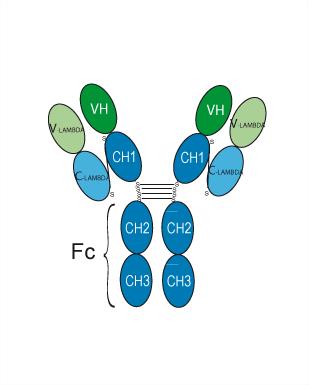
Dusigitumab is a fully human IgG2λ monoclonal antibody that targets IGF-1 and IGF-2 with picomolar binding affinity. Dusigitumab blocks IGF-1 and IGF-2-induced phosphorylation of IGF-1R as well as activation of IR-A, thereby inhibiting the activation of downstream signaling pathways. Dusigitumab is applicable to research related to advanced solid tumors and hormone receptor-positive metastatic breast cancer.
For research use only. We do not sell to patients.
- Purity : 96.37%
- CAS No.: 1204390-13-5
- Molecular Weight:144.24 kDa
-
Storage:
Please store the product under the recommended conditions in the Certificate of Analysis.


To place orders, for customer services and technical support, please contact: MedChemExpress USA
Tel: 609-228-6898 E-mail: [email protected] [email protected]


-
 Biological Activity
-
Chemical Information
- Protocols
- Purity & Documentation
- References
-
 Help & FAQs


-

HY-P99274
XentuzumabIGF1/IGF2 Antagonist -

HY-P99294
GanitumabAnti-IGF1R Antibody -

HY-P99165
TeprotumumabIGF-1R Inhibitor -

HY-P99197
FigitumumabAnti-IGF1R Antibody -

HY-P99218
RobatumumabAnti-human IGF-1R Antibody -

HY-P2093
S961Insulin Receptor Antagonist -

HY-P990084
VeligrotugIGF-1 Receptor Antagonist -

HY-135680
I-OMe-Tyrphostin AG 538IGF-1R/PI5P4Kα Inhibitor -
HY-18785
Indirubin Derivative E804IGF-1R Inhibitor


Biological Activity
Description
Isotype
Human IgG2 lambda
Recommend Isotype Controls
Species Reactivity
Human
IC50 & Target
IGF-1&IGF-2
In Vitro
Gene ID
Accession
P01344-1
Conjugated
Unconjugated
Reconsititution
The product can be reconstituted/diluted with sterile PBS or saline.
Format
-
Human IgG2 lambda
Application
ELISA, FACS, Functional assay
Verified Bioactivity
-
 Immobilized IGF-I/IGF-1 Protein, Human can bind Dusigitumab. The EC50 for this effect is 145.7 ng/mL.
 -
 Flow cytometric analysis of 1X10⁶ HepG2 cells with Dusigitumab (HY-P99867, red). Cells were fixed with 4% paraformaldehyde. Then stained with the primary antibody at 1/200 dilution for an hour at 4℃. AF 488-conjugated AffiniPure Goat Anti-Human IgG H&L (HY-P83776) was used as the secondary antibody at 1/1,000 dilution for 30 minutes at 4℃. Human IgG2 lambda (HY-P991206, blue) was used as the isotype control, cells without incubation with primary antibody were used as the unlabeled control (black).


Chemical Information
-
CAS No. 1204390-13-5
-
Appearance Liquid
-
Molecular Weight 144.24 kDa
-
Color Colorless to light yellow
-
SMILES
[Dusigitumab]
-
Synonyms
MEDI 573
-
Shipping
Shipping with dry ice.
-
Formulation
Please refer to the lot-specific COA for specific buffer information.
-
Storage
Please store the product under the recommended conditions in the Certificate of Analysis.

Protocols
-
Kinase activity and phosphorylation assays
Kinase activity assays measure the ability of kinases to transfer phosphate groups from ATP to specific substrates, while phosphorylation assays detect the presence and levels of phosphorylated proteins. Common methods include radiolabeled ATP incorporation (e. g. ,), ADP release detection via bioluminescence (e. g. ,[3]), enzyme-linked immunosorbent assays (ELISA) for phospho-specific epitopes (e. g. ,[6]), and microtiter-based formats for high-throughput screening (e. g. ,[8]). The ADP-Glo assay quantifies kinase activity by measuring ADP produced during phosphorylation using a luciferase-based system. Radiometric assays involve autoradiography or scintillation counting after incorporation of 32P-labeled ATP into substrate proteins. ELISA-based approaches rely on phospho-specific antibodies to detect activated kinases in cell lysates or purified samples.
-
Western Blot
Western blotting (WB) is a commonly used experimental method in molecular biology, biochemistry, and immunogenetics for identifying and quantifying target proteins. It combines gel electrophoresis with immunoassay, enabling researchers to analyze protein expression, post-translational modifications, and molecular weight.
-
Research Protocol for Endocrine Diseases
Endocrine diseases often arise from disrupted hormone production, hormone signaling, or target-tissue responsiveness; for diabetes-focused endocrine disease models, insulin signaling regulates glucose uptake, hepatic glucose output, lipid metabolism, and β-cell compensation. Type 2 diabetes develops through interacting defects in insulin resistance, β-cell dysfunction, adipose inflammation, hepatic glucose overproduction, altered incretin signaling, and ectopic lipid metabolism. A major unresolved question is whether endocrine dysfunction is driven primarily by target-tissue insulin resistance, intrinsic β-cell failure, immune/inflammatory stress, or combined multi-organ failure that differs by disease stage.
-
Protocol for Kinase activity and phosphorylation assays
Kinase activity assays measure transfer of phosphate from ATP to a protein or peptide substrate, generating phosphorylated substrate, ADP, or incorporated radiolabeled phosphate as the readout; phosphorylation assays measure site-specific phosphorylation in cells or tissues as a proxy for kinase-pathway activation, inhibition, or substrate regulation. Phosphorylation can be detected by phospho-specific Western blot, immunoprecipitation kinase assay, phospho-immunofluorescence, phospho-flow cytometry, luminescent ADP detection, radiolabeled ATP incorporation, or reporter-based pathway assays, and these readouts can be applied to cancer cells, primary neurons, mouse tumors, organoids, inflammatory macrophages, ferroptosis studies, and mitophagy studies when the kinase target is biologically relevant.
-
Breast Cancer Modeling
Breast cancer is a heterogeneous cancer, and it has been distinguished into four subtypes: luminal A, luminal B, HER2-positive and basal-like. Molecular mutations, epigenetic alterations, hormone exposure and immune microenvironment are related to the progression of breast cancer.


Purity & Documentation
-
Data Sheet (256 KB)
-
SDS (251 KB)
- English - EN (251 KB)
- Français - FR (251 KB)
- Deutsch - DE (251 KB)
- Norwegian - NO (251 KB)
- Español - ES (251 KB)
- Swedish - SV (251 KB)
- Italian - IT (251 KB)
- Korean - KR (251 KB)
- Portuguese - PT (251 KB)
-
Inhibitory Antibodies User Guide (603 KB)
References
[1]. de Bono J, et al. Two first-in-human studies of xentuzumab, a humanised insulin-like growth factor (IGF)-neutralising antibody, in patients with advanced solid tumours. Br J Cancer. 2020;122(9):1324-1332. [Content Brief]
[2]. Osher E, et al. Therapeutic Targeting of the IGF Axis. Cells. 2019;8(8):895. Published 2019 Aug 14. [Content Brief]

Calculators
Concentration (start) × Volume (start) = Concentration (final) × Volume (final)


Dusigitumab Related Classifications
HY-P99867 Related Classifications
 Powered by Bioz