



ENMD-1068 hydrochloride
Based on 3 publication(s) in Google Scholar
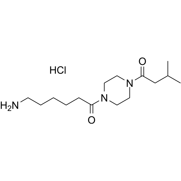
ENMD-1068 hydrochloride is a selective protease-activated receptor 2 (PAR2) antagonist. ENMD-1068 hydrochloride reduces hepatic stellate cell activation and collagen expression by inhibiting TGF-β1/Smad signaling. ENMD-1068 hydrochloride also inhibits the proliferation of endometrial cells and induces apoptosis of epithelial cells in the lesion. ENMD-1068 hydrochloride can be used in the study of endometriosis and liver fibrosis.
For research use only. We do not sell to patients.
- Purity : 98.33%
- CAS No.: 2703451-51-6
- Formula: C15H30ClN3O2
- Molecular Weight:319.87
-
Storage:
4°C, stored under nitrogen
* In solvent : -80°C, 6 months; -20°C, 1 month (stored under nitrogen)

To place orders, for customer services and technical support, please contact: MedChemExpress USA
Tel: 609-228-6898 E-mail: [email protected] [email protected]


-
 Biological Activity
-
Chemical Information
-
Solvent & Solubility
- Protocols
- Purity & Documentation
- References
-
 Help & FAQs


-
HY-10119
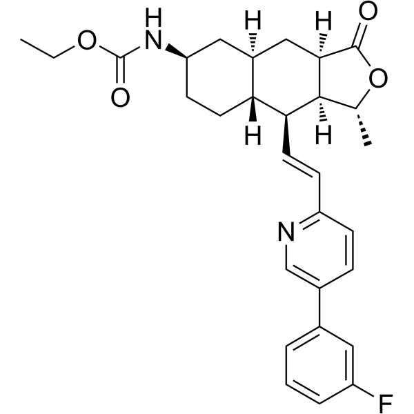
VorapaxarPAR-1 Antagonist -
HY-14993
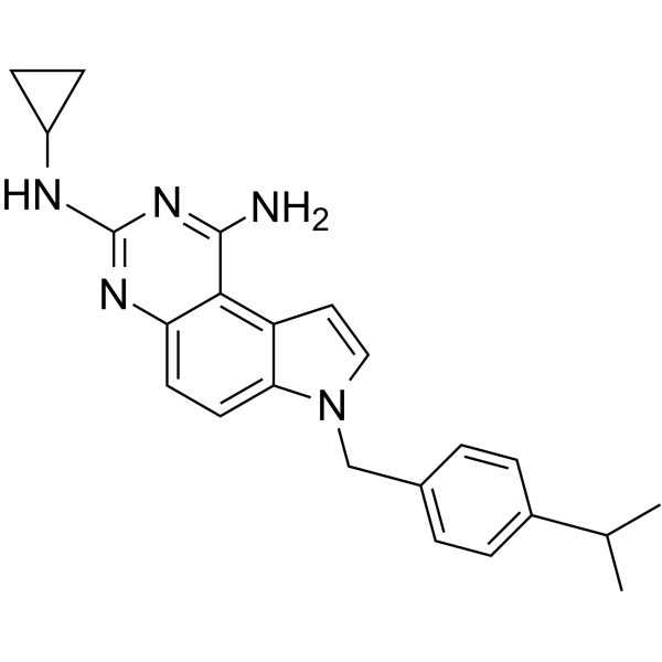
SCH79797PAR1 Antagonist -
HY-124061
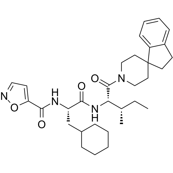
GB83PAR2 Antagonist -
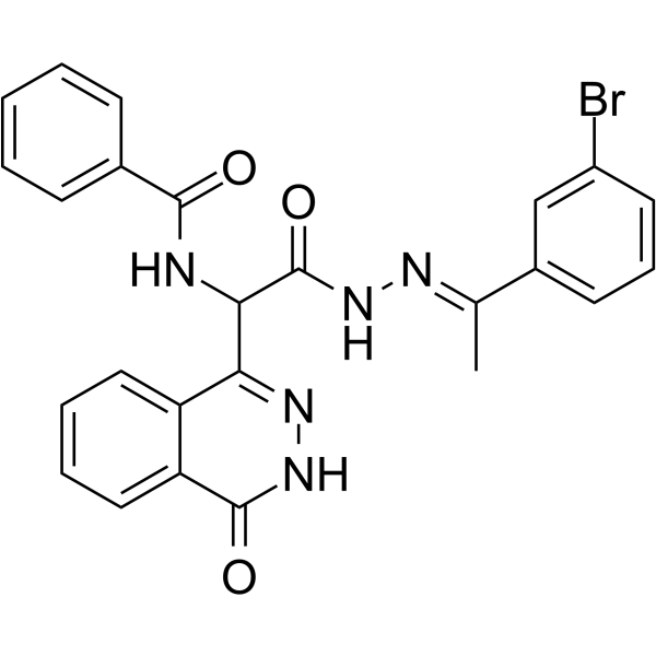
HY-14350
AC-55541PAR Agonist -
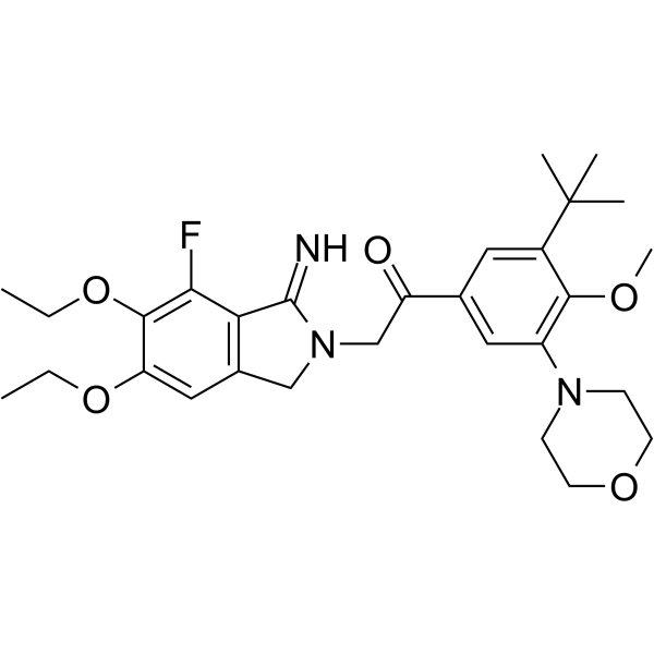
HY-18200
AtopaxarPAR-1 Antagonist -
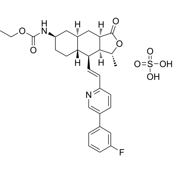
HY-10119A
Vorapaxar sulfatePAR-1 Antagonist -
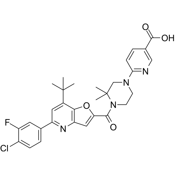
HY-148016
I-287PAR2 Inhibitor -

HY-10119R
Vorapaxar (Standard)PAR-1 Antagonist -

HY-10119AR
Vorapaxar sulfate (Standard)PAR-1 Antagonist -
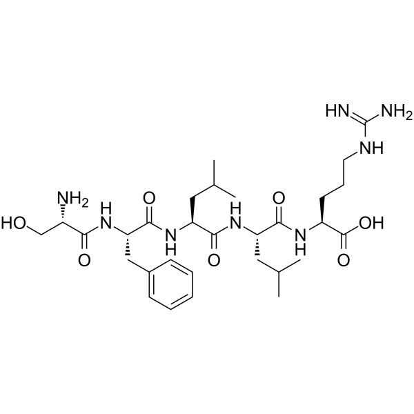
HY-P1536
Thrombin Receptor Activator for Peptide 5 (TRAP-5) -
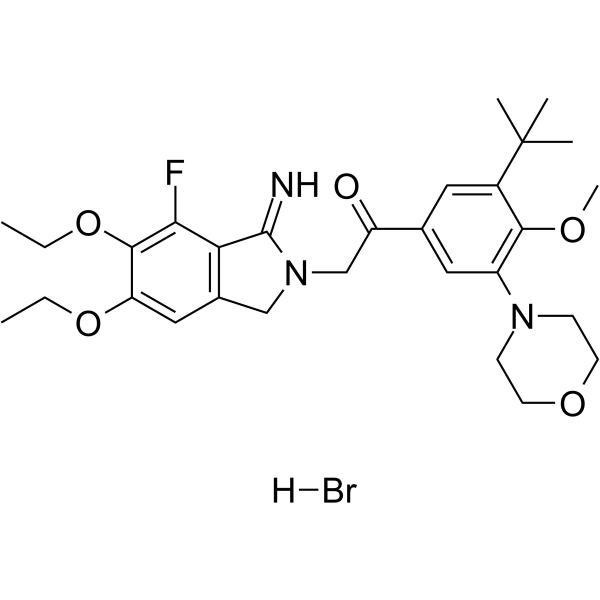
HY-18200B
Atopaxar hydrobromidePAR-1 Antagonist -
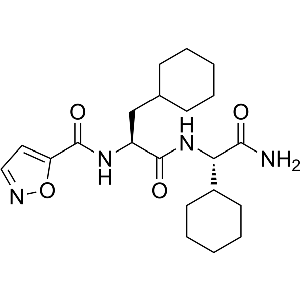
HY-138951
AY77PAR2 Agonist
Publications Citing Use of MedChemExpress (MCE) ENMD-1068 hydrochloride
More

Biological Activity
Description
IC50 & Target
|
PAR2 |
In Vitro
ENMD-1068 (10 mM; 24 h) blocks TGF-β1/Smad signaling in primary mouse HSCs (TGF-β1/Smad signal pathway plays a crucial role in HSCs activation and collagen production)[1].
ENMD-1068 (10 mM) inhibits trypsin or SLIGRL-NH2 stimulated calcium release in HSCs[1].
MedChemExpress (MCE) has not independently confirmed the accuracy of these methods. They are for reference only. Further protocols information, click here.
-
Cell Line:Hepatic stellate cells (HSCs) (TGF-β1-stimulated)
-
Concentration:10 mM
-
Incubation Time:24 h
-
Result:Inhibited TGF-β1-induced expression of α-SMA, Col α1(І),Col α1(III), and Smad2/3 C-terminal phosphorylation.
In Vivo
ENMD-1068 (25, 50 mg/kg; i.p.; single daily for 5 days) inhibits endometriosis growth and suppresses the levels of IL-6 and MCP-1 in a dose-dependent manner[2].
ENMD-1068 (25, 50 mg/kg; i.p.; single daily for 5 days) causes a decrease in epithelial cell proliferation and an increase in the apoptotic index in mice[2].
MedChemExpress (MCE) has not independently confirmed the accuracy of these methods. They are for reference only.
-
Animal Model:ICR mice (8-week-old; CCl4-induced liver fibrosis model)[1].
-
Dosage:25, 50 mg/kg
-
Administration:Intraperitoneal injection; twice per week for 4 weeks
-
Result:Markedly attenuated collagen deposition.
-
Animal Model:Mice with surgically induced endometriosis[2].
-
Dosage:25, 50 mg/kg
-
Administration:Intraperitoneal injection; single daily for 5 days
-
Result:Reduced the volume of observed lesions in a dose-dependent manner.
Inhibited the expression of IL-6 and MCP-1.
Decreased the proliferation rate of endometriotic cells and increased the percentage of apoptotic epithelial cells in the lesions.


Chemical Information
-
CAS No. 2703451-51-6
-
Appearance Solid
-
Molecular Weight 319.87
-
Formula C15H30ClN3O2
-
Color White to off-white
-
SMILES
O=C(N1CCN(C(CC(C)C)=O)CC1)CCCCCN.Cl
-
Shipping
Room temperature in continental US; may vary elsewhere.
-
Storage
4°C, stored under nitrogen
* In solvent : -80°C, 6 months; -20°C, 1 month (stored under nitrogen)


Publications (3)
-
Journal Impact Factor
-
Most Recent
-
Cancer Cell
Coagulation factor X promotes resistance to androgen-deprivation therapy in prostate cancer. [Abstract]2024 Oct 14;42(10):1676-1692.e11. PMID: 39303726 -
J Inflamm Res
Astragalus membranaceus and Salvia miltiorrhiza Inhibit Tryptase/PAR2-Mediated Mast Cells - Peritoneal Mesothelial Cells Crosstalk to Ameliorate Peritoneal Fibrosis. [Abstract]2026 May 11:19:577472. PMID: 42147796 -


Solvent & Solubility
In Vitro:
DMSO : 200 mg/mL (625.25 mM; Need ultrasonic; Hygroscopic DMSO has a significant impact on the solubility of product, please use newly opened DMSO)
H2O : 100 mg/mL (312.63 mM; Need ultrasonic)
Please refer to the solubility information to select the appropriate solvent. Once prepared, please aliquot and store the solution to prevent product inactivation from repeated freeze-thaw cycles.
Storage method and period of stock solution: -80°C, 6 months; -20°C, 1 month (stored under nitrogen). When stored at -80°C, please use it within 6 months. When stored at -20°C, please use it within 1 month.
* Note: If you choose water as the stock solution, please dilute it to the working solution, then filter and sterilize it with a 0.22 μm filter before use.
Please refer to the solubility information to select the appropriate solvent. Once prepared, please aliquot and store the solution to prevent product inactivation from repeated freeze-thaw cycles.
Storage method and period of stock solution: -80°C, 6 months; -20°C, 1 month (stored under nitrogen). When stored at -80°C, please use it within 6 months. When stored at -20°C, please use it within 1 month.
* Note: If you choose water as the stock solution, please dilute it to the working solution, then filter and sterilize it with a 0.22 μm filter before use.
Concentration (start) × Volume (start) = Concentration (final) × Volume (final)
In Vivo:
Select the appropriate dissolution method based on your experimental animal and administration route.
- For the following dissolution methods, please ensure to first prepare a clear stock solution using an In Vitro approach and then sequentially add co-solvents:
- To ensure reliable experimental results, the clarified stock solution can be appropriately stored based on storage conditions. As for the working solution for In Vivo experiments, it is recommended to prepare freshly and use it on the same day.
- The percentages shown for the solvents indicate their volumetric ratio in the final prepared solution. If precipitation or phase separation occurs during preparation, heat and/or sonication can be used to aid dissolution.
Add each solvent one by one: 10% DMSO 40% PEG300 5% Tween-80 45% Saline
Solubility: 5 mg/mL (15.63 mM); Suspended solution; Need ultrasonic
This protocol yields a suspended solution of 5 mg/mL. Suspended solution can be used for oral and intraperitoneal injection.
Taking 1 mL working solution as an example, add 100 μL DMSO stock solution (50.0 mg/mL) to 400 μL PEG300, and mix evenly; then add 50 μL Tween-80 and mix evenly; then add 450 μL Saline to adjust the volume to 1 mL.
Preparation of Saline: Dissolve 0.9 g sodium chloride in ddH₂O and dilute to 100 mL to obtain a clear Saline solution.
Add each solvent one by one: 10% DMSO 90% (20% SBE-β-CD in Saline)
Solubility: ≥ 5 mg/mL (15.63 mM); Clear solution
This protocol yields a clear solution of ≥ 5 mg/mL (saturation unknown).
Taking 1 mL working solution as an example, add 100 μL DMSO stock solution (50.0 mg/mL) to 900 μL 20% SBE-β-CD in Saline, and mix evenly.
Preparation of 20% SBE-β-CD in Saline (4°C, storage for one week): 2 g SBE-β-CD powder is dissolved in 10 mL Saline, completely dissolve until clear.
For the following dissolution methods, please prepare the working solution directly:
It is recommended to prepare fresh solutions and use them promptly within a short period of time.
The percentages shown for the solvents indicate their volumetric ratio in the final prepared solution. If precipitation or phase separation occurs during preparation, heat and/or sonication can be used to aid dissolution.
Add each solvent one by one: PBS
Solubility: 100 mg/mL (312.63 mM); Clear solution; Need ultrasonic
In Vivo Dissolution Calculator
Please enter the basic information of animal experiments:
-
-
-
-
Recommended: Prepare an additional quantity of animals to account for potential losses during experiments.
Working solution concentration: 0.22 mg/mL
This product has good water solubility, please refer to the measured solubility data in water/PBS/Saline for details.

Protocols
-
Fibrosis/Collagen Morphometry
Fibrosis and collagen morphometry is based on the quantitative visualization of fibrillar collagen deposition in tissue sections using histochemical stains such as Sirius Red (Picrosirius Red) or Masson's trichrome, followed by image-based or polarization-enhanced analysis to estimate collagen proportional area as a surrogate of extracellular matrix accumulation during fibrotic remodeling. Sirius Red combined with polarized light microscopy enhances detection of collagen fibers due to birefringence properties, enabling more specific visualization of collagen type I and III fibrils compared to conventional bright-field histology, while whole-section or region-restricted digital morphometry reduces field-selection bias in fibrosis assessment. Alternative quantitative approaches include second harmonic generation (SHG) and two-photon excited fluorescence microscopy, which enable label-free detection of fibrillar collagen and have been validated against histological staining and biochemica
-
RNA extraction experimental
By lysing cells, releasing RNA, and removing impurities such as proteins and DNA, high-purity RNA products are finally obtained. The commonly used traditional method is the guanidine isothiocyanate/phenol/chloroform method (Trizol), which is suitable for a variety of animal materials including animal tissues, microorganisms, cultured cells, etc., and most plant materials.
-
Collagen: Sirius Red Staining
Sirius Red or picrosirius red staining is a histochemical method for visualizing collagen-rich extracellular matrix in tissue sections, and collagen fibers are detected as red-stained structures under bright-field microscopy with enhanced birefringence under polarized light. Picrosirius red is useful for assessing total collagen organization, distribution, and fibrosis burden, but polarized color should not be interpreted as a definitive collagen type I versus type III readout because color is affected by fiber orientation, thickness, and packing.
-
Apoptosis
Apoptosis, also called programmed cell death, is generally characterized by distinct morphological characteristics.
-
TUNEL staining for apoptotic DNA fragmentation
TUNEL staining detects DNA strand breaks by using terminal deoxynucleotidyl transferase to add labeled nucleotides to exposed 3′-OH DNA termini, generating either microscopic staining in fixed cells or tissue sections, or fluorescence/cytometric signal in cell suspensions. TUNEL positivity reflects DNA fragmentation but should not be interpreted alone as definitive apoptosis, because TUNEL can also label necrotic, autolytic, mechanically damaged, or DNA-repair-associated DNA breaks.
-
Annexin V plus membrane-impermeant dye apoptosis staining
Annexin V-based apoptosis assays rely on the detection of phosphatidylserine (PS) externalization from the inner leaflet of the plasma membrane to the outer leaflet, an early biochemical hallmark of apoptosis. Fluorescently labeled Annexin V binds PS in a calcium-dependent manner, enabling identification of early apoptotic cells by flow cytometry or fluorescence microscopy. When combined with a membrane-impermeant DNA-binding dye (e. g. , propidium iodide), this approach allows discrimination between viable (Annexin V−/dye−), early apoptotic (Annexin V+/dye−), and late apoptotic or necrotic (Annexin V+/dye+) cell populations by assessing membrane integrity and PS exposure.
-
Connective Tissue: Masson's Trichrome/Collagen Trichrome Staining
Masson’s Trichrome (collagen/trichrome staining) is a histological technique that differentially stains tissue compartments using sequential acidic dyes to distinguish collagen from muscle and cytoplasmic components based on dye affinity and tissue permeability differences, enabling visualization of fibrosis and connective tissue architecture in histological sections. The classical formulation typically uses Weigert's iron hematoxylin for nuclear staining, Biebrich scarlet-acid fuchsin for cytoplasm and muscle, and aniline blue (or light green variants) for collagen, producing a characteristic blue/green collagen signal contrasted against red cytoplasm and dark nuclei. The staining principle relies on selective displacement of smaller dye molecules by larger anionic dyes in collagen-rich regions under controlled acidified conditions, which enhances collagen-specific dye retention. This property makes the method widely used for fibrosis assessment in organs such as heart, liver, lung, a
-
Apoptosis Solutions
Apoptosis is a regulated, generally non-lytic cell-death pathway that removes unwanted, damaged, infected, or abnormal cells through coordinated morphological changes, caspase activation, DNA fragmentation, and membrane remodeling. The intrinsic apoptosis pathway is controlled mainly by mitochondrial outer membrane permeabilization, BCL-2 family proteins, cytochrome c release, apoptosome formation, caspase-9 activation, and downstream executioner caspase-3/7 activation. The extrinsic apoptosis pathway is initiated by death receptors such as Fas, TNFR, and TRAIL receptors, which recruit adaptor proteins and activate caspase-8 before engaging executioner caspases or mitochondrial amplification through BID cleavage. Apoptosis is linked to many phenotypes, including cancer cell killing, tissue homeostasis, immune regulation, neurodegeneration, infection response, and treatment-induced cytotoxicity; unresolved questions include how apoptosis interacts with necroptosis, pyroptosis, ferroptos
-
Cell Viability Determination by MTT Colorimetric Assay
The following protocol uses the MTT colorimetric assay as a classic literature-established method for assessing cell viability/metabolic activity in cultured mammalian cells. MTT[3-(4,5-dimethylthiazol-2-yl)-2,5-diphenyltetrazolium bromide] is reduced by metabolically active cells to a colored formazan product; the amount of formazan is quantified spectrophotometrically and provides an indirect measure of metabolically active viable cells. Importantly, MTT reduction reflects cellular oxidoreductase/metabolic activity rather than an absolute direct count of living cells, so changes in cellular metabolism can alter the signal independently of cell number.
-
Liver Histomorphometry
Liver histomorphometry is a quantitative histological approach used to measure structural alterations in hepatic tissue, including parenchymal loss, steatosis, fibrosis, and vascular remodeling, by combining stained tissue section analysis with stereological or computerized image-based measurements. Classical morphometric frameworks quantify volume fractions of liver compartments and fibrotic regions using systematic sampling and image analysis, enabling objective comparison of pathological changes across experimental groups. These approaches are widely applied in liver cirrhosis and fibrosis studies to reduce subjectivity in histological scoring and improve reproducibility of tissue evaluation. Recent methodological advances integrate automated image analysis and radiomics-based extraction of histological features from standard liver stains (e. g. , H&E and fibrotic stains), enabling quantitative correlation between morphometric features and fibrosis stages in non-alcoholic fatty live


Purity & Documentation
-
Data Sheet (275 KB)
-
SDS (392 KB)
- English - EN (392 KB)
- Français - FR (392 KB)
- Deutsch - DE (392 KB)
- Norwegian - NO (392 KB)
- Español - ES (392 KB)
- Swedish - SV (392 KB)
- Italian - IT (392 KB)
- Korean - KR (392 KB)
- Portuguese - PT (392 KB)
-
Handling Instructions (2659 KB)
References
[1]. Sun Q, et al. ENMD-1068 inhibits liver fibrosis through attenuation of TGF-β1/Smad2/3 signaling in mice. Sci Rep. 2017 Jul 14;7(1):5498. doi: 10.1038/s41598-017-05190-7. Erratum in: Sci Rep. 2019 Dec 10;9(1):19125. [Content Brief]
[2]. Wang Y, et al. ENMD-1068, a protease-activated receptor 2 antagonist, inhibits the development of endometriosis in a mouse model. Am J Obstet Gynecol. 2014 Jun;210(6):531.e1-8. [Content Brief]


Complete Stock Solution Preparation Table
Please refer to the solubility information to select the appropriate solvent. Once prepared, please aliquot and store the solution to prevent product inactivation from repeated freeze-thaw cycles.
Storage method and period of stock solution: -80°C, 6 months; -20°C, 1 month (stored under nitrogen). When stored at -80°C, please use it within 6 months. When stored at -20°C, please use it within 1 month.
| Optional Solvent | Concentration Solvent Mass | 1 mg | 5 mg | 10 mg | 25 mg |
|---|---|---|---|---|---|
| H2O / DMSO | 1 mM | 3.1263 mL | 15.6314 mL | 31.2627 mL | 78.1568 mL |
| 5 mM | 0.6253 mL | 3.1263 mL | 6.2525 mL | 15.6314 mL | |
| 10 mM | 0.3126 mL | 1.5631 mL | 3.1263 mL | 7.8157 mL | |
| 15 mM | 0.2084 mL | 1.0421 mL | 2.0842 mL | 5.2105 mL | |
| 20 mM | 0.1563 mL | 0.7816 mL | 1.5631 mL | 3.9078 mL | |
| 25 mM | 0.1251 mL | 0.6253 mL | 1.2505 mL | 3.1263 mL | |
| 30 mM | 0.1042 mL | 0.5210 mL | 1.0421 mL | 2.6052 mL | |
| 40 mM | 0.0782 mL | 0.3908 mL | 0.7816 mL | 1.9539 mL | |
| 50 mM | 0.0625 mL | 0.3126 mL | 0.6253 mL | 1.5631 mL | |
| 60 mM | 0.0521 mL | 0.2605 mL | 0.5210 mL | 1.3026 mL | |
| 80 mM | 0.0391 mL | 0.1954 mL | 0.3908 mL | 0.9770 mL | |
| 100 mM | 0.0313 mL | 0.1563 mL | 0.3126 mL | 0.7816 mL |
* Note: If you choose water as the stock solution, please dilute it to the working solution, then filter and sterilize it with a 0.22 μm filter before use.


ENMD-1068 hydrochloride Related Classifications
HY-124748A Related Classifications
 Powered by Bioz