



CAM833
Based on 1 publication(s) in Google Scholar
CAM833 is a potent orthosteric inhibitor of the interaction between BRCA2 and RAD51 with a Kd of 366 nM against the ChimRAD51 protein. CAM833 also inhibits RAD51 oligomerization. CAM833 increases the progression of G2/M-arrested cells into apoptosis.
For research use only. We do not sell to patients.
- Purity : 99.83%
- CAS No.: 2758364-02-0
- Formula: C26H26ClFN4O5
- Molecular Weight:528.96
-
Storage:Powder -20°C, 3 years , 4°C, 2 years ; In solvent -80°C, 6 months , -20°C, 1 month

To place orders, for customer services and technical support, please contact: MedChemExpress USA
Tel: 609-228-6898 E-mail: [email protected] [email protected]


-
 Biological Activity
-
Chemical Information
-
Solvent & Solubility
- Protocols
- Purity & Documentation
- References
-
 Help & FAQs


-
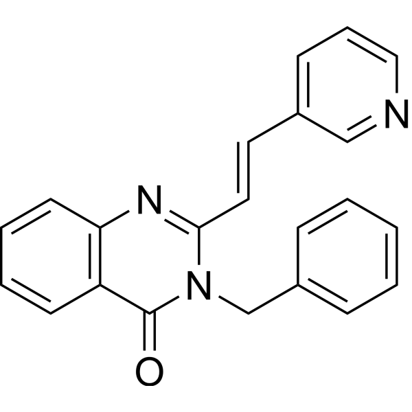
HY-101462
RAD51 Inhibitor B02RAD51 Inhibitor -
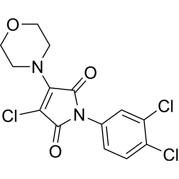
HY-15317
RI-1RAD51 Inhibitor -
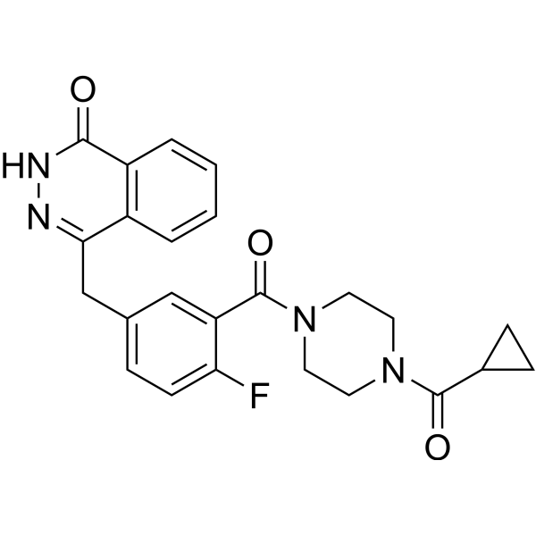
HY-10162
OlaparibPARP Inhibitor -
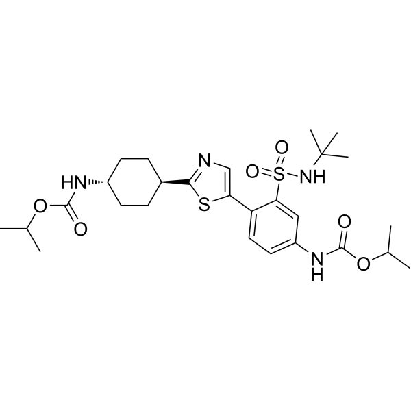
HY-111887
EmzadiribRAD51 Inhibitor -
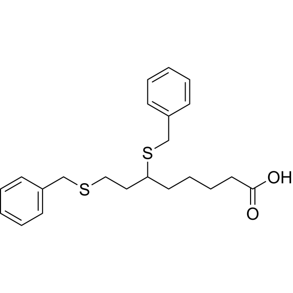
HY-15453
DevimistatMitochondrial Metabolism Inhibitor -
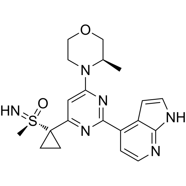
HY-19323
CeralasertibATR Inhibitor -
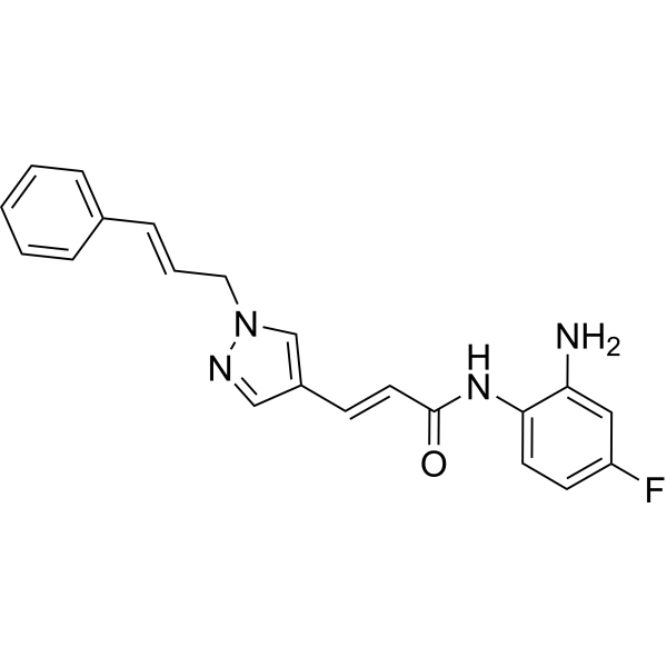
HY-13909
RGFP966HDAC3 Inhibitor -
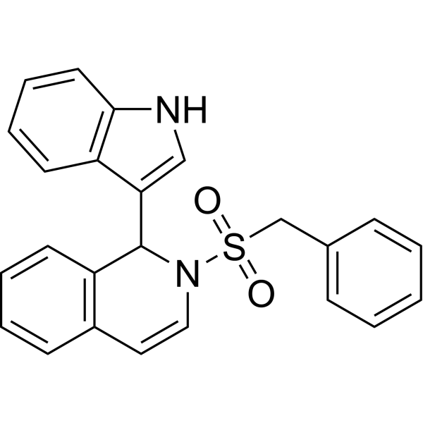
HY-103710
IBR2RAD51 Inhibitor -
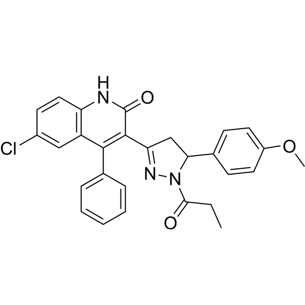
HY-148183
Homologous recombination-IN-1RAD51-BRCA2 Inhibitor -
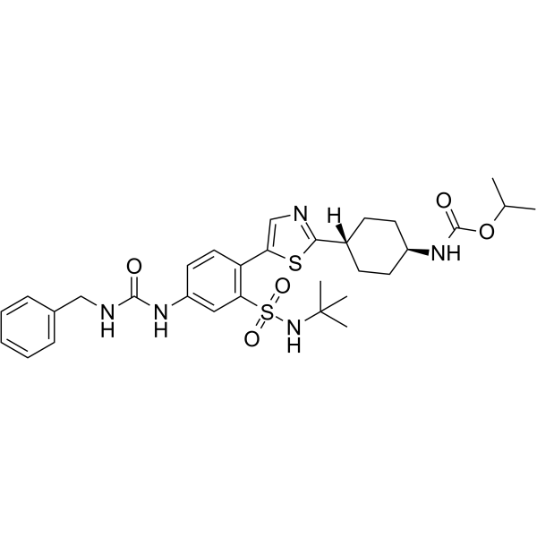
HY-136604
RAD51-IN-3RAD51 Inhibitor -
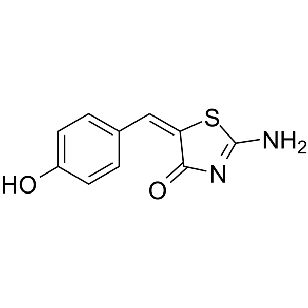
HY-117693
(E/Z)-MirinMre11-Rad50-Nbs1 Complex Inhibitor -
HY-128729
DNA2 inhibitor C5DNA2 Nuclease Inhibitor
Publications Citing Use of MedChemExpress (MCE) CAM833
More

Biological Activity
Description
IC50 & Target
Kd: 355 nM (ChimRAD51, measured by FP), 366 nM (ChimRAD51, measured by ITC)[1]
In Vitro
CAM833 (3.125-50 μM; 24 h) causes a concentration-dependent decrease in RAD51 foci and subsequent increase in DNA damage[1].
CAM833 (25 μM) inhibits RAD51 molecular clustering at DNA damage sites and suppresses extended RAD51 filament assembly[1].
CAM833 (0-50 μM) inhibits DNA repair by homologous recombination[1].
CAM833 (20 μM; 0-72 h) potentiates radiation-induced cell-cycle arrest and increases apoptosis over time in HCT116 cells[1].
CAM833 (0.1-100 μM; 96 h) causes a dose-dependent growth inhibition of multiple cancer-derived human cell lines that is enhanced when combined with ionizing radiation[1].
CAM833 (20 μM; 96 h) potentiates the growth suppressive effect of PARP1 inhibition in BRCA2 wild-type cells[1].
CAM833 (96 h) alone inhibits the growth of HCT116 colon carcinoma cells with a GI50 (50% growth inhibition) of 38 μM, when combined with 3 Gy IR, CAM833 suppresses the growth of HCT116 cells with a GI50 of 14 μM[1].
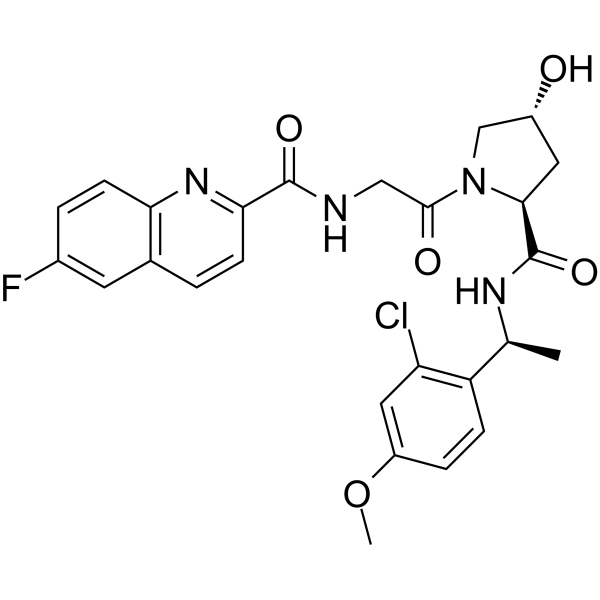
The quinoline of CAM833 occupies a hotspot, the Phe-binding pocket on RAD51 and the methyl of the substituted α-methylbenzyl group occupies the Ala-binding pocket[1].
MedChemExpress (MCE) has not independently confirmed the accuracy of these methods. They are for reference only.
-
Cell Line:HCT116 cells
-
Concentration:20 μM
-
Incubation Time:0-72 h
-
Result:In the control the percentage of cells in the apoptotic subG1 fraction remains below 5% throughout, in the compound-treated cells this rises progressively to peak at 15% at 48 hours.


Chemical Information
-
CAS No. 2758364-02-0
-
Appearance Solid
-
Molecular Weight 528.96
-
Formula C26H26ClFN4O5
-
Color White to off-white
-
SMILES
C[C@@H](C1=C(C=C(C=C1)OC)Cl)NC([C@H]2N(C[C@@H](C2)O)C(CNC(C3=NC4=C(C=C(C=C4)F)C=C3)=O)=O)=O
-
Shipping
Room temperature in continental US; may vary elsewhere.
-
Storage
Powder -20°C 3 years 4°C 2 years In solvent -80°C 6 months -20°C 1 month


Publications (1)
-
Journal Impact Factor
-
Most Recent
-
Nature
2025 Apr;640(8060):1103-1111. PMID: 40140565


Solvent & Solubility
In Vitro:
DMSO : 125 mg/mL (236.31 mM; Need ultrasonic; Hygroscopic DMSO has a significant impact on the solubility of product, please use newly opened DMSO)
Please refer to the solubility information to select the appropriate solvent. Once prepared, please aliquot and store the solution to prevent product inactivation from repeated freeze-thaw cycles.
Storage method and period of stock solution: -80°C, 6 months; -20°C, 1 month. When stored at -80°C, please use it within 6 months. When stored at -20°C, please use it within 1 month.
Please refer to the solubility information to select the appropriate solvent. Once prepared, please aliquot and store the solution to prevent product inactivation from repeated freeze-thaw cycles.
Storage method and period of stock solution: -80°C, 6 months; -20°C, 1 month. When stored at -80°C, please use it within 6 months. When stored at -20°C, please use it within 1 month.
Concentration (start) × Volume (start) = Concentration (final) × Volume (final)


Purity & Documentation
-
Data Sheet (278 KB)
-
SDS (251 KB)
- English - EN (251 KB)
- Français - FR (251 KB)
- Deutsch - DE (251 KB)
- Norwegian - NO (251 KB)
- Español - ES (251 KB)
- Swedish - SV (251 KB)
- Italian - IT (251 KB)
- Korean - KR (251 KB)
- Portuguese - PT (251 KB)
-
Handling Instructions (2659 KB)
References


Complete Stock Solution Preparation Table
Please refer to the solubility information to select the appropriate solvent. Once prepared, please aliquot and store the solution to prevent product inactivation from repeated freeze-thaw cycles.
Storage method and period of stock solution: -80°C, 6 months; -20°C, 1 month. When stored at -80°C, please use it within 6 months. When stored at -20°C, please use it within 1 month.
| Optional Solvent | Concentration Solvent Mass | 1 mg | 5 mg | 10 mg | 25 mg |
|---|---|---|---|---|---|
| DMSO | 1 mM | 1.8905 mL | 9.4525 mL | 18.9050 mL | 47.2626 mL |
| 5 mM | 0.3781 mL | 1.8905 mL | 3.7810 mL | 9.4525 mL | |
| 10 mM | 0.1891 mL | 0.9453 mL | 1.8905 mL | 4.7263 mL | |
| 15 mM | 0.1260 mL | 0.6302 mL | 1.2603 mL | 3.1508 mL | |
| 20 mM | 0.0945 mL | 0.4726 mL | 0.9453 mL | 2.3631 mL | |
| 25 mM | 0.0756 mL | 0.3781 mL | 0.7562 mL | 1.8905 mL | |
| 30 mM | 0.0630 mL | 0.3151 mL | 0.6302 mL | 1.5754 mL | |
| 40 mM | 0.0473 mL | 0.2363 mL | 0.4726 mL | 1.1816 mL | |
| 50 mM | 0.0378 mL | 0.1891 mL | 0.3781 mL | 0.9453 mL | |
| 60 mM | 0.0315 mL | 0.1575 mL | 0.3151 mL | 0.7877 mL | |
| 80 mM | 0.0236 mL | 0.1182 mL | 0.2363 mL | 0.5908 mL | |
| 100 mM | 0.0189 mL | 0.0945 mL | 0.1891 mL | 0.4726 mL |


CAM833 Related Classifications
HY-150147 Related Classifications
 Powered by Bioz