



FabH-IN-2
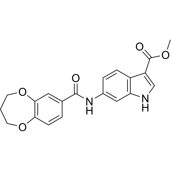
FabH-IN-2 (25), an antimicrobial agent, exhibits remarkable potential as an agent with an MIC range of 1.25-3.13 μg/mL against the tested bacterial strains and an IC50 of 2.0 μM against E. coli-derived FabH.
For research use only. We do not sell to patients.
- CAS No.: 3040281-87-3
- Formula: C20H18N2O5
- Molecular Weight:366.37
-
Storage:
Please store the product under the recommended conditions in the Certificate of Analysis.

To place orders, for customer services and technical support, please contact: MedChemExpress USA
Tel: 609-228-6898 E-mail: [email protected] [email protected]


-
 Biological Activity
-
Chemical Information
- Protocols
- Purity & Documentation
- References
-
 Help & FAQs




Biological Activity
Description
Cellular Effect
|
Cell Line

|
Type | Value | Description | References |
|---|---|---|---|---|
| NIH3T3 | IC50 |
141.6 μM
Compound: 25
|
Cytotoxicity against mouse NIH3T3 cells incubated for 24 hrs by MTT analysis
Cytotoxicity against mouse NIH3T3 cells incubated for 24 hrs by MTT analysis
|
[PMID: 38159483] |


Chemical Information
-
CAS No. 3040281-87-3
-
Molecular Weight 366.37
-
Formula C20H18N2O5
-
SMILES
O=C(NC1=CC(NC=C2C(OC)=O)=C2C=C1)C3=CC(OCCCO4)=C4C=C3
-
Shipping
Room temperature in continental US; may vary elsewhere.
-
Storage
Please store the product under the recommended conditions in the Certificate of Analysis.

Protocols
-
Research Protocol for Infectious Diseases
Infectious-disease experiments test how pathogens interact with host barriers, innate immune receptors, inflammatory signaling, pathogen replication, and tissue injury; pattern-recognition receptors such as TLRs, RIG-I-like receptors, NOD-like receptors, and inflammasomes detect microbial molecules and activate NF-κB, interferon, and cytokine responses. The central hypothesis is that infection severity reflects the balance between pathogen burden and host response: protective inflammation restricts pathogen growth, whereas excessive or mislocalized inflammation contributes to tissue damage and disease phenotype. Unresolved questions include which host pathways are protective versus pathogenic, why some infection models fail to translate to human disease, and which combined readouts best predict clinically relevant infection outcomes.
-
Cell Cytotoxicity Assay
Cytotoxicity assays are usually based on the assessment of cell membrane damage, which can also be indirectly detected by measuring cell viability. Detection methods include MTT assay, CKK-8 assay, LDH assay and ATP assay, etc.


Purity & Documentation
References

Calculators
Concentration (start) × Volume (start) = Concentration (final) × Volume (final)


FabH-IN-2 Related Classifications
HY-162087 Related Classifications
 Powered by Bioz