



FGFR4-IN-9
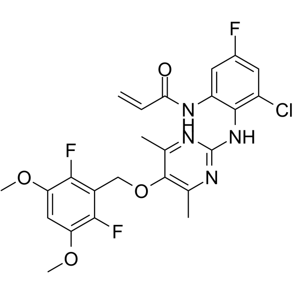
FGFR4-IN-9 (Compound 6O) is a selective FGFR4 inhibitor with an IC50 of 75.3 nM. FGFR4-IN-9 effectively inhibits both the growth and angiogenesis of HCC.
For research use only. We do not sell to patients.
- CAS No.: 3033043-66-9
- Formula: C24H22ClF3N4O4
- Molecular Weight:522.90
-
Storage:
Please store the product under the recommended conditions in the Certificate of Analysis.

To place orders, for customer services and technical support, please contact: MedChemExpress USA
Tel: 609-228-6898 E-mail: [email protected] [email protected]


-
 Biological Activity
-
Chemical Information
- Protocols
- Purity & Documentation
- References
-
 Help & FAQs




Biological Activity
Description
IC50 & Target
|
FGFR4 75.3 nM (IC50) |
Cellular Effect
|
Cell Line

|
Type | Value | Description | References |
|---|---|---|---|---|
| Hep 3B2 | IC50 |
0.9 μM
Compound: 20
|
Antiproliferative activity against human Hep3B cells expressing FGFR2 assessed as reduction in cell viability incubated for 48 hrs by MTT assay
Antiproliferative activity against human Hep3B cells expressing FGFR2 assessed as reduction in cell viability incubated for 48 hrs by MTT assay
|
[PMID: 37976704] |
| Hep 3B2 | IC50 |
4.5 μM
Compound: 20
|
Antiproliferative activity against human Hep3B cells expressing FGFR3 assessed as reduction in cell viability incubated for 48 hrs by MTT assay
Antiproliferative activity against human Hep3B cells expressing FGFR3 assessed as reduction in cell viability incubated for 48 hrs by MTT assay
|
[PMID: 37976704] |
| Huh-7 | IC50 |
12.6 μM
Compound: 20
|
Antiproliferative activity against human Huh-7 cells expressing FGFR3 assessed as reduction in cell viability incubated for 48 hrs by MTT assay
Antiproliferative activity against human Huh-7 cells expressing FGFR3 assessed as reduction in cell viability incubated for 48 hrs by MTT assay
|
[PMID: 37976704] |
| Huh-7 | IC50 |
7.1 μM
Compound: 20
|
Antiproliferative activity against human Huh-7 cells expressing FGFR2 assessed as reduction in cell viability incubated for 48 hrs by MTT assay
Antiproliferative activity against human Huh-7 cells expressing FGFR2 assessed as reduction in cell viability incubated for 48 hrs by MTT assay
|
[PMID: 37976704] |


Chemical Information
-
CAS No. 3033043-66-9
-
Molecular Weight 522.90
-
Formula C24H22ClF3N4O4
-
SMILES
C=CC(NC1=CC(F)=CC(Cl)=C1NC2=NC(C)=C(C(C)=N2)OCC3=C(C(OC)=CC(OC)=C3F)F)=O
-
Shipping
Room temperature in continental US; may vary elsewhere.
-
Storage
Please store the product under the recommended conditions in the Certificate of Analysis.

Protocols
-
Human pluripotent stem cell endothelial-cell differentiation
Human pluripotent stem cell endothelial differentiation is based on stepwise developmental patterning: early activation of WNT/GSK3β inhibition promotes mesodermal or vascular progenitor entry, followed by endothelial specification using VEGF-related signaling, BMP4, FGF2, Notch modulation, or cAMP depending on the published protocol. Endothelial differentiation is read out by acquisition of CD31, CD34, VE-cadherin/CD144, KDR/VEGFR2, vWF, Tie2, NOS3, acetylated LDL uptake, tube/network formation, barrier function, and in vivo vessel-forming capacity where tested.
-
Cell Cytotoxicity Assay
Cytotoxicity assays are usually based on the assessment of cell membrane damage, which can also be indirectly detected by measuring cell viability. Detection methods include MTT assay, CKK-8 assay, LDH assay and ATP assay, etc.
-
Vascular/Branching Fractal Analysis
Vascular/branching fractal analysis quantifies the geometric complexity of vessel trees or vascular networks from segmented 2D images, commonly by converting vessels into binary and/or skeletonized maps and estimating fractal dimension using box-counting or related approaches. Fractal dimension is interpreted as an image-derived readout of vascular branching complexity, space filling, or density, and has been applied to retinal photographs, fluorescein angiography, OCT angiography, capillary perfusion maps, and in vitro Matrigel angiogenesis networks. The assay readout is generated from vessel-positive pixels after image preprocessing, vessel segmentation, binarization, and optional skeletonization; reported outputs include fractal dimension, vessel density, branchpoint density, endpoint density, vessel length density, tortuosity, and generation-based branching metrics when VESGEN-style analysis is used. The biological interpretation is limited to quantitative vascular patterning and s
-
Endothelial Tube Formation Assay
Endothelial tube formation assay evaluates the ability of endothelial cells to attach, migrate, align, and organize into capillary-like networks when cultured on gelled basement membrane extract or Matrigel; the readout is the morphology and quantity of tube-like networks, which reflects an in vitro endothelial morphogenesis step related to angiogenesis. Basement membrane extract/Matrigel provides laminin-rich extracellular matrix cues that support endothelial differentiation into capillary-like structures, but it can contain biologically active growth factors, so growth-factor-reduced matrix is preferred when testing defined angiogenic stimulators or inhibitors.
-
Liver Cancer Modeling
Liver cancer can be classified into primary liver cancer and secondary liver cancer. Secondary liver cancer is the metastatic liver cancer. Primary liver cancer includes hepatocellular carcinoma (HCC), intrahepatic cholangiocarcinoma (ICC) and fibrolamellar HCC, of which HCC is the most common form, accounting for approximately 90% of primary liver cancers[1]. HCC mouse models include chemical agent-induced models, transplanted tumor models, and genetic engineered models.
-
Endothelial Cell Migration/Angiogenic Sprouting Assay
Endothelial cell migration and angiogenic sprouting assays are in vitro (and partially ex vivo-adapted) functional models that quantify the ability of endothelial cells to undergo coordinated migration, extracellular matrix invasion, and multicellular organization into capillary-like sprouts in response to pro-angiogenic stimuli such as VEGF, bFGF, or conditioned microenvironments. These assays are used to model early angiogenic events including tip-cell formation, directional migration, and lumen-like sprout extension, which collectively reflect angiogenic activation and vascular morphogenesis processes observed in vivo.


Purity & Documentation
References
[1]. Chaudhary CL, et al. 6-Amino-2,4,5-trimethylpyridin-3-ol and 2-amino-4,6-dimethylpyrimidin-5-ol derivatives as selective fibroblast growth factor receptor 4 inhibitors: design, synthesis, molecular docking, and anti-hepatocellular carcinoma efficacy evaluation. J Enzyme Inhib Med Chem. 2022 Dec;37(1):844-856. [Content Brief]

Calculators
Concentration (start) × Volume (start) = Concentration (final) × Volume (final)


FGFR4-IN-9 Related Classifications
HY-144759 Related Classifications
 Powered by Bioz