



Glyphosate
Based on 1 publication(s) in Google Scholar
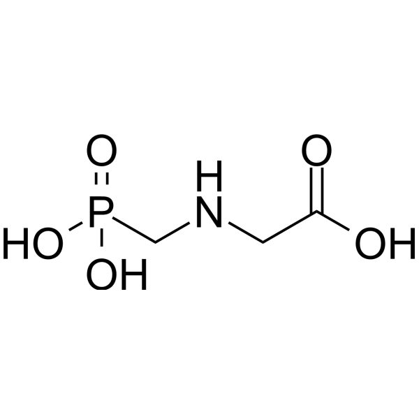
Glyphosate, a non-selective systemic biocide with broad-spectrum activity, is an herbicidal derivative of the amino acid glycine. Glyphosate inhibits the enzymatic activity of the 5-endopyruvylshikimate 3-phosphate synthase (EPSPS) in the shikimic acid pathway, preventing the synthesis of the aromatic amino acids tyrosine, phenylalanine, and tryptophan. Glyphosate induces oxidative stress, neuroinflammation, and mitochondrial dysfunction, processes that lead to neuronal death by autophagia, necrosis, or apoptosis, as well as the appearance of behavioral and motor disorders.
For research use only. We do not sell to patients.
- Purity : 99.89%
- CAS No.: 1071-83-6
- Formula: C3H8NO5P
- Molecular Weight:169.07
-
Storage:
4°C, protect from light
* In solvent : -80°C, 6 months; -20°C, 1 month (protect from light)

To place orders, for customer services and technical support, please contact: MedChemExpress USA
Tel: 609-228-6898 E-mail: [email protected] [email protected]


-
 Biological Activity
-
Chemical Information
-
Solvent & Solubility
- Protocols
- Purity & Documentation
- References
-
 Help & FAQs


-
HY-B0815
ChlorpyrifosMuscarinic Antagonist -
HY-B0673
PirfenidoneAntifibrotic Agent -
HY-B1945
DEHPEndogenous Metabolite -
HY-W018392
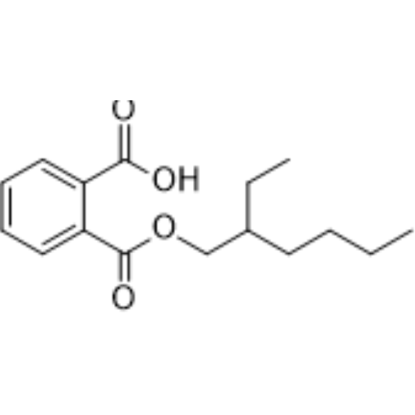
Mono-(2-ethylhexyl) phthalateEndogenous Metabolite -
HY-16561
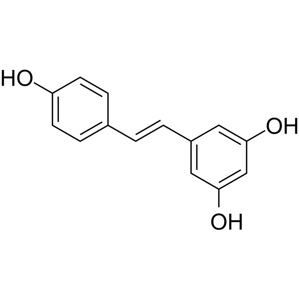
ResveratrolPolyphenolic Phytoalexin -
HY-W019870
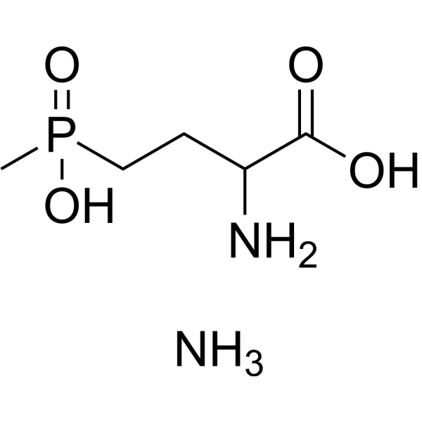
Glufosinate ammoniumHerbicide -
HY-N7091
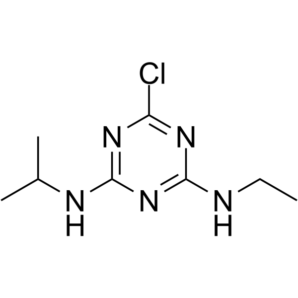
AtrazinePhotophosphorylation Inhibitor -
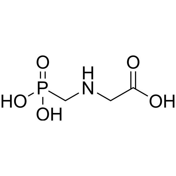
HY-B0863R
Glyphosate (Standard)Herbicide -
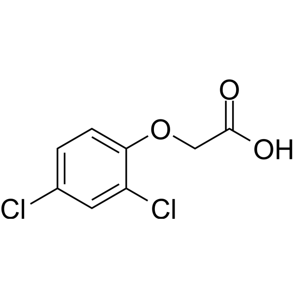
HY-18572
2,4-DHerbicide -
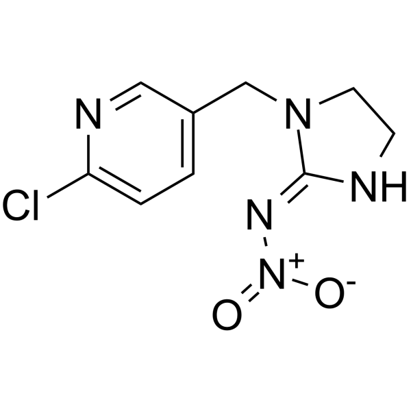
HY-B0838
ImidaclopridInsecticide -
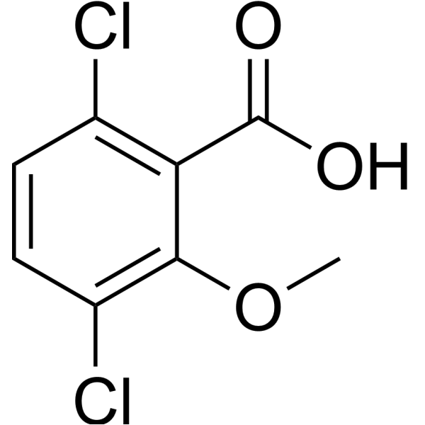
HY-121267
DicambaHerbicide -
HY-W009861
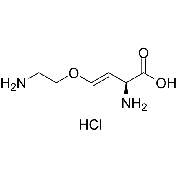
Aviglycine hydrochlorideEthylene Biosynthesis Inhibitor
Publications Citing Use of MedChemExpress (MCE) Glyphosate
More

Biological Activity
Description
In Vitro
Glyphosate exposure can interfere with mouse oocyte maturation by generating oxidative stress and early apoptosis.Glyphosate induces early apoptosis and autophagy in mouse oocytes[2].
MedChemExpress (MCE) has not independently confirmed the accuracy of these methods. They are for reference only.


Chemical Information
-
CAS No. 1071-83-6
-
Appearance Solid
-
Molecular Weight 169.07
-
Formula C3H8NO5P
-
Color White to off-white
-
SMILES
O=C(O)CNCP(O)(O)=O
-
Shipping
Room temperature in continental US; may vary elsewhere.
-
Storage
4°C, protect from light
* In solvent : -80°C, 6 months; -20°C, 1 month (protect from light)


Publications (1)
-
Journal Impact Factor
-
Most Recent
-
Environ Toxicol Pharmacol
Environmental factors influence goat immune response to PFOS and glyphosate: Difference between grazing and stall-feeding. [Abstract]2025 Aug:117:104754. PMID: 40619046


Solvent & Solubility
In Vitro:
H2O : 13.33 mg/mL (78.84 mM; ultrasonic and warming and heat to 60°C)
Please refer to the solubility information to select the appropriate solvent. Once prepared, please aliquot and store the solution to prevent product inactivation from repeated freeze-thaw cycles.
Storage method and period of stock solution: -80°C, 6 months; -20°C, 1 month (protect from light). When stored at -80°C, please use it within 6 months. When stored at -20°C, please use it within 1 month.
* Note: If you choose water as the stock solution, please dilute it to the working solution, then filter and sterilize it with a 0.22 μm filter before use.
Please refer to the solubility information to select the appropriate solvent. Once prepared, please aliquot and store the solution to prevent product inactivation from repeated freeze-thaw cycles.
Storage method and period of stock solution: -80°C, 6 months; -20°C, 1 month (protect from light). When stored at -80°C, please use it within 6 months. When stored at -20°C, please use it within 1 month.
* Note: If you choose water as the stock solution, please dilute it to the working solution, then filter and sterilize it with a 0.22 μm filter before use.
Concentration (start) × Volume (start) = Concentration (final) × Volume (final)
In Vivo:
For the following dissolution methods, please prepare the working solution directly:
It is recommended to prepare fresh solutions and use them promptly within a short period of time.
The percentages shown for the solvents indicate their volumetric ratio in the final prepared solution. If precipitation or phase separation occurs during preparation, heat and/or sonication can be used to aid dissolution.
Add each solvent one by one: PBS
Solubility: 6.67 mg/mL (39.45 mM); Clear solution; Need ultrasonic and warming and heat to 60°C


Purity & Documentation
-
Data Sheet (271 KB)
-
SDS (758 KB)
- English - EN (758 KB)
- Français - FR (758 KB)
- Deutsch - DE (758 KB)
- Norwegian - NO (758 KB)
- Español - ES (758 KB)
- Swedish - SV (758 KB)
- Italian - IT (758 KB)
- Korean - KR (758 KB)
- Portuguese - PT (758 KB)
-
Handling Instructions (2659 KB)
References
[1]. Greim H, et al. Evaluation of carcinogenic potential of the herbicide glyphosate, drawing on tumor incidence data from fourteen chronic/carcinogenicity rodent studies. Crit Rev Toxicol. 2015;45(3):185-208. [Content Brief]
[2]. Zhang JW, et al The toxic effects and possible mechanisms of glyphosate on mouse oocytes. Chemosphere. 2019 Dec;237:124435. [Content Brief]
[3]. Carmen Costas-Ferreira, et al. Toxic Effects of Glyphosate on the Nervous System: A Systematic Review. Int J Mol Sci. 2022 Apr 21;23(9):4605. [Content Brief]


Complete Stock Solution Preparation Table
Please refer to the solubility information to select the appropriate solvent. Once prepared, please aliquot and store the solution to prevent product inactivation from repeated freeze-thaw cycles.
Storage method and period of stock solution: -80°C, 6 months; -20°C, 1 month (protect from light). When stored at -80°C, please use it within 6 months. When stored at -20°C, please use it within 1 month.
| Optional Solvent | Concentration Solvent Mass | 1 mg | 5 mg | 10 mg | 25 mg |
|---|---|---|---|---|---|
| H2O | 1 mM | 5.9147 mL | 29.5735 mL | 59.1471 mL | 147.8677 mL |
| 5 mM | 1.1829 mL | 5.9147 mL | 11.8294 mL | 29.5735 mL | |
| 10 mM | 0.5915 mL | 2.9574 mL | 5.9147 mL | 14.7868 mL | |
| 15 mM | 0.3943 mL | 1.9716 mL | 3.9431 mL | 9.8578 mL | |
| 20 mM | 0.2957 mL | 1.4787 mL | 2.9574 mL | 7.3934 mL | |
| 25 mM | 0.2366 mL | 1.1829 mL | 2.3659 mL | 5.9147 mL | |
| 30 mM | 0.1972 mL | 0.9858 mL | 1.9716 mL | 4.9289 mL | |
| 40 mM | 0.1479 mL | 0.7393 mL | 1.4787 mL | 3.6967 mL | |
| 50 mM | 0.1183 mL | 0.5915 mL | 1.1829 mL | 2.9574 mL | |
| 60 mM | 0.0986 mL | 0.4929 mL | 0.9858 mL | 2.4645 mL |
* Note: If you choose water as the stock solution, please dilute it to the working solution, then filter and sterilize it with a 0.22 μm filter before use.


Glyphosate Related Classifications
HY-B0863 Related Classifications
 Powered by Bioz
Keywords
- Glyphosate
- 1071-83-6
- Environmental Pollutants
- Autophagy
- Apoptosis
- Necroptosis
- Herbicidal
- amino acid glycine
- 5-endopyruvylshikimate 3-phosphate synthase
- EPSPS
- shikimic acid pathway
- aromatic amino acids tyrosine
- phenylalanine
- tryptophan
- oxidative stress
- neuroinflammation
- mitochondrial dysfunction
- Inhibitor
- inhibitor
- inhibit