



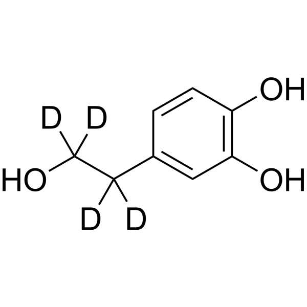
Hydroxytyrosol-d4

Based on 1 Customer Validation
Hydroxytyrosol-d4 (DOPET-d4) is the deuterium labeled Hydroxytyrosol (HY-N0570). Hydroxytyrosol is an orally active, blood-brain barrier-permeable multi-active compound with multiple effects including antibacterial, antioxidant, anti-platelet aggregation, and neuroprotective activities. Hydroxytyrosol not only inhibits the growth of Vibrio by increasing bacterial membrane permeability, but also interacts with DNA and mediates supercoiled DNA relaxation. Meanwhile, Hydroxytyrosol effectively reduces thrombosis and inhibits lipid oxidation by inhibiting COX activity and promoting vascular nitric oxide production. In terms of neuroprotection, Hydroxytyrosol significantly alleviates neuronal apoptosis and inflammatory responses by up-regulating the expression level of ERβ, thereby improving cognitive function in Alzheimer's disease models. Hydroxytyrosol has been widely used in scientific research related to Vibrio infection, arterial thrombosis, Alzheimer's disease and other related fields.
For research use only. We do not sell to patients.
- Purity : 96.15%
- CAS No.: 1330260-89-3
- Formula: C8H6D4O3
- Molecular Weight:158.19
-
Storage:
4°C, protect from light
* In solvent : -80°C, 6 months; -20°C, 1 month (protect from light)

To place orders, for customer services and technical support, please contact: MedChemExpress USA
Tel: 609-228-6898 E-mail: [email protected] [email protected]


-
 Biological Activity
-
Chemical Information
-
Solvent & Solubility
- Protocols
- Purity & Documentation
- References
-
 Help & FAQs




Biological Activity
Description
In Vitro
Stable heavy isotopes of hydrogen, carbon, and other elements have been incorporated into drug molecules, largely as tracers for quantitation during the drug development process. Deuteration has gained attention because of its potential to affect the pharmacokinetic and metabolic profiles of drugs[1].
MedChemExpress (MCE) has not independently confirmed the accuracy of these methods. They are for reference only. Further protocols information, click here.
Application
1. This compound can be used as a tracer
2. This compound can be used as an internal standard for quantitative analysis by NMR, GC-MS, or LC-MS.


Chemical Information
-
CAS No. 1330260-89-3
-
Unlabeled CAS 10597-60-1
-
Appearance Solid
-
Molecular Weight 158.19
-
Formula C8H6D4O3
-
Color White to off-white
-
SMILES
OC([2H])([2H])C([2H])([2H])C1=CC(O)=C(C=C1)O
-
Synonyms
DOPET-d4; 3,4-Dihydroxyphenethyl alcohol-d4; 3-Hydroxytyrosol-d4
-
Shipping
Room temperature in continental US; may vary elsewhere.
-
Storage
4°C, protect from light
* In solvent : -80°C, 6 months; -20°C, 1 month (protect from light)


Solvent & Solubility
In Vitro:
H2O : 100 mg/mL (632.15 mM; Need ultrasonic)
Ethanol : 50 mg/mL (316.08 mM; Need ultrasonic)
Please refer to the solubility information to select the appropriate solvent. Once prepared, please aliquot and store the solution to prevent product inactivation from repeated freeze-thaw cycles.
Storage method and period of stock solution: -80°C, 6 months; -20°C, 1 month (protect from light). When stored at -80°C, please use it within 6 months. When stored at -20°C, please use it within 1 month.
* Note: If you choose water as the stock solution, please dilute it to the working solution, then filter and sterilize it with a 0.22 μm filter before use.
Please refer to the solubility information to select the appropriate solvent. Once prepared, please aliquot and store the solution to prevent product inactivation from repeated freeze-thaw cycles.
Storage method and period of stock solution: -80°C, 6 months; -20°C, 1 month (protect from light). When stored at -80°C, please use it within 6 months. When stored at -20°C, please use it within 1 month.
* Note: If you choose water as the stock solution, please dilute it to the working solution, then filter and sterilize it with a 0.22 μm filter before use.
Concentration (start) × Volume (start) = Concentration (final) × Volume (final)

Protocols
-
Research Protocol for Infectious Diseases
Infectious-disease experiments test how pathogens interact with host barriers, innate immune receptors, inflammatory signaling, pathogen replication, and tissue injury; pattern-recognition receptors such as TLRs, RIG-I-like receptors, NOD-like receptors, and inflammasomes detect microbial molecules and activate NF-κB, interferon, and cytokine responses. The central hypothesis is that infection severity reflects the balance between pathogen burden and host response: protective inflammation restricts pathogen growth, whereas excessive or mislocalized inflammation contributes to tissue damage and disease phenotype. Unresolved questions include which host pathways are protective versus pathogenic, why some infection models fail to translate to human disease, and which combined readouts best predict clinically relevant infection outcomes.
-
RNA extraction experimental
By lysing cells, releasing RNA, and removing impurities such as proteins and DNA, high-purity RNA products are finally obtained. The commonly used traditional method is the guanidine isothiocyanate/phenol/chloroform method (Trizol), which is suitable for a variety of animal materials including animal tissues, microorganisms, cultured cells, etc., and most plant materials.
-
Apoptosis
Apoptosis, also called programmed cell death, is generally characterized by distinct morphological characteristics.
-
TUNEL staining for apoptotic DNA fragmentation
TUNEL staining detects DNA strand breaks by using terminal deoxynucleotidyl transferase to add labeled nucleotides to exposed 3′-OH DNA termini, generating either microscopic staining in fixed cells or tissue sections, or fluorescence/cytometric signal in cell suspensions. TUNEL positivity reflects DNA fragmentation but should not be interpreted alone as definitive apoptosis, because TUNEL can also label necrotic, autolytic, mechanically damaged, or DNA-repair-associated DNA breaks.
-
Annexin V plus membrane-impermeant dye apoptosis staining
Annexin V-based apoptosis assays rely on the detection of phosphatidylserine (PS) externalization from the inner leaflet of the plasma membrane to the outer leaflet, an early biochemical hallmark of apoptosis. Fluorescently labeled Annexin V binds PS in a calcium-dependent manner, enabling identification of early apoptotic cells by flow cytometry or fluorescence microscopy. When combined with a membrane-impermeant DNA-binding dye (e. g. , propidium iodide), this approach allows discrimination between viable (Annexin V−/dye−), early apoptotic (Annexin V+/dye−), and late apoptotic or necrotic (Annexin V+/dye+) cell populations by assessing membrane integrity and PS exposure.
-
Protocol for Water Maze
The Morris Water Maze is a rodent spatial learning and memory assay in which a mouse or rat swims in opaque water to find an escape platform; in the hidden-platform version, the animal cannot see the platform and must use distal extra-maze cues to learn its fixed spatial location. The assay primarily measures hippocampus-dependent spatial learning during acquisition trials and spatial reference memory during probe trials after platform removal; readouts include escape latency, swim path length, swim speed, quadrant occupancy, platform-site crossings, and proximity to the former platform location.
-
Transepithelial/transendothelial electrical resistance assay
TEER measures electrical resistance across epithelial or endothelial monolayers cultured on permeable supports, and the readout reflects ionic conductance through the cell barrier, especially the paracellular pathway regulated by junctional integrity. TEER can be measured without destroying the monolayer and is commonly used before or during transport, permeability, barrier-disruption, and barrier-maturation experiments. TEER values are influenced by biological maturation and technical conditions; reported factors include temperature, medium formulation, passage number, electrode geometry, membrane properties, and junctional length during early monolayer maturation. Therefore, TEER should be interpreted with blank-insert subtraction, area normalization, repeated readings, and, when possible, orthogonal barrier readouts such as FITC-dextran flux or tight-junction staining.
-
Apoptosis Solutions
Apoptosis is a regulated, generally non-lytic cell-death pathway that removes unwanted, damaged, infected, or abnormal cells through coordinated morphological changes, caspase activation, DNA fragmentation, and membrane remodeling. The intrinsic apoptosis pathway is controlled mainly by mitochondrial outer membrane permeabilization, BCL-2 family proteins, cytochrome c release, apoptosome formation, caspase-9 activation, and downstream executioner caspase-3/7 activation. The extrinsic apoptosis pathway is initiated by death receptors such as Fas, TNFR, and TRAIL receptors, which recruit adaptor proteins and activate caspase-8 before engaging executioner caspases or mitochondrial amplification through BID cleavage. Apoptosis is linked to many phenotypes, including cancer cell killing, tissue homeostasis, immune regulation, neurodegeneration, infection response, and treatment-induced cytotoxicity; unresolved questions include how apoptosis interacts with necroptosis, pyroptosis, ferroptos
-
Alzheimer’s Disease Modeling
Alzheimer’s Disease (AD) is a neurodegenerative disorder characterized by a progressive decline in cognitive functions and loss of specific types of neurons and synapses. Alzheimer's symptoms can be simulated in mice by injecting drugs (such as Aβ) or genetically modified.


Purity & Documentation
-
Data Sheet (276 KB)
-
SDS (616 KB)
- English - EN (616 KB)
- Français - FR (616 KB)
- Deutsch - DE (616 KB)
- Norwegian - NO (616 KB)
- Español - ES (616 KB)
- Swedish - SV (616 KB)
- Italian - IT (616 KB)
- Korean - KR (616 KB)
- Portuguese - PT (616 KB)
-
Handling Instructions (2659 KB)
References
[1]. Wei J, et al. Antibacterial Activity of Hydroxytyrosol Acetate from Olive Leaves (Olea Europaea L.). Nat Prod Res. 2018;32(16):1967-1970. [Content Brief]
[2]. González-Correa JA, et al. Effects of hydroxytyrosol and hydroxytyrosol acetate administration to rats on platelet function compared to acetylsalicylic acid. J Agric Food Chem. 2008;56(17):7872-7876. [Content Brief]
[3]. Gordon MH, et al. Antioxidant activity of hydroxytyrosol acetate compared with that of other olive oil polyphenols. J Agric Food Chem. 2001;49(5):2480-2485. [Content Brief]


Complete Stock Solution Preparation Table
Please refer to the solubility information to select the appropriate solvent. Once prepared, please aliquot and store the solution to prevent product inactivation from repeated freeze-thaw cycles.
Storage method and period of stock solution: -80°C, 6 months; -20°C, 1 month (protect from light). When stored at -80°C, please use it within 6 months. When stored at -20°C, please use it within 1 month.
| Optional Solvent | Concentration Solvent Mass | 1 mg | 5 mg | 10 mg | 25 mg |
|---|---|---|---|---|---|
| Ethanol / H2O | 1 mM | 6.3215 mL | 31.6076 mL | 63.2151 mL | 158.0378 mL |
| 5 mM | 1.2643 mL | 6.3215 mL | 12.6430 mL | 31.6076 mL | |
| 10 mM | 0.6322 mL | 3.1608 mL | 6.3215 mL | 15.8038 mL | |
| 15 mM | 0.4214 mL | 2.1072 mL | 4.2143 mL | 10.5359 mL | |
| 20 mM | 0.3161 mL | 1.5804 mL | 3.1608 mL | 7.9019 mL | |
| 25 mM | 0.2529 mL | 1.2643 mL | 2.5286 mL | 6.3215 mL | |
| 30 mM | 0.2107 mL | 1.0536 mL | 2.1072 mL | 5.2679 mL | |
| 40 mM | 0.1580 mL | 0.7902 mL | 1.5804 mL | 3.9509 mL | |
| 50 mM | 0.1264 mL | 0.6322 mL | 1.2643 mL | 3.1608 mL | |
| 60 mM | 0.1054 mL | 0.5268 mL | 1.0536 mL | 2.6340 mL | |
| 80 mM | 0.0790 mL | 0.3951 mL | 0.7902 mL | 1.9755 mL | |
| 100 mM | 0.0632 mL | 0.3161 mL | 0.6322 mL | 1.5804 mL |
* Note: If you choose water as the stock solution, please dilute it to the working solution, then filter and sterilize it with a 0.22 μm filter before use.


Hydroxytyrosol-d4 Related Classifications
HY-N0570S Related Classifications
 Powered by Bioz