



Gol-NTR

Based on 1 Customer Validation
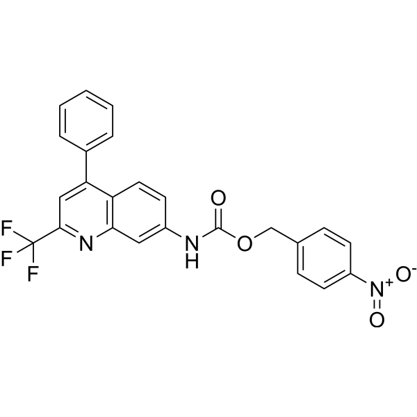
Gol-NTR is a Golgi-targetable probe with high selectivity and sensitivity. Gol-NTR is Nitroreductase (NTR)-activated and has visualization acute lung injury (ALI) and repair function. Gol-NTR has a low detection limit of 54.8 ng/mL. Gol-NTR can be used for the research for monitoring and assessing research response of sepsis-induced ALI.
Nos produits utilisent uniquement pour la recherche. Nous ne vendons pas aux patients.
- Pureté : 99.44%
- CAS No.: 2968461-58-5
- Formule: C24H16F3N3O4
- Masse moléculaire:467.40
-
Stockage:
4°C, sealed storage, away from moisture and light
* In solvent : -80°C, 6 months; -20°C, 1 month (sealed storage, away from moisture and light)

To place orders, for customer services and technical support, please contact: MedChemExpress USA
Tel: 609-228-6898 E-mail: [email protected] [email protected]


-
 Activité biologique
-
Chemical Information
- Protocole
- Pureté et documentation
- Références
-
 Help & FAQs




Activité biologique
Description
In Vitro
Guide (Following is our recommended protocol. This protocol only provides a guideline, and should be modified according to your specific needs).
Fluorescent labeling of NTR by Gol-NTR[1]
(1) Prepare 1.0 mM Gol-NTR stock solution with DMSO solution.
(2) Dilute the stock solution with DMSO solution to prepare 5.0 μM Gol-NTR working solution.
(3) Mix 5.0 μM Gol-NTR with 50 μM NADH in PBS buffer (10 mM, pH 7.4) containing 5% DMSO, and then add appropriate NTR.
(4) After incubation at 37°C for 30 min, the spectra was recorded at 405 nm (slit width: dex/dem=5/5 nm).
Fluorescence labeling of NTR in A549 cells by Gol-NTR[1]
(1) A549 cells were cultured at different oxygen concentrations (1%, 5%, 10%, 15% and 20% O2) for 8 h.
(2) A549 cells were washed with phosphate buffered saline (PBS).
(3) A549 cells were treated with 5.0 μM Gol-NTR for 1 h.
(4) Fluorescence images of A549 cells were observed using confocal fluorescence imaging.
MedChemExpress (MCE) has not independently confirmed the accuracy of these methods. They are for reference only. Further protocols information, click here.
In Vivo
(1) C57BL/6 male mice (6-8 weeks old, weight 20-22 g) were pre injected with 300 μL DMOG (25 mg/mL), after 24 h, intraperitoneal injection of 300 μL LPS (10 mg/kg) for 6 h.
(2) Mice were killed by cervical vertebra dislocation and lung organs were collected.
(3) After washing with PBS, incubate with 50 μM Gol-NTR in PBS for 1 h.
(4) After washing with PBS, fluorescence imaging was performed on a small animal imaging system (excitation wavelength of 420 nm and emission wavelength of 510 nm).
MedChemExpress (MCE) has not independently confirmed the accuracy of these methods. They are for reference only.
Emission (Em)
730
Excitation (Ex)
680


Chemical Information
-
CAS No. 2968461-58-5
-
Appearance Solid
-
Masse moléculaire 467.40
-
Formule C24H16F3N3O4
-
Color White to off-white
-
SMILES
FC(F)(C1=NC(C=C(NC(OCC2=CC=C([N+]([O-])=O)C=C2)=O)C=C3)=C3C(C4=CC=CC=C4)=C1)F
-
Livraison
Room temperature in continental US; may vary elsewhere.
-
Stockage
4°C, sealed storage, away from moisture and light
* In solvent : -80°C, 6 months; -20°C, 1 month (sealed storage, away from moisture and light)

Protocole
-
LPS-Induced Endotoxemia/Systemic Inflammation
Lipopolysaccharide (LPS)-induced endotoxemia is a widely used in vivo model of acute systemic inflammation in which LPS, a Gram-negative bacterial endotoxin, activates innate immune signaling primarily through TLR4, leading to rapid and transient induction of pro-inflammatory cytokines such as TNF-α, IL-6, and IL-1β in circulation and tissues. This cytokine surge is commonly used as a measurable readout of systemic inflammatory activation and immune dysregulation, and is typically assessed within hours after intraperitoneal LPS administration in mouse models of endotoxemia. The model captures key features of systemic inflammatory response syndrome, including cytokine release, immune cell activation, and downstream tissue responses, and has been used to evaluate anti-inflammatory interventions such as cytokine modulation, lipid mediators, and immune cell-targeting therapies.
-
Inhalation Toxicity Study
Inhalation toxicity studies expose rodents to a controlled aerosol, vapor, gas, or smoke atmosphere and assess respiratory and systemic toxicity using exposure-atmosphere characterization, clinical observations, body and organ weights, bronchoalveolar lavage fluid, histopathology, blood chemistry, hematology, and, when included, molecular endpoints such as transcriptomics, proteomics, lipidomics, or tissue burden analysis. The primary biological readouts are airway irritation, pulmonary inflammation, cytotoxicity, altered surfactant or lipid homeostasis, impaired particle clearance, and tissue remodeling, reflected by BALF cell differentials, BALF protein, LDH, phosphatase activities, cytokines, lung weight, microscopic respiratory-tract lesions, and retained lung burden.


Pureté et documentation
-
Fiche technique (271 KB)
-
SDS (251 KB)
- English - EN (251 KB)
- Français - FR (251 KB)
- Deutsch - DE (251 KB)
- Norwegian - NO (251 KB)
- Español - ES (251 KB)
- Swedish - SV (251 KB)
- Italian - IT (251 KB)
- Korean - KR (251 KB)
- Portuguese - PT (251 KB)
-
Instruction de manipulation (2659 KB)
Références

Calculators
Concentration (start) × Volume (start) = Concentration (final) × Volume (final)


Gol-NTR Related Classifications
HY-151537 Related Classifications
 Powered by Bioz