



HG106
Based on 2 publication(s) in Google Scholar
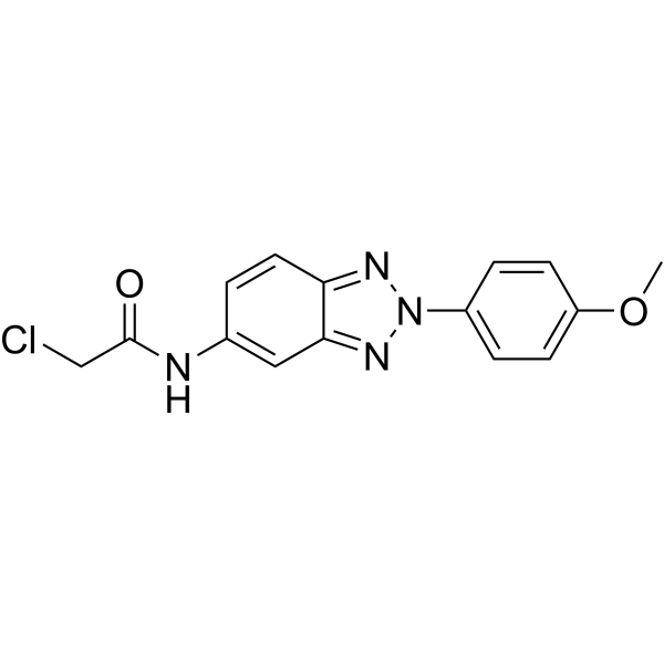
HG106 is an effective SLC7A11 inhibitor. HG106 mediates apoptosis by increasing oxidative stress and endoplasmic reticulum stress, and it has antitumor activity.
For research use only. We do not sell to patients.
- Purity : 99.90%
- CAS No.: 928712-10-1
- Formula: C15H13ClN4O2
- Molecular Weight:316.74
-
Storage:
-20°C, sealed storage, away from moisture and light
* In solvent : -80°C, 6 months; -20°C, 1 month (sealed storage, away from moisture and light)

To place orders, for customer services and technical support, please contact: MedChemExpress USA
Tel: 609-228-6898 E-mail: [email protected] [email protected]


-
 Biological Activity
-
Chemical Information
-
Solvent & Solubility
- Protocols
- Purity & Documentation
- References
-
 Help & FAQs


-

HY-15763
ErastinFerroptosis Inducer -

HY-100579
Ferrostatin-1Ferroptosis Inhibitor -
HY-138170
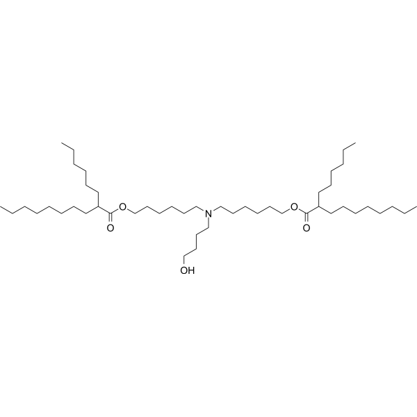
ALC-0315Lipid Nanoparticle Component -

HY-100558
Bafilomycin A1V-ATPase Inhibitor -
HY-106376
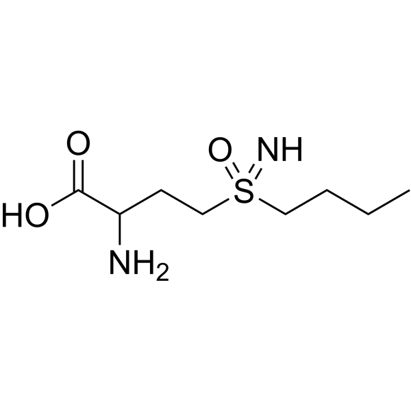
DL-Buthionine-(S,R)-sulfoximineGlutamylcysteine Synthetase Inhibitor -
HY-100218A
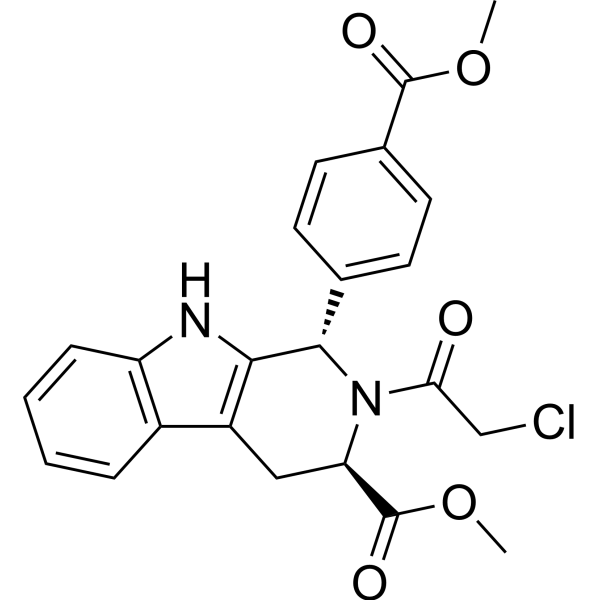
RSL3GPX4 Inhibitor -
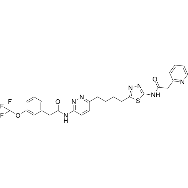
HY-12248
TelaglenastatGLS1 Inhibitor -
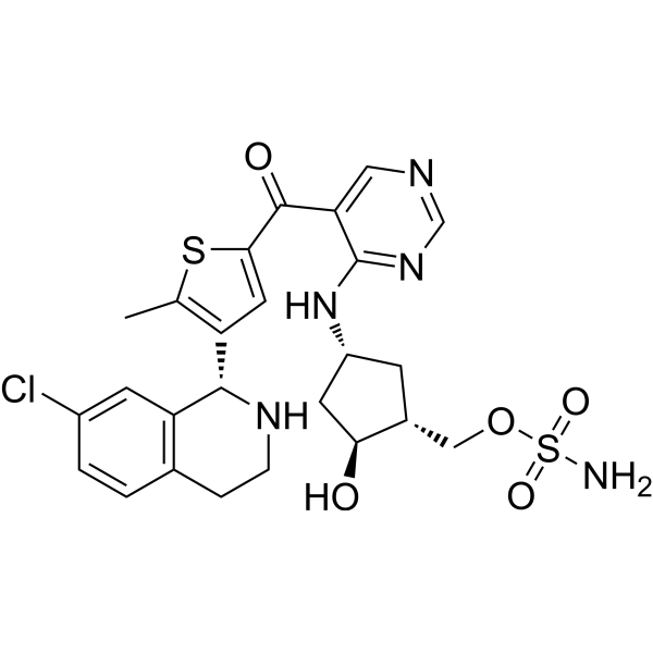
HY-111789
SubasumstatSUMOylation Inhibitor -
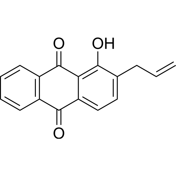
HY-103096
R162GDH1 Inhibitor -
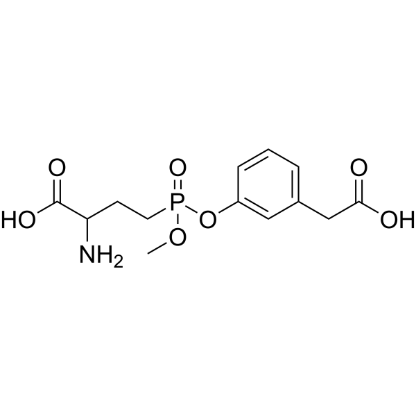
HY-108467
GGsTopγ GGT Inhibitor -

HY-K0301
Cell Counting Kit-8 -

HY-W402682
SXC2023Cystine-glutamate Antiporter Modulator
Publications Citing Use of MedChemExpress (MCE) HG106
More Customer Validation & Images

-
Flow Cytometry


Biological Activity
Description
IC50 & Target
SLC7A11
In Vitro
HG106 (1.25-10 μM, 3 min) shows a concentration-dependent inhibition of [14C] cystine consumption and glutathione production[1].
HG106 (0.1-100 μM, 72 h) has a stronger cytotoxic effect on KRAS mutant cell lines[1].
HG106 (0-10 μM, 6 h) dose-dependently increases the total ROS levels in A549 cells[1].
HG106 (0-5 μM, 24 h) causes mitochondrial dysfunction and endoplasmic reticulum stress in A549 cells[1].
HG106 (0-10 μM, 72 h) significantly induces apoptosis in KRAS mutant LUAD cells and inhibits colony formation[1].
MedChemExpress (MCE) has not independently confirmed the accuracy of these methods. They are for reference only.
-
Cell Line:H441 KRAS(G12V), HPNE, HPNE/KRAS
-
Concentration:0.1, 1, 10, 100 μM
-
Incubation Time:72 h
-
Result:Inhibited cell proliferation, with greater effects on KRAS mutant cell lines.
-
Cell Line:A549
-
Concentration:0, 1.25, 2.5, 5 μM
-
Incubation Time:24 h
-
Result:Increased activation of ER stress-related markers IRE1α, PERK and GRP78.
-
Cell Line:KRAS mutant LUAD
-
Concentration:0, 1.25, 2.5, 5, 10; 0, 0.5, 1, 2 μM
-
Incubation Time:72 h
-
Result:Did not cause cell autophagy, induced cell apoptosis, and inhibited colony formation.
In Vivo
HG106 (0-4 mg/kg, once a day, intraperitoneal injection, 20 days) inhibits tumor growth in mice in a xenograft model and induces apoptosis by triggering endoplasmic membrane stress[1].
MedChemExpress (MCE) has not independently confirmed the accuracy of these methods. They are for reference only.
-
Animal Model:A549 mouse transplant model[1]
-
Dosage:0, 1, 2, 4 mg/kg; daily; 26 days
-
Administration:Intraperitoneal injection (i.p.)
-
Result:Inhibited tumor growth and prolonged the inhibition time of tumor growth.
-
Animal Model:LUAD patient-derived xenograft (PDX) models harboring the G12V mutation in KRAS[1]
-
Dosage:0, 1, 2, 4 mg/kg; daily; 20 days
-
Administration:Intraperitoneal injection (i.p.)
-
Result:Inhibited tumor growth, increased ROS generation and TUNEL signal in patient-derived xenografts validated that HG106 triggered endoplasmic membrane stress-induced apoptosis in vivo.


Chemical Information
-
CAS No. 928712-10-1
-
Appearance Solid
-
Molecular Weight 316.74
-
Formula C15H13ClN4O2
-
Color Off-white to light yellow
-
SMILES
O=C(NC1=CC2=NN(C3=CC=C(OC)C=C3)N=C2C=C1)CCl
-
Shipping
Room temperature in continental US; may vary elsewhere.
-
Storage
-20°C, sealed storage, away from moisture and light
* In solvent : -80°C, 6 months; -20°C, 1 month (sealed storage, away from moisture and light)


Publications (2)
-
Journal Impact Factor
-
Most Recent
-
J Control Release
Apoptotic neutrophil membrane-modified nanoparticles promote wound healing by enhancing efferocytotic capacity of dendritic cells. [Abstract]2025 Oct 10:386:114127. PMID: 40816639
HG106 purchased from MedChemExpress. Usage Cited in: J Control Release. 2025 Oct 10:386:114127. [Abstract]
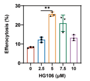
Flow cytometry analysis of HG106 (2.5-10 μM) in promoting BMDCs phagocytosis efficiency in vitro. The results showed that HG106 was effective at promoting BMDCs efferocytosis in vitro, with the highest phagocytic rate observed at a concentration of 5 μM.
-
Cell Chem Biol
Electrochemical sensor toolkit for simultaneous glutamate detection at edge of cleft and peri-soma. [Abstract]2025 Jun 19;32(6):885-898.e11. PMID: 40466640


Solvent & Solubility
In Vitro:
DMSO : 125 mg/mL (394.65 mM; Need ultrasonic; Hygroscopic DMSO has a significant impact on the solubility of product, please use newly opened DMSO)
Please refer to the solubility information to select the appropriate solvent. Once prepared, please aliquot and store the solution to prevent product inactivation from repeated freeze-thaw cycles.
Storage method and period of stock solution: -80°C, 6 months; -20°C, 1 month (sealed storage, away from moisture and light). When stored at -80°C, please use it within 6 months. When stored at -20°C, please use it within 1 month.
Please refer to the solubility information to select the appropriate solvent. Once prepared, please aliquot and store the solution to prevent product inactivation from repeated freeze-thaw cycles.
Storage method and period of stock solution: -80°C, 6 months; -20°C, 1 month (sealed storage, away from moisture and light). When stored at -80°C, please use it within 6 months. When stored at -20°C, please use it within 1 month.
Concentration (start) × Volume (start) = Concentration (final) × Volume (final)


Purity & Documentation
-
Data Sheet (275 KB)
-
SDS (614 KB)
- English - EN (614 KB)
- Français - FR (614 KB)
- Deutsch - DE (614 KB)
- Norwegian - NO (614 KB)
- Español - ES (614 KB)
- Swedish - SV (614 KB)
- Italian - IT (614 KB)
- Korean - KR (614 KB)
- Portuguese - PT (614 KB)
-
Handling Instructions (2659 KB)
References


Complete Stock Solution Preparation Table
Please refer to the solubility information to select the appropriate solvent. Once prepared, please aliquot and store the solution to prevent product inactivation from repeated freeze-thaw cycles.
Storage method and period of stock solution: -80°C, 6 months; -20°C, 1 month (sealed storage, away from moisture and light). When stored at -80°C, please use it within 6 months. When stored at -20°C, please use it within 1 month.
| Optional Solvent | Concentration Solvent Mass | 1 mg | 5 mg | 10 mg | 25 mg |
|---|---|---|---|---|---|
| DMSO | 1 mM | 3.1572 mL | 15.7858 mL | 31.5716 mL | 78.9291 mL |
| 5 mM | 0.6314 mL | 3.1572 mL | 6.3143 mL | 15.7858 mL | |
| 10 mM | 0.3157 mL | 1.5786 mL | 3.1572 mL | 7.8929 mL | |
| 15 mM | 0.2105 mL | 1.0524 mL | 2.1048 mL | 5.2619 mL | |
| 20 mM | 0.1579 mL | 0.7893 mL | 1.5786 mL | 3.9465 mL | |
| 25 mM | 0.1263 mL | 0.6314 mL | 1.2629 mL | 3.1572 mL | |
| 30 mM | 0.1052 mL | 0.5262 mL | 1.0524 mL | 2.6310 mL | |
| 40 mM | 0.0789 mL | 0.3946 mL | 0.7893 mL | 1.9732 mL | |
| 50 mM | 0.0631 mL | 0.3157 mL | 0.6314 mL | 1.5786 mL | |
| 60 mM | 0.0526 mL | 0.2631 mL | 0.5262 mL | 1.3155 mL | |
| 80 mM | 0.0395 mL | 0.1973 mL | 0.3946 mL | 0.9866 mL | |
| 100 mM | 0.0316 mL | 0.1579 mL | 0.3157 mL | 0.7893 mL |


HG106 Related Classifications
HY-W451275 Related Classifications
 Powered by Bioz