



MitoPerOx
Based on 6 publication(s) in Google Scholar
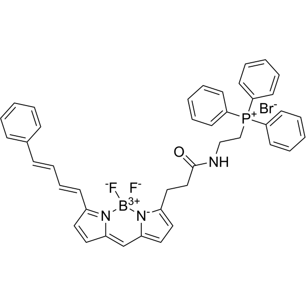
MitoPerOx is a mitochondrial-targeted, lipid peroxidation-indicating fluorescent probe with BODIPY581/591 fluorophores. The triphenylphosphine cation (TPP+) of MitoPerOx can be selectively enriched in mitochondria (depending on membrane potential) and can be used to detect lipid peroxidation in the inner mitochondrial membrane. Under the action of lipid peroxides, the BODIPY581/591 fluorophores of MitoPerOx shift their emission wavelength from 590 nm (reduced state) to 520 nm (oxidized state), and ratiometric detection can be performed at an excitation wavelength of 488 nm. MitoPerOx can specifically monitor the peroxidation of mitochondrial phospholipids (especially cardiolipin) and is used in the study of oxidative stress-related diseases (such as aging, neurodegenerative diseases, and mitochondrial dysfunction)[1][2].
For research use only. We do not sell to patients.
- Purity : 95.01%
- CAS No.: 1392820-50-6
- Formula: C42H38BBrF2N3OP
- Molecular Weight:760.46
-
Storage:
-20°C, sealed storage, away from moisture and light
* In solvent : -80°C, 6 months; -20°C, 1 month (sealed storage, away from moisture and light)

To place orders, for customer services and technical support, please contact: MedChemExpress USA
Tel: 609-228-6898 E-mail: [email protected] [email protected]


-
 Biological Activity
-
Chemical Information
-
Solvent & Solubility
- Protocols
- Purity & Documentation
- References
-
 Help & FAQs


-

HY-100579
Ferrostatin-1Ferroptosis Inhibitor -
HY-D0940
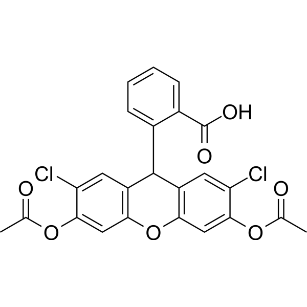
H2DCFDAROS-Sensitive Probe -

HY-15763
ErastinFerroptosis Inducer -
HY-112879
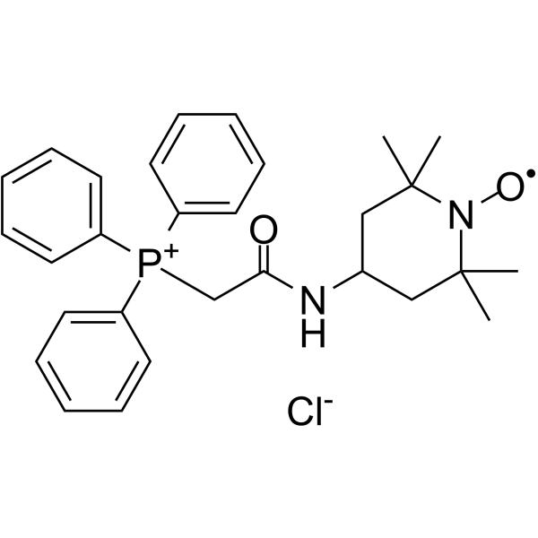
Mito-TEMPOAntioxidant -
HY-15534
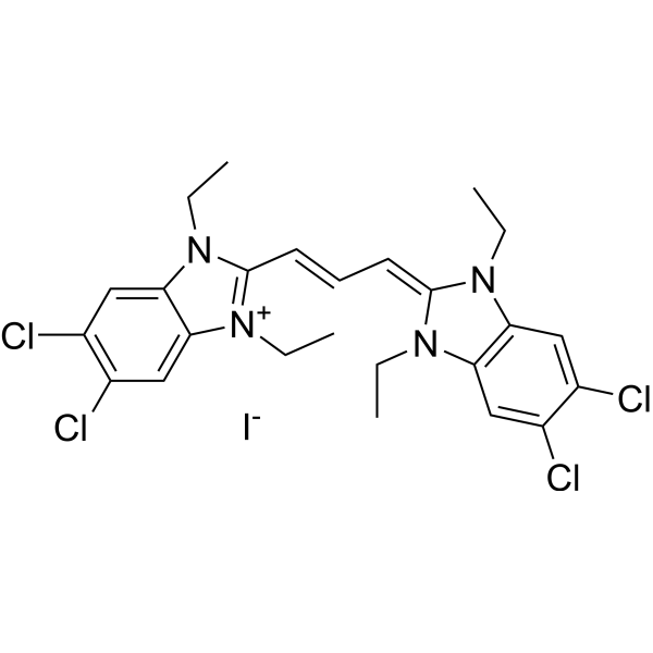
JC-1Mitochondrial Membrane Potential Probe -
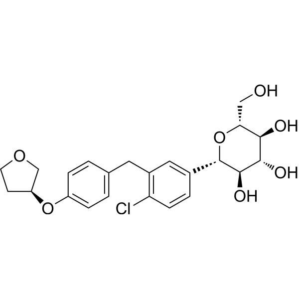
HY-15409
EmpagliflozinSGLT2 Inhibitor -

HY-10219
RapamycinmTOR Inhibitor -

HY-DY1073
MitoPerOx (solution)Fluorescent Probe -
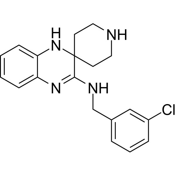
HY-12726
Liproxstatin-1Ferroptosis Inhibitor -
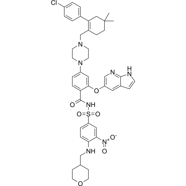
HY-15531
VenetoclaxBCL-2 Inhibitor -
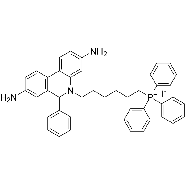
HY-D1055
MitoSOX RedMitochondrial ROS Indicator -
HY-14164
Zileuton5-Lipoxygenase Inhibitor
Publications Citing Use of MedChemExpress (MCE) MitoPerOx
More- Signal Transduct Target Ther. 2025 Dec 26;10(1):420. [Abstract]
- Redox Biol. 2026 May:92:104132. [Abstract]
- Adv Sci (Weinh). 2026 Apr;13(22):e21997. [Abstract]
- Cell Rep. 2026 Jul 27;45(8):117704.
- Chem Biol Interact. 2026 Jul 25:435:112163. [Abstract]
- Blood Neoplasia. 2026 Apr 16;3(3):100236. [Abstract]


Biological Activity
Description
In Vitro
Guide (Following is our recommended protocol. This protocol only provides a guideline, and should be modified according to your specific needs)[2].
1. Solution preparation
1.1 Preparation of stock solution
Solvent: DMSO
Concentration: 1 mM.
1.2 Preparation of working solution
Dilute to 100 nM-1 μM with PBS or cell culture medium (optimized according to the experiment).
Note: The working solution should be prepared and used immediately. Keep it away from light.
2. MitoPerOx Assay Protocol Cell Culture:
2.1 Cells (e.g., HEK293 or fibroblasts) are plated on coverslips in 35 mm dishes and cultured at 37 °C, 5% CO2.
2.2 Add 100 nM MitoPerOx to phenol red-free medium and incubate at 37 °C for 30 min in the dark.
2.3 Washing: Discard medium and wash cells three times with PBS.
2.4 Induce oxidative stress: Treat cells with 500 μM H2O2 in Hepes-Tris buffer for 15-30 min.
2.5 Fluorescence imaging: Confocal microscopy (excitation wavelength 488 nm) was used to collect emission light at 520 nm (oxidized state) and 590 nm (reduced state), and the extent of lipid peroxidation was quantified by calculating the 520/590 nm ratio.
MedChemExpress (MCE) has not independently confirmed the accuracy of these methods. They are for reference only.
Emission (Em)
520/590
Excitation (Ex)
490


Chemical Information
-
CAS No. 1392820-50-6
-
Appearance Solid
-
Molecular Weight 760.46
-
Formula C42H38BBrF2N3OP
-
Color Dark purple to black
-
SMILES
O=C(CCC1=CC=C2[N-]1[B+3]([F-])([N]3=C(C=CC3=C2)/C=C/C=C/C4=CC=CC=C4)[F-])NCC[P+](C5=CC=CC=C5)(C6=CC=CC=C6)C7=CC=CC=C7.[Br-]
-
Shipping
Room temperature in continental US; may vary elsewhere.
-
Storage
-20°C, sealed storage, away from moisture and light
* In solvent : -80°C, 6 months; -20°C, 1 month (sealed storage, away from moisture and light)


Publications (6)
-
Journal Impact Factor
-
Most Recent
-
Signal Transduct Target Ther
Inhibin beta A drives colorectal cancer progression through macrophage M2 polarization and mitochondria-dependent ferroptosis suppression. [Abstract]2025 Dec 26;10(1):420. PMID: 41449244 -
Redox Biol
CTPS1 modulates mitophagy to propel diffuse large B-cell lymphoma via reshaping CEPT1-mediated phospholipid metabolism. [Abstract]2026 May:92:104132. PMID: 41865720 -
Adv Sci (Weinh)
TrxR2 Lactylation Facilitates Mitochondrial Protection and Endothelial Ferroptosis Resistance in Diabetic Cardiomyopathy. [Abstract]2026 Apr;13(22):e21997. PMID: 41704008 -
-
Chem Biol Interact
PM2.5 induces mitochondrial damage-triggered ferroptosis in renal tubular cells leading to renal injury. [Abstract]2026 Jul 25:435:112163. PMID: 42173391 -
Blood Neoplasia
The KDM-family inhibitor JIB-04 sensitizes AML cells to venetoclax by inducing a ferroptosis-like phenotype. [Abstract]2026 Apr 16;3(3):100236. PMID: 42221838


Solvent & Solubility
In Vitro:
DMSO : 125 mg/mL (164.37 mM; Need ultrasonic; Hygroscopic DMSO has a significant impact on the solubility of product, please use newly opened DMSO)
Please refer to the solubility information to select the appropriate solvent. Once prepared, please aliquot and store the solution to prevent product inactivation from repeated freeze-thaw cycles.
Storage method and period of stock solution: -80°C, 6 months; -20°C, 1 month (sealed storage, away from moisture and light). When stored at -80°C, please use it within 6 months. When stored at -20°C, please use it within 1 month.
Please refer to the solubility information to select the appropriate solvent. Once prepared, please aliquot and store the solution to prevent product inactivation from repeated freeze-thaw cycles.
Storage method and period of stock solution: -80°C, 6 months; -20°C, 1 month (sealed storage, away from moisture and light). When stored at -80°C, please use it within 6 months. When stored at -20°C, please use it within 1 month.
Concentration (start) × Volume (start) = Concentration (final) × Volume (final)


Purity & Documentation
-
Data Sheet (280 KB)
-
SDS (252 KB)
- English - EN (252 KB)
- Français - FR (252 KB)
- Deutsch - DE (252 KB)
- Norwegian - NO (252 KB)
- Español - ES (252 KB)
- Swedish - SV (252 KB)
- Italian - IT (252 KB)
- Korean - KR (252 KB)
- Portuguese - PT (252 KB)
-
Handling Instructions (2659 KB)
References
[1]. Lyamzaev KG, Sumbatyan NV, Nesterenko AM, Kholina EG, Voskoboynikova N, Steinhoff HJ, Mulkidjanian AY, Chernyak BV. MitoCLox: A Novel Mitochondria-Targeted Fluorescent Probe for Tracing Lipid Peroxidation. Oxid Med Cell Longev. 2019 Nov 13;2019:9710208. [Content Brief]
[2]. Prime TA, et, al. A ratiometric fluorescent probe for assessing mitochondrial phospholipid peroxidation within living cells. Free Radic Biol Med. 2012 Aug 1;53(3):544-53. [Content Brief]


Complete Stock Solution Preparation Table
Please refer to the solubility information to select the appropriate solvent. Once prepared, please aliquot and store the solution to prevent product inactivation from repeated freeze-thaw cycles.
Storage method and period of stock solution: -80°C, 6 months; -20°C, 1 month (sealed storage, away from moisture and light). When stored at -80°C, please use it within 6 months. When stored at -20°C, please use it within 1 month.
| Optional Solvent | Concentration Solvent Mass | 1 mg | 5 mg | 10 mg | 25 mg |
|---|---|---|---|---|---|
| DMSO | 1 mM | 1.3150 mL | 6.5750 mL | 13.1499 mL | 32.8748 mL |
| 5 mM | 0.2630 mL | 1.3150 mL | 2.6300 mL | 6.5750 mL | |
| 10 mM | 0.1315 mL | 0.6575 mL | 1.3150 mL | 3.2875 mL | |
| 15 mM | 0.0877 mL | 0.4383 mL | 0.8767 mL | 2.1917 mL | |
| 20 mM | 0.0657 mL | 0.3287 mL | 0.6575 mL | 1.6437 mL | |
| 25 mM | 0.0526 mL | 0.2630 mL | 0.5260 mL | 1.3150 mL | |
| 30 mM | 0.0438 mL | 0.2192 mL | 0.4383 mL | 1.0958 mL | |
| 40 mM | 0.0329 mL | 0.1644 mL | 0.3287 mL | 0.8219 mL | |
| 50 mM | 0.0263 mL | 0.1315 mL | 0.2630 mL | 0.6575 mL | |
| 60 mM | 0.0219 mL | 0.1096 mL | 0.2192 mL | 0.5479 mL | |
| 80 mM | 0.0164 mL | 0.0822 mL | 0.1644 mL | 0.4109 mL | |
| 100 mM | 0.0131 mL | 0.0657 mL | 0.1315 mL | 0.3287 mL |


MitoPerOx Related Classifications
HY-125623 Related Classifications
 Powered by Bioz