



Lipid Peroxidation (MDA) Assay Kit
Based on 10 publication(s) in Google Scholar
MCE Lipid Peroxidation (MDA) Assay Kit is suitable for measuring MDA levels in a variety of samples including plasma, serum, urine, tissues or cell lysates.
-
Storage :
-20℃, 1 year. Keep away from light and avoid repeated freezing and thawing.


To place orders, for customer services and technical support, please contact: MedChemExpress USA
Tel: 609-228-6898 E-mail: [email protected] [email protected]



-

HY-K0311
GSH/GSSG Assay Kit -

HY-100579
Ferrostatin-1Ferroptosis Inhibitor -

HY-K0320
ROS Assay Kit -
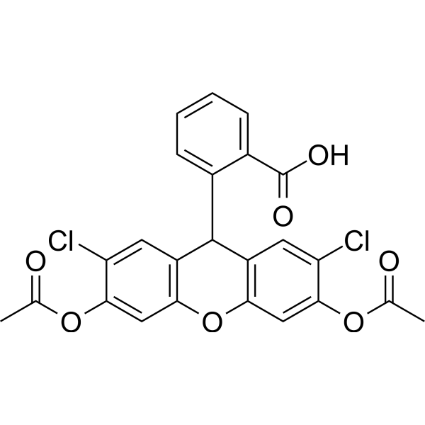
HY-D0940
H2DCFDAROS-Sensitive Probe -
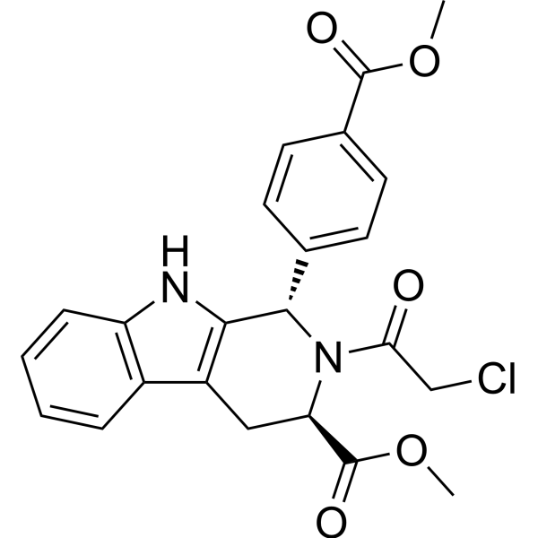
HY-100218A
RSL3GPX4 Inhibitor -
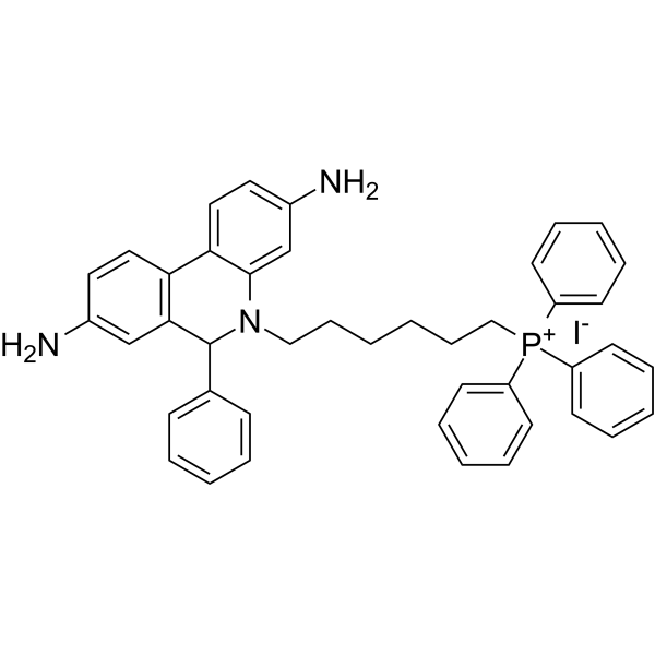
HY-D1055
MitoSOX RedMitochondrial ROS Indicator -

HY-15763
ErastinFerroptosis Inducer -
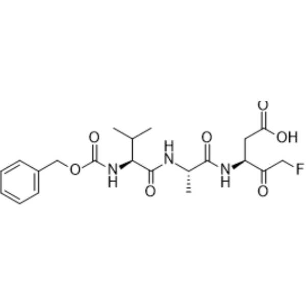
HY-16658B
Z-VAD-FMKPan-Caspase Inhibitor -

HY-K0322
Cell Ferrous Iron (Fe2+) Assay Kit (Fluorometric) -

HY-K0314
ATP Assay Kit -
HY-K1090
Cytotoxicity LDH Assay Kit -
HY-12726
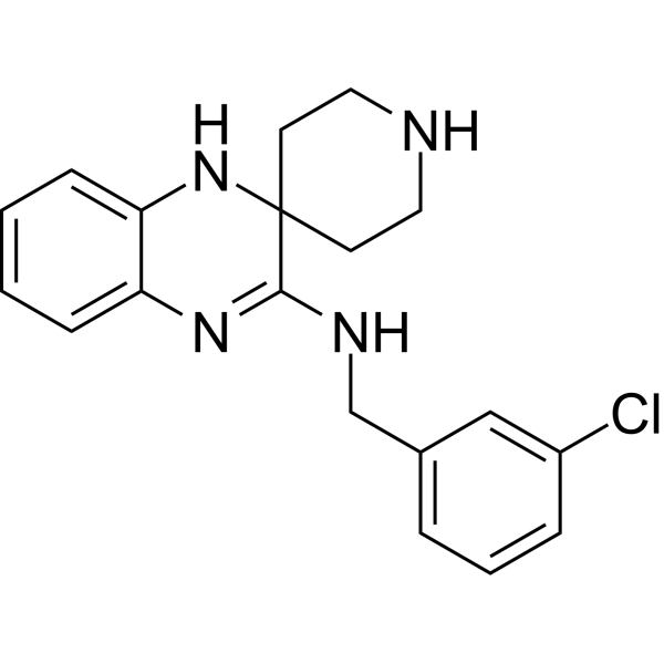
Liproxstatin-1Ferroptosis Inhibitor


Description & Advantages
Lipid peroxidation generally refers to the oxidative degradation of cellular lipids by reactive oxygen species. Peroxidation of unsaturated lipids affects cell membrane properties, signal transduction pathways, apoptosis, and the deterioration of foods and other biological compounds. Lipid peroxidation degradation forms reactive aldehydes such as malondialdehyde (MDA) and 4-hydroxynonenal (4-HNE). In this process free radicals take electrons from the lipids (generally in cell membranes), resulting in cell damage. Therefore MDA and 4-HNE are often used as markers of lipid peroxidation, and to assay for oxidative damage / oxidative stress.
MCE Lipid Peroxidation (MDA) Assay Kit is suitable for measuring MDA levels in a variety of samples including plasma, serum, urine, tissues or cell lysates. The detection principle is that the MDA in the sample reacts with thiobarbituric acid (TBA) to generate a MDA-TBA adduct (The reaction principle is illustrated in the following figure). The MDA-TBA adduct can be quantified colorimetrically (OD = 535 nm) fluorescent (Ex/Em = 532/553 nm) detection.


Protocols
Instructions for Use (Using a Cell Sample as an Example)
Sample Preparation
1. Cell Collection: Since MDA content in cell samples is relatively low, a larger quantity of cells (≥ 106) should be prepared.
2. Lyse the cells using RIPA Lysis Buffer (containing 0.5% antioxidant), and quantify protein content by the Bradford assay to facilitate subsequent normalization. Note: Add 0.5% antioxidant to the RIPA buffer before lysis.
Standard Solution Preparation
Prepare a series of standard solutions at different concentrations by diluting with cell lysis buffer (see table below):
| Group | Lysis Buffer Volume | Standard Volume | Final Standard Concentration |
|---|---|---|---|
| A | 307.2 μL | 12.8 μL of 1 mM MDA | 40 μM |
| B | 100 μL | 100 μL from A | 20 μM |
| C | 100 μL | 100 μL from B | 10 μM |
| D | 100 μL | 100 μL from C | 5 μM |
| E | 100 μL | 100 μL from D | 2.5 μM |
| F | 100 μL | 100 μL from E | 1.25 μM |
| G | 100 μL | 100 μL from F | 0.63 μM |
| H | 100 μL | 100 μL from G, discard 100 μL | 0.31 μM |
| I | 100 μL | 0 | 0 |
Detection
1. Ensure that the volume of both the standard solutions and the samples is 100 μL.
2. In advance, warm the MDA Detection Reagent until fully dissolved, then cool to room temperature down to 30-40°C. Dissolve the antioxidant at 30-40°C and add it to the MDA Detection Reagent at a final concentration of 0.1%. Note: Do not allow the MDA Detection Reagent to cool below this range or place it on ice.
3. Add 200 μL of the antioxidant-containing MDA Detection Reagent to each standard tube and sample tube. The reaction mixture may become turbid upon addition of the detection reagent; do not vortex vigorously — mix gently, then heat in a 95°C water bath for 30 min.
4. After heating, cool on ice to 4°C, then centrifuge at 13,000 rpm for 10 min at 4°C.
5. Transfer 200 μL of the supernatant to a


Publications
-
Journal Impact Factor
-
Most Recent
-
Clin Exp Hypertens2026 Dec 31;48(1):2607404. PMID: 41447318
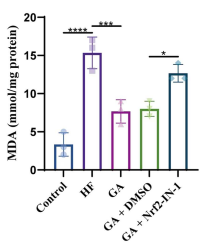
Lipid Peroxidation (MDA) Assay Kit purchased from MedChemExpress. Usage Cited in: Clin Exp Hypertens. 2026 Dec 31;48(1):2607404. [Abstract]
Quantification of intracellular MDA levels in H9c2 cells. Malondialdehyde (MDA) content was quantified according to the manufacturer's instructions of the Lipid Peroxidation (MDA) Assay Kit.
-
Mol Med RepIdentification of the clinical value and biological effects of TTN mutation in liver cancer. [Abstract]2025 Jun;31(6):165. PMID: 40242970
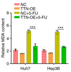
Lipid Peroxidation (MDA) Assay Kit purchased from MedChemExpress. Usage Cited in: Mol Med Rep. 2025 Jun;31(6):165. [Abstract]
MDA content in Huh7 and Hep3B cells after TTN overexpression and 5‑FU treatment.Malondialdehyde (MDA) content was quantified according to the manufacturer's instructions of the Lipid Peroxidation (MDA) Assay Kit.
-
Pestic Biochem PhysiolChaetoglobosin D isolated from the endophytic fungus MG2 exhibited a good control to apple tree Valsa canker. [Abstract]2025 Sep:213:106563. PMID: 40744597
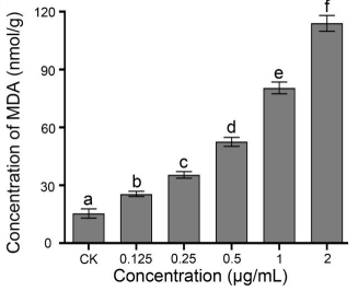
Lipid Peroxidation (MDA) Assay Kit purchased from MedChemExpress. Usage Cited in: Pestic Biochem Physiol. 2025 Sep:213:106563. [Abstract]
Effects of varying chaetoglobosin D concentrations on content in C. mali mycelia (For lipid peroxidation analysis, 100 mg of ground mycelia were homogenized in 1 mL of 10 % (w/v) trichloroacetic acid (TCA) containing 0.1 g quartz sand using a chilled mortar and pestle. The homogenate was centrifuged at 10,000 ×g for 15 min at 4 ◦C, and the supernatant was collected. Malondialdehyde (MDA) content was quantified according to the manufacturer’s instructions of the lipid peroxidation (MDA) assay kit)
-
Mol Cell Endocrinol2026 Mar:613:112721. PMID: 41422938
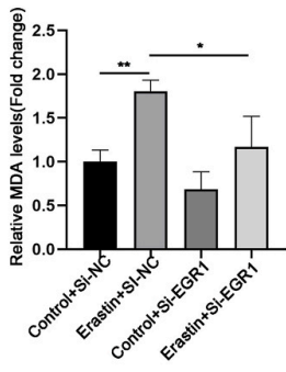
Lipid Peroxidation (MDA) Assay Kit purchased from MedChemExpress. Usage Cited in: Mol Cell Endocrinol. 2026 Mar:613:112721. [Abstract]
Detection of MDA concentration of indicated cells in each group. Malondialdehyde (MDA) content was quantified according to the manufacturer's instructions of the Lipid Peroxidation (MDA) Assay Kit.
-
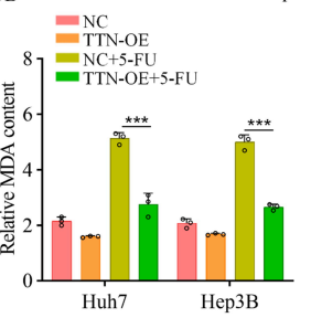
Lipid Peroxidation (MDA) Assay Kit purchased from MedChemExpress. Usage Cited in: Research Square Preprint. 2024 Nov 06.
The content of malondialdehyde (MDA) in Huh7 and Hep3B cells after TTN overexpression and 5-FU treatment. Malondialdehyde (MDA) content was quantified according to the manufacturer's instructions of the Lipid Peroxidation (MDA) Assay Kit.
Storage
-20℃, 1 year.
Keep away from light and avoid repeated freezing and thawing.


Components
| Components | HY-K0319-100 T |
|---|---|
| MDA Detection Solution | 20 mL |
| Antioxidant | 200 μL |
| MDA Standard Solution (1 mM) | 500 μL |


 Please enter the correct Lot Number
