



Citrinin

Based on 1 Customer Validation
Citrinin (NSC 186) is a mycotoxin that causes food contamination and has different toxic effects. Citrinin is an effective oral anticancer agent. Citrinin has various regulatory effects on mouse immune system, including regulating the number of immune cells, inducing apoptosis and autophagy of immune cells, altering toll-like receptor expression and cytokine production. Citrinin can induce oxidative stress and lead to early apoptosis of oocytes. Low doses of Citrinin have neuroprotective effects against glutamate-induced excitotoxicity in rat cortical neurons. In addition, Citrinin also has antibacterial activity.
For research use only. We do not sell to patients.
- Purity : 99.94%
- CAS No.: 518-75-2
- Formula: C13H14O5
- Molecular Weight:250.25
-
Storage:
-20°C, protect from light, stored under nitrogen
* In solvent : -80°C, 6 months; -20°C, 1 month (protect from light, stored under nitrogen)

To place orders, for customer services and technical support, please contact: MedChemExpress USA
Tel: 609-228-6898 E-mail: [email protected] [email protected]


-
 Biological Activity
-
Chemical Information
-
Solvent & Solubility
- Protocols
- Purity & Documentation
- References
-
 Help & FAQs


-
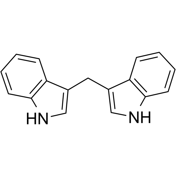
HY-15758
3,3'-DiindolylmethaneAndrogen Receptor Antagonist -
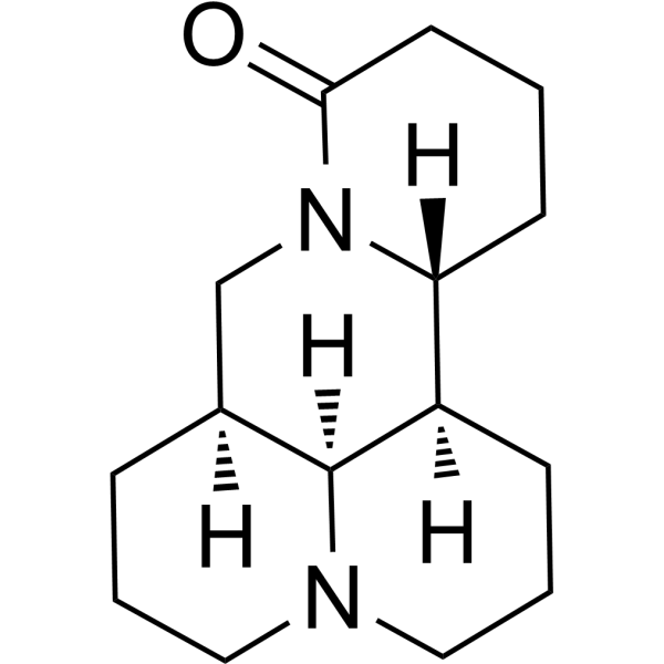
HY-N0164
MatrineAnticancer Agent -

HY-N6746R
Citrinin (Standard)Bacterial Inhibitor -
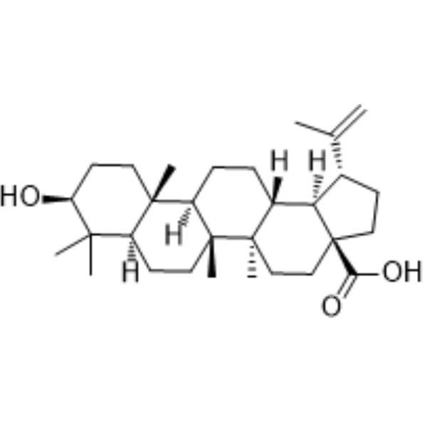
HY-10529
Betulinic acidApoptosis Inducer -
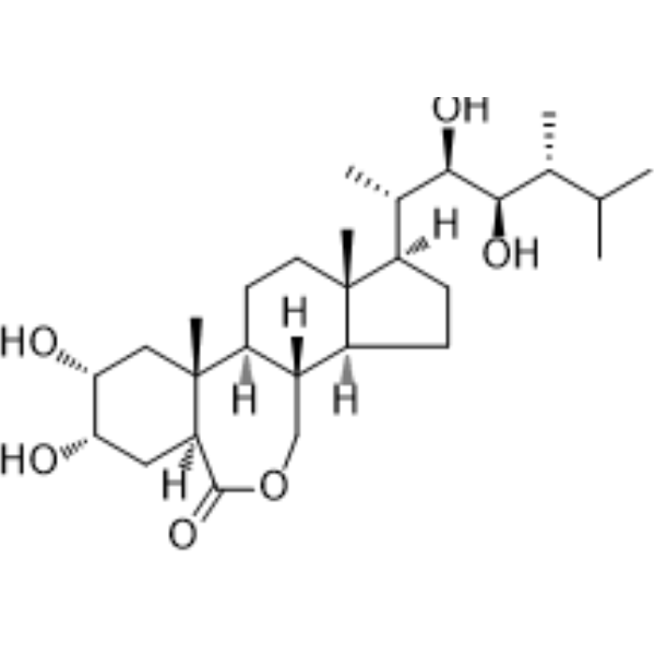
HY-N0848
EpibrassinolideApoptosis Inducer -
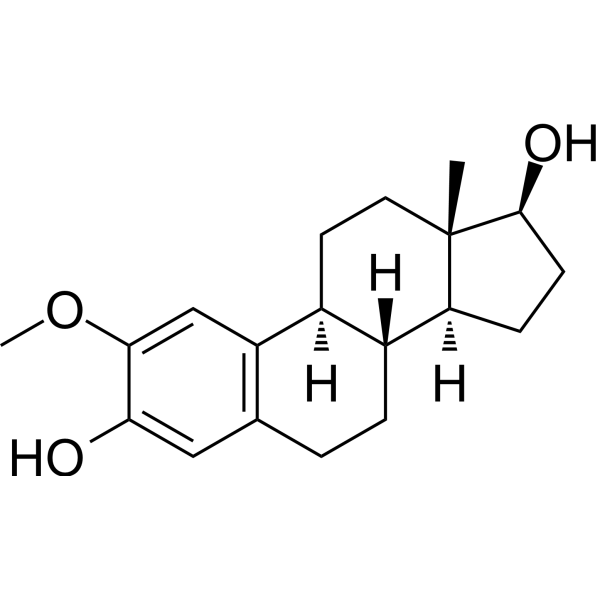
HY-12033
2-MethoxyestradiolSteroid Metabolite -
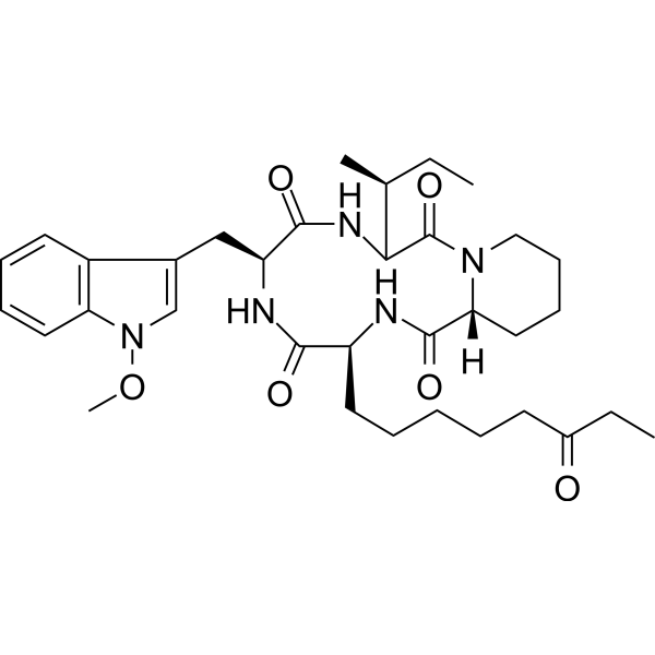
HY-N6735
ApicidinHDAC Inhibitor -
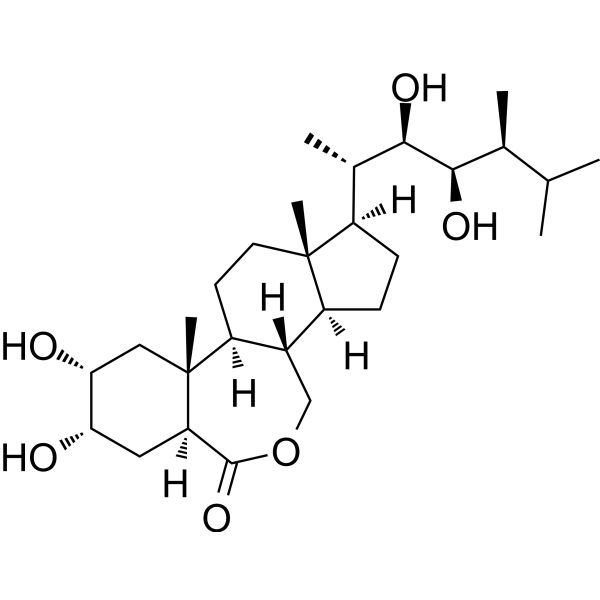
HY-N0273
BrassinolideApoptosis Inducer -
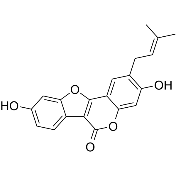
HY-N0232
PsoralidinCOX-2/5-LOX Inhibitor -
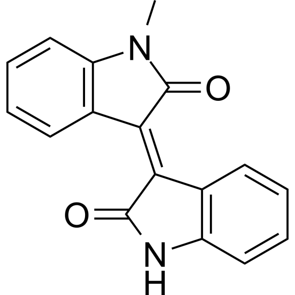
HY-13680
MeisoindigoApoptosis Inducer
All Endogenous Metabolite Isoforms
More

Biological Activity
Description
IC50 & Target
|
Microbial Metabolite |
Cellular Effect
|
Cell Line

|
Type | Value | Description | References |
|---|---|---|---|---|
| HCT-116 | IC50 |
>10 μM
Compound: 13
|
Cytotoxicity against human HCT116 cells after 72 hrs by SRB assay
Cytotoxicity against human HCT116 cells after 72 hrs by SRB assay
|
[PMID: 21761866] |
| HL-60 | IC50 |
>10 μM
Compound: 13
|
Cytotoxicity against human HL60 cells after 72 hrs by MTT assay
Cytotoxicity against human HL60 cells after 72 hrs by MTT assay
|
[PMID: 21761866] |
| KB | IC50 |
>10 μM
Compound: 13
|
Cytotoxicity against human KB cells after 72 hrs by SRB assay
Cytotoxicity against human KB cells after 72 hrs by SRB assay
|
[PMID: 21761866] |
In Vitro
Citrinin (0.1-1000 nM; 24 h) has neuroprotective effects in glutamate-treated rat cortical neurons (at low doses)[2].
Citrinin (1-10 μg/mL; 6 h) induces late apoptosis of immune cells[3].
Citrinin (1-10 μg/mL; 6 h) increases the rate of Bax/Bcl-2 expression in freshly isolated spleen cells[3].
MedChemExpress (MCE) has not independently confirmed the accuracy of these methods. They are for reference only.
-
Cell Line:Splenocytes and MLN cells from a SPF mouse ages 7 seven weeks old
-
Concentration:1, 5 and 10 μg/mL
-
Incubation Time:6 h
-
Result:Significantly increased the number of late apoptotic cells, but not in early apoptotic cells.
-
Cell Line:Splenocytes
-
Concentration:1, 5 and 10 μg/mL
-
Incubation Time:6 h
-
Result:Decreased the level of Bcl-2, and the expression of Bax was not affected.
In Vivo
Citrinin (2 mg/kg; Oral administration; 3 weeks) has an antitumor effect in a mouse breast cancer model induced by DMBA (HY-W011845)[4].
MedChemExpress (MCE) has not independently confirmed the accuracy of these methods. They are for reference only.
-
Animal Model:Female BALB/c mice aged seven weeks old[3]
-
Dosage:1, 5, or 10 mg/kg
-
Administration:Oral gavage (i.g.); 14 days
-
Result:Significantly reduced the populations of F4/80+ cells and CD19+ cells in spleen and MLN.
Increased the populations of CD4+, CD8+ and CD4+CD25+Foxp3+ cells in MLN.
Increased the populations of CD8+ cells and decreased the populations of CD19+ cells in intra-epithelial, lamina propria and Peyer’s patches lymphocytes.
Reduced IgM antibody production in serum.
Increased the ratios of Bax/Bcl-2 in the spleen.
Up-regulated the expression of Atg5, Beclin-1 and TLR 2 in the spleen.
-
Animal Model:DMBA (HY-W011845) treated swiss albino female mice aged 6-7 weeks old (25-30 g) [4]
-
Dosage:2 mg/kg
-
Administration:Oral administration (p.o.); 3 weeks
-
Result:Reduced the weight of both breasts, and reduced cell proliferation in the breast.
Did not cause necrosis in the cells, the characteristics of the carcinoma such as intraductal calcification, injured tissue, fibrosed with ductal hyperplasia, and histopathological characteristics indicative of preneoplasia still remained.
Decreased genotoxic damage, damage index and damage frequency in breast and non-tumor cells induced by DMBA (HY-W011845).
Did not increase micronuclei or chromosomal bridge formation in hepatocytes of the animals.
Showed apoptotic effects of induction of binucleated cells in hepatic tissue.
- RNA extraction experimental
- Research Protocol for Infectious Diseases
- Annexin V plus membrane-impermeant dye apoptosis staining
- ROS/oxidative-stress fluorescent staining
- Primary Embryonic Cortical Neuron Culture
- Research Protocol for Inflammation-related Diseases
- Bacterial live/dead nucleic-acid viability staining
- Detection of Apoptosis
- TUNEL staining for apoptotic DNA fragmentation
- Autophagy
- Apoptosis Solutions
- Lysosome and acidic-vesicle live-cell staining
- Macroautophagy Solutions


Chemical Information
-
CAS No. 518-75-2
-
Appearance Solid
-
Molecular Weight 250.25
-
Formula C13H14O5
-
Color Light yellow to yellow
-
SMILES
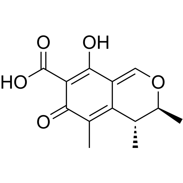
OC1=C(C(C(C)=C([C@@H]2C)C1=CO[C@@H]2C)=O)C(O)=O
-
Synonyms
NSC 186
-
Structure Classification
-
Initial Source
Monascus sp.
-
Shipping
Room temperature in continental US; may vary elsewhere.
-
Storage
-20°C, protect from light, stored under nitrogen
* In solvent : -80°C, 6 months; -20°C, 1 month (protect from light, stored under nitrogen)


Solvent & Solubility
In Vitro:
DMSO : 50 mg/mL (199.80 mM; Need ultrasonic; Hygroscopic DMSO has a significant impact on the solubility of product, please use newly opened DMSO)
Please refer to the solubility information to select the appropriate solvent. Once prepared, please aliquot and store the solution to prevent product inactivation from repeated freeze-thaw cycles.
Storage method and period of stock solution: -80°C, 6 months; -20°C, 1 month (protect from light, stored under nitrogen). When stored at -80°C, please use it within 6 months. When stored at -20°C, please use it within 1 month.
Please refer to the solubility information to select the appropriate solvent. Once prepared, please aliquot and store the solution to prevent product inactivation from repeated freeze-thaw cycles.
Storage method and period of stock solution: -80°C, 6 months; -20°C, 1 month (protect from light, stored under nitrogen). When stored at -80°C, please use it within 6 months. When stored at -20°C, please use it within 1 month.
Concentration (start) × Volume (start) = Concentration (final) × Volume (final)
In Vivo:
Select the appropriate dissolution method based on your experimental animal and administration route.
- For the following dissolution methods, please ensure to first prepare a clear stock solution using an In Vitro approach and then sequentially add co-solvents:
- To ensure reliable experimental results, the clarified stock solution can be appropriately stored based on storage conditions. As for the working solution for In Vivo experiments, it is recommended to prepare freshly and use it on the same day.
- The percentages shown for the solvents indicate their volumetric ratio in the final prepared solution. If precipitation or phase separation occurs during preparation, heat and/or sonication can be used to aid dissolution.
Add each solvent one by one: 10% DMSO 40% PEG300 5% Tween-80 45% Saline
Solubility: ≥ 1.25 mg/mL (5.00 mM); Clear solution
This protocol yields a clear solution of ≥ 1.25 mg/mL (saturation unknown).
Taking 1 mL working solution as an example, add 100 μL DMSO stock solution (12.5 mg/mL) to 400 μL PEG300, and mix evenly; then add 50 μL Tween-80 and mix evenly; then add 450 μL Saline to adjust the volume to 1 mL.
Preparation of Saline: Dissolve 0.9 g sodium chloride in ddH₂O and dilute to 100 mL to obtain a clear Saline solution.
Add each solvent one by one: 10% DMSO 90% (20% SBE-β-CD in Saline)
Solubility: 1.25 mg/mL (5.00 mM); Suspended solution; Need ultrasonic
This protocol yields a suspended solution of 1.25 mg/mL. Suspended solution can be used for oral and intraperitoneal injection.
Taking 1 mL working solution as an example, add 100 μL DMSO stock solution (12.5 mg/mL) to 900 μL 20% SBE-β-CD in Saline, and mix evenly.
Preparation of 20% SBE-β-CD in Saline (4°C, storage for one week): 2 g SBE-β-CD powder is dissolved in 10 mL Saline, completely dissolve until clear.
In Vivo Dissolution Calculator
Please enter the basic information of animal experiments:
-
-
-
-
Recommended: Prepare an additional quantity of animals to account for potential losses during experiments.
Please enter your animal formula composition:
-
%DMSO +
Recommended: Keep the proportion of DMSO in working solution below 2% if your animal is weak.
-
%+
-
+%Tween-80 + +
-
%Saline +
The co-solvents required include: DMSO, . All of co-solvents are available by MedChemExpress (MCE). , Tween 80. All of co-solvents are available by MedChemExpress (MCE).
Working solution concentration: 0.22 mg/mL
Method for preparing stock solution: mg drug dissolved in μL DMSO. Stock solution concentration: mg/mL. * In solvent : -80°C, 6 months; -20°C, 1 month (protect from light, stored under nitrogen)
1. Take μL DMSO stock solution;
2. Add μL .
μL , mix evenly;
3. Then add μL Tween 80, mix evenly;
4. Then add μL
Please ensure that the stock solution in the first step is dissolved to a clear state, and add co-solvents in sequence. You can use ultrasonic heating (ultrasonic cleaner, recommended frequency 20-40 kHz), vortexing, etc. to assist dissolution.


Purity & Documentation
-
Data Sheet (292 KB)
-
SDS (585 KB)
- English - EN (585 KB)
- Français - FR (585 KB)
- Deutsch - DE (585 KB)
- Norwegian - NO (585 KB)
- Español - ES (585 KB)
- Swedish - SV (585 KB)
- Italian - IT (585 KB)
- Korean - KR (585 KB)
- Portuguese - PT (585 KB)
-
Handling Instructions (2659 KB)
References
[1]. de Oliveira Filho JWG, et al, A comprehensive review on biological properties of citrinin. Food Chem Toxicol. 2017 Dec;110:130-141. [Content Brief]
[2]. Nakajima Y, et al. Low doses of the mycotoxin citrinin protect cortical neurons against glutamate-induced excitotoxicity. J Toxicol Sci. 2016 Apr;41(2):311-9. [Content Brief]
[3]. de Oliveira Filho JWG, et al. Citrinin against breast cancer: A cytogenotoxicological study. Phytother Res. 2021 Jan;35(1):504-516. [Content Brief]
[4]. Islam MR, et al. Immune modulatory effects of the foodborne contaminant citrinin in mice. Food Chem Toxicol. 2012 Oct;50(10):3537-47. [Content Brief]
[5]. Wu Y, et al. Citrinin exposure affects oocyte maturation and embryo development by inducing oxidative stress-mediated apoptosis. Oncotarget. 2017 May 23;8(21):34525-34533. [Content Brief]


Complete Stock Solution Preparation Table
Please refer to the solubility information to select the appropriate solvent. Once prepared, please aliquot and store the solution to prevent product inactivation from repeated freeze-thaw cycles.
Storage method and period of stock solution: -80°C, 6 months; -20°C, 1 month (protect from light, stored under nitrogen). When stored at -80°C, please use it within 6 months. When stored at -20°C, please use it within 1 month.
| Optional Solvent | Concentration Solvent Mass | 1 mg | 5 mg | 10 mg | 25 mg |
|---|---|---|---|---|---|
| DMSO | 1 mM | 3.9960 mL | 19.9800 mL | 39.9600 mL | 99.9001 mL |
| 5 mM | 0.7992 mL | 3.9960 mL | 7.9920 mL | 19.9800 mL | |
| 10 mM | 0.3996 mL | 1.9980 mL | 3.9960 mL | 9.9900 mL | |
| 15 mM | 0.2664 mL | 1.3320 mL | 2.6640 mL | 6.6600 mL | |
| 20 mM | 0.1998 mL | 0.9990 mL | 1.9980 mL | 4.9950 mL | |
| 25 mM | 0.1598 mL | 0.7992 mL | 1.5984 mL | 3.9960 mL | |
| 30 mM | 0.1332 mL | 0.6660 mL | 1.3320 mL | 3.3300 mL | |
| 40 mM | 0.0999 mL | 0.4995 mL | 0.9990 mL | 2.4975 mL | |
| 50 mM | 0.0799 mL | 0.3996 mL | 0.7992 mL | 1.9980 mL | |
| 60 mM | 0.0666 mL | 0.3330 mL | 0.6660 mL | 1.6650 mL | |
| 80 mM | 0.0500 mL | 0.2498 mL | 0.4995 mL | 1.2488 mL | |
| 100 mM | 0.0400 mL | 0.1998 mL | 0.3996 mL | 0.9990 mL |


Citrinin Related Classifications
HY-N6746 Related Classifications
 Powered by Bioz