



Ethynylferrocene

Based on 1 Customer Validation
Ethynylferrocene (Ferrocenylacetylene) is a click chemistry agent. Ethynylferrocene can be used for labeling of hairpin DNA probes to monitor the DNA hybridization via the Cu(I)-catalyzed azide-alkyne cycloaddition (CuAAC).
商品は「研究用試薬」です。人や動物の医療用・臨床診断用・食品用の製品ではありません。
研究用途以外に使用した場合、当社は一切の責任を負いかねます。
- CAS 番号: 1271-47-2
- 分子式: C12H10Fe
- 分子量:210.05
-
保管条件:
4°C, protect from light, stored under nitrogen
* In solvent : -80°C, 6 months; -20°C, 1 month (protect from light, stored under nitrogen)


To place orders, for customer services and technical support, please contact: MedChemExpress USA
Tel: 609-228-6898 E-mail: [email protected] [email protected]


-
 生物活性
生物活性
-
化学情報
- プロトコル
- 純度とドキュメンテーション
- 参考文献
-
 Help & FAQs


-
![[Ir(cod)Cl]2](https://file.medchemexpress.com/product_pic/hy-w008260.gif)
HY-W008260
[Ir(cod)Cl]2Biochemical Reagent -

HY-79504
FerroceneOrganometallic Compound -

HY-W020958
Ru(acac)3Caspase-3 Activator -

HY-Y1109
Dichloro(p-cymene)ruthenium(II) dimerPrecursor of Metal Complexes -

HY-Y0416
Potassium ferrocyanide trihydrate, 98.5%Biochemical Reagent -

HY-W051271
Titanium(IV) oxidePhotocatalyst -
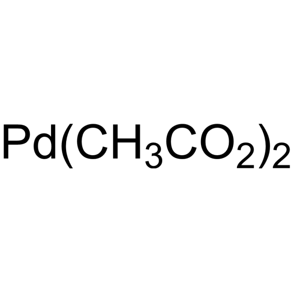
HY-W002072
Palladium (II) acetateCatalyst -
![Dichloro[1,1'-bis(diphenylphosphino)ferrocene]palladium(II) dichloromethane adduct](https://file.medchemexpress.com/product_pic/hy-21623.gif)
HY-21623
Dichloro[1,1'-bis(diphenylphosphino)ferrocene]palladium(II) dichloromethane adductCatalyst -

HY-Y1237
DPPFMetal Ligand -

HY-Y0998
Potassium hexacyanoferrate(III), 99.5%Chelating Agent -
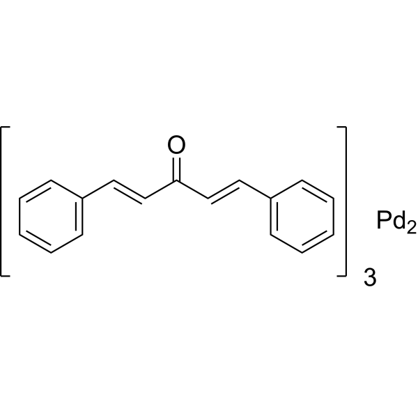
HY-W018864
Tris(dibenzylideneacetonyl)bis-palladiumCatalyst


生物活性
製品説明


化学情報
-
CAS 番号 1271-47-2
-
性状 Solid
-
分子量 210.05
-
分子式 C12H10Fe
-
Color Orange to reddish brown
-
SMILES
[][Fe][].C#Cc1cccc1.c2cccc2
-
別名
Ferrocenylacetylene; Ferrocenylethyne
-
輸送条件
Room temperature in continental US; may vary elsewhere.
-
保管条件
4°C, protect from light, stored under nitrogen
* In solvent : -80°C, 6 months; -20°C, 1 month (protect from light, stored under nitrogen)

プロトコル
-
EdU Incorporation Assay (Click Chemistry-Based DNA Synthesis Measurement)
The EdU incorporation assay measures DNA synthesis by adding the thymidine analog 5-ethynyl-2′-deoxyuridine to cells or tissues, where it is incorporated into newly synthesized DNA during S phase. Incorporated EdU is detected by copper-catalyzed azide-alkyne cycloaddition, in which a fluorescent azide covalently reacts with the ethynyl group on EdU, allowing S-phase cells to be detected by fluorescence microscopy, flow cytometry, or high-content imaging. EdU detection does not require DNA denaturation or anti-BrdU antibody access, which preserves sample structure and improves compatibility with immunostaining and multiparameter cytometry compared with BrdU-based detection. EdU can be cytotoxic in a cell-type- and exposure-dependent manner, so pulse duration, concentration, and continuous-labeling designs should be validated for each cell type.
-
Protocol for Southern Blot
Southern blot is a DNA hybridization assay used to detect a defined DNA sequence within restriction-digested or otherwise fragmented genomic DNA. The method separates DNA fragments by agarose gel electrophoresis, transfers the size-resolved DNA pattern onto a solid support, denatures the DNA to permit base pairing, and detects fragments that hybridize with a complementary labeled probe; the readout is a band, smear, or fragment-size distribution corresponding to the target sequence and its restriction-fragment context. The assay reflects sequence presence, restriction fragment length, gene copy pattern, structural rearrangement, insertion or deletion affecting restriction sites, and some repeat-length or terminal restriction fragment applications when the experimental design links the probe to those genomic features. Classic applications include Southern blot-based telomere terminal restriction fragment analysis and minisatellite-based DNA fingerprinting, which illustrate how the same


純度とドキュメンテーション
-
データシート (270 KB)
-
SDS (393 KB)
- English - EN (393 KB)
- Français - FR (393 KB)
- Deutsch - DE (393 KB)
- Norwegian - NO (393 KB)
- Español - ES (393 KB)
- Swedish - SV (393 KB)
- Italian - IT (393 KB)
- Korean - KR (393 KB)
- Portuguese - PT (393 KB)
-
取扱説明書 (2659 KB)
参考文献

Calculators
濃度 (開始) × 体積 (開始) = 濃度 (終了) × 体積 (終了)


Ethynylferrocene 関連分類
HY-W018008 関連分類
 Powered by Bioz