



FPR-A14
Based on 1 publication(s) in Google Scholar
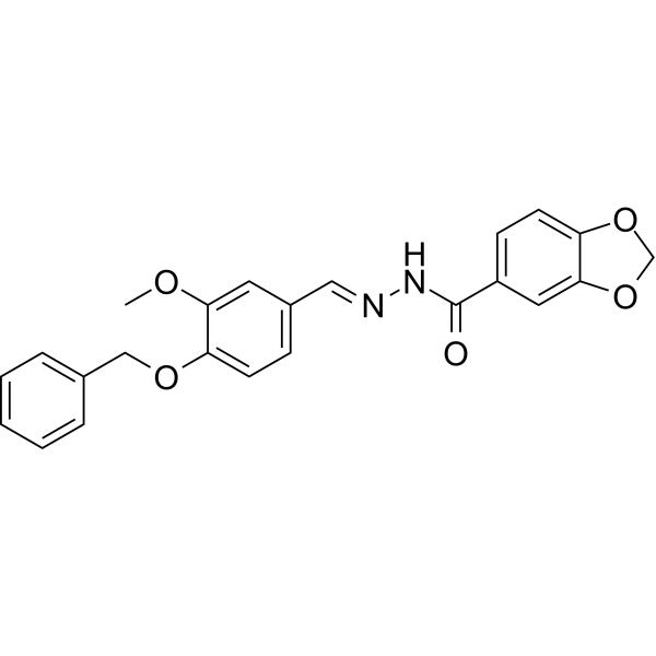
FPR-A14 is a potent formyl peptide receptor (FPR) agonist. FPR-A14 is a potent activator of neutrophil Ca2+ mobilization and chemotaxis with EC50s of 630 nM and 42 nM, respectively. FPR-A14 induces cell differentiation.
商品は「研究用試薬」です。人や動物の医療用・臨床診断用・食品用の製品ではありません。
研究用途以外に使用した場合、当社は一切の責任を負いかねます。
- 純度 : 98.58%
- CAS 番号: 329691-12-5
- 分子式: C23H20N2O5
- 分子量:404.42
-
保管条件:
4°C, protect from light
* In solvent : -80°C, 6 months; -20°C, 1 month (protect from light)

To place orders, for customer services and technical support, please contact: MedChemExpress USA
Tel: 609-228-6898 E-mail: [email protected] [email protected]


-
 生物活性
生物活性
-
化学情報
-
溶剤 & 溶解度
- プロトコル
- 純度とドキュメンテーション
- 参考文献
-
 Help & FAQs





MedChemExpress(MCE)の使用を引用している文献 FPR-A14
More

生物活性
製品説明
体外実験
FPR-A14 (compound 14) activates Ca2+ release with an EC50 of 630 nM in human neutrophils. FPR-A14 is a neutrophil chemoattractant and dose-dependently induces neutrophil migration with an EC50 value of 42 nM[1].
FPR-A14 (1-10 μM; 48h) elicites a significant increase in % cell differentiation versus controls at concentrations of 4μM (32.0%), 6μM (64.9%), 8μM (89.1%) and 10μM (93.3%) in mouse neuroblastoma N2a cells. FPRa14 (100μM) produces similar effects in IMR-32 and SH-SY5Y cells[2].
MedChemExpress (MCE) has not independently confirmed the accuracy of these methods. They are for reference only.


化学情報
-
CAS 番号 329691-12-5
-
性状 Solid
-
分子量 404.42
-
分子式 C23H20N2O5
-
Color White to off-white
-
SMILES
O=C(C1=CC=C(OCO2)C2=C1)N/N=C/C3=CC=C(OCC4=CC=CC=C4)C(OC)=C3
-
輸送条件
Room temperature in continental US; may vary elsewhere.
-
保管条件
4°C, protect from light
* In solvent : -80°C, 6 months; -20°C, 1 month (protect from light)


Publications (1)
-
Journal Impact Factor
-
Most Recent


溶剤 & 溶解度
体外:
DMSO : 125 mg/mL (309.08 mM; ultrasonic and warming and heat to 60°C; Hygroscopic DMSO has a significant impact on the solubility of product, please use newly opened DMSO)
Please refer to the solubility information to select the appropriate solvent. Once prepared, please aliquot and store the solution to prevent product inactivation from repeated freeze-thaw cycles.
Storage method and period of stock solution: -80°C, 6 months; -20°C, 1 month (protect from light). When stored at -80°C, please use it within 6 months. When stored at -20°C, please use it within 1 month.
Please refer to the solubility information to select the appropriate solvent. Once prepared, please aliquot and store the solution to prevent product inactivation from repeated freeze-thaw cycles.
Storage method and period of stock solution: -80°C, 6 months; -20°C, 1 month (protect from light). When stored at -80°C, please use it within 6 months. When stored at -20°C, please use it within 1 month.
濃度 (開始) × 体積 (開始) = 濃度 (終了) × 体積 (終了)


純度とドキュメンテーション
-
データシート (271 KB)
-
SDS (393 KB)
- English - EN (393 KB)
- Français - FR (393 KB)
- Deutsch - DE (393 KB)
- Norwegian - NO (393 KB)
- Español - ES (393 KB)
- Swedish - SV (393 KB)
- Italian - IT (393 KB)
- Korean - KR (393 KB)
- Portuguese - PT (393 KB)
-
取扱説明書 (2659 KB)
参考文献
[1]. Igor A Schepetkin, et al. High-throughput screening for small-molecule activators of neutrophils: identification of novel N-formyl peptide receptor agonists. Mol Pharmacol. 2007 Apr;71(4):1061-74. [Content Brief]
[2]. Peter J G Cussell, et al. The formyl peptide receptor agonist FPRa14 induces differentiation of Neuro2a mouse neuroblastoma cells into multiple distinct morphologies which can be specifically. inhibited with FPR antagonists and FPR knockdown using siRNA. PLoS One. 2019 Jun 6;14(6):e0217815. [Content Brief]


Complete Stock Solution Preparation Table
Please refer to the solubility information to select the appropriate solvent. Once prepared, please aliquot and store the solution to prevent product inactivation from repeated freeze-thaw cycles.
Storage method and period of stock solution: -80°C, 6 months; -20°C, 1 month (protect from light). When stored at -80°C, please use it within 6 months. When stored at -20°C, please use it within 1 month.
| Optional Solvent | Concentration Solvent Mass | 1 mg | 5 mg | 10 mg | 25 mg |
|---|---|---|---|---|---|
| DMSO | 1 mM | 2.4727 mL | 12.3634 mL | 24.7268 mL | 61.8169 mL |
| 5 mM | 0.4945 mL | 2.4727 mL | 4.9454 mL | 12.3634 mL | |
| 10 mM | 0.2473 mL | 1.2363 mL | 2.4727 mL | 6.1817 mL | |
| 15 mM | 0.1648 mL | 0.8242 mL | 1.6485 mL | 4.1211 mL | |
| 20 mM | 0.1236 mL | 0.6182 mL | 1.2363 mL | 3.0908 mL | |
| 25 mM | 0.0989 mL | 0.4945 mL | 0.9891 mL | 2.4727 mL | |
| 30 mM | 0.0824 mL | 0.4121 mL | 0.8242 mL | 2.0606 mL | |
| 40 mM | 0.0618 mL | 0.3091 mL | 0.6182 mL | 1.5454 mL | |
| 50 mM | 0.0495 mL | 0.2473 mL | 0.4945 mL | 1.2363 mL | |
| 60 mM | 0.0412 mL | 0.2061 mL | 0.4121 mL | 1.0303 mL | |
| 80 mM | 0.0309 mL | 0.1545 mL | 0.3091 mL | 0.7727 mL | |
| 100 mM | 0.0247 mL | 0.1236 mL | 0.2473 mL | 0.6182 mL |


FPR-A14 関連分類
HY-103472 関連分類
 Powered by Bioz