



Dehydroleucodine
Based on 1 publication(s) in Google Scholar
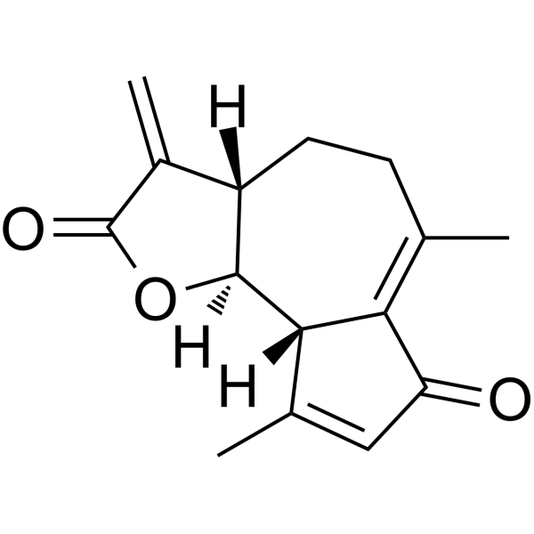
Dehydroleucodine is a sesquiterpene lactone isolated from Artemisia douglasiana. Dehydroleucodine is a mast cell stabilizer that inhibits tmast cell degranulation induced by compound 48/80. Dehydroleucodine inudces cells apoptosis, and has gastric ulcer inhibition and antileukemic effects.
For research use only. We do not sell to patients.
- CAS No.: 36150-07-9
- Formula: C15H16O3
- Molecular Weight:244.29
-
Storage:
-20°C, sealed storage, away from moisture
* In solvent : -80°C, 6 months; -20°C, 1 month (sealed storage, away from moisture)

To place orders, for customer services and technical support, please contact: MedChemExpress USA
Tel: 609-228-6898 E-mail: [email protected] [email protected]


-
 Biological Activity
-
Chemical Information
- Protocols
- Purity & Documentation
- References
-
 Help & FAQs


Publications Citing Use of MedChemExpress (MCE) Dehydroleucodine
More

Biological Activity
Description
Cellular Effect
|
Cell Line

|
Type | Value | Description | References |
|---|---|---|---|---|
| HeLa S3 | IC50 |
10 μM
Compound: 1
|
Cytotoxicity against human HeLaS3 cells assessed as decrease in cell viability after 72 to 96 hrs by MTT assay
Cytotoxicity against human HeLaS3 cells assessed as decrease in cell viability after 72 to 96 hrs by MTT assay
|
[PMID: 27057812] |
| KB | IC50 |
5.3 μM
Compound: 1
|
Cytotoxicity against human KB cells assessed as decrease in cell viability after 72 hrs by MTT assay
Cytotoxicity against human KB cells assessed as decrease in cell viability after 72 hrs by MTT assay
|
[PMID: 27057812] |
| MCF7 | IC50 |
5 μM
Compound: 1
|
Cytotoxicity against human MCF7 cells assessed as decrease in cell viability after 72 to 96 hrs by MTT assay
Cytotoxicity against human MCF7 cells assessed as decrease in cell viability after 72 to 96 hrs by MTT assay
|
[PMID: 27057812] |


Chemical Information
-
CAS No. 36150-07-9
-
Appearance Solid
-
Molecular Weight 244.29
-
Formula C15H16O3
-
Color White to off-white
-
SMILES
O=C(O[C@@]1([H])[C@@]2([H])CCC(C)=C3C(C=C(C)[C@@]31[H])=O)C2=C
-
Initial Source
-
Shipping
Room temperature in continental US; may vary elsewhere.
-
Storage
-20°C, sealed storage, away from moisture
* In solvent : -80°C, 6 months; -20°C, 1 month (sealed storage, away from moisture)


Publications (1)
-
Journal Impact Factor
-
Most Recent


Purity & Documentation
-
Data Sheet (274 KB)
-
SDS (252 KB)
- English - EN (252 KB)
- Français - FR (252 KB)
- Deutsch - DE (252 KB)
- Norwegian - NO (252 KB)
- Español - ES (252 KB)
- Swedish - SV (252 KB)
- Italian - IT (252 KB)
- Korean - KR (252 KB)
- Portuguese - PT (252 KB)
-
Handling Instructions (2659 KB)
References
[1]. Paola E Ordóñez, et al. Dehydroleucodine, a Sesquiterpene Lactone from Gynoxys verrucosa, Demonstrates Cytotoxic Activity against Human Leukemia Cells. J Nat Prod. 2016 Apr 22;79(4):691-6. [Content Brief]
[2]. Mariano E Vera, et al. Activation of human leukemic mast cell line LAD2 is modulated by dehydroleucodine and xanthatin. Leuk Lymphoma. 2012 Sep;53(9):1795-803. [Content Brief]
[3]. Valeria V Costantino, et al. Dehydroleucodine inhibits tumor growth in a preclinical melanoma model by inducing cell cycle arrest, senescence and apoptosis. Cancer Lett. 2016 Mar 1;372(1):10-23. [Content Brief]

Calculators
Concentration (start) × Volume (start) = Concentration (final) × Volume (final)


Dehydroleucodine Related Classifications
HY-122295 Related Classifications
 Powered by Bioz