



BMV109
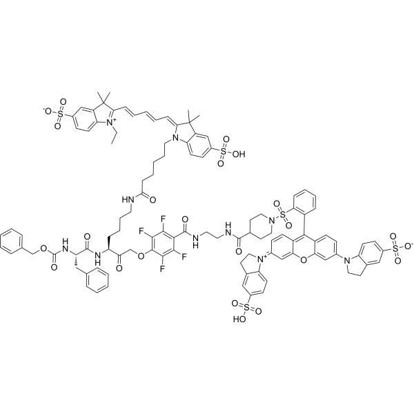
BMV109 is a quenched probe that becomes fluorescent when cleaved and covalently bound by active cathepsin proteases. BMV109 can be exploited for tumor imaging.
연구목적의 판매만을 진행합니다. 환자를 대상으로 한 판매는 하지 않습니다.
- CAS No.: 1458731-58-2
- 화학식: C107H108F4N10O23S5
- 분자량:2138.37
-
보관:
Please store the product under the recommended conditions in the Certificate of Analysis.

To place orders, for customer services and technical support, please contact: MedChemExpress USA
Tel: 609-228-6898 E-mail: [email protected] [email protected]


-
 Biological Activity
-
Chemical Information
- Protocol
- 순도&문서
- References
-
 Help & FAQs




Biological Activity
제품 설명
In Vitro
BMV109 (compound 8; 0.05 µM, 0.1 µM, 0.5 µM, 1 µM, 5 µM) labels all target cysteine cathepsins (B, S, L, X) with equal intensity in living RAW cells[1].
MedChemExpress (MCE) has not independently confirmed the accuracy of these methods. They are for reference only.
In Vivo
MedChemExpress (MCE) has not independently confirmed the accuracy of these methods. They are for reference only.
-
Animal Model:Balb/c mice bearing 4T1 cells[1]
-
Dosage:20 nmol
-
Administration:Tail vein
-
Result:Robust tumor specific activation of fluorescence with high overall intensity could be observed.


Chemical Information
-
CAS No. 1458731-58-2
-
분자량 2138.37
-
화학식 C107H108F4N10O23S5
-
SMILES
O=S(C1=C(C=CC=C1)C(C2=CC=C(N3C4=CC=C(S(=O)([O-])=O)C=C4CC3)C=C2O5)=C(C=C/6)C5=CC6=[N+]7C8=CC=C(S(=O)(O)=O)C=C8CC/7)(N9CCC(CC9)C(NCCNC(C%10=C(C(F)=C(C(F)=C%10F)OCC([C@H](CCCCNC(CCCCCN%11C%12=CC=C(S(=O)(O)=O)C=C%12C(C)(/C%11=C/C=C/C=C/C%13=[N+](C%14=CC=C(S(=O)([O-])=O)C=C%14C%13(C)C)CC)C)=O)NC([C@@H](NC(OCC%15=CC=CC=C%15)=O)CC%16=CC=CC=C%16)=O)=O)F)=O)=O)=O
-
선적
Room temperature in continental US; may vary elsewhere.
-
보관
Please store the product under the recommended conditions in the Certificate of Analysis.


순도&문서
References
[1]. Martijn Verdoes, et al. Improved quenched fluorescent probe for imaging of cysteine cathepsin activity. J Am Chem Soc. 2013 Oct 2;135(39):14726-30. [Content Brief]
[2]. Steven Sensarn, et al. A Clinical Wide-Field Fluorescence Endoscopic Device for Molecular Imaging Demonstrating Cathepsin Protease Activity in Colon Cancer. Mol Imaging Biol. 2016 Dec;18(6):820-829. [Content Brief]

Calculators
Concentration (start) × Volume (start) = Concentration (final) × Volume (final)


BMV109 Related Classifications
HY-149931 Related Classifications
 Powered by Bioz