



Fibronectin Adhesion-promoting Peptide TFA
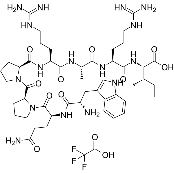
Fibronectin Adhesion-promoting Peptide (Heparin Binding Peptide) is one of the heparin-binding amino acid sequences found in the carboxy-terminal heparin-binding domain of fibronectin. It promotes assembly of mesenchymal stem cell (MSC) spheroids into larger aggregates. Fibronectin Adhesion-promoting Peptide directly promotes the adhesion, spreading, and migration of endothelial cells by reacting with heparin binding domains of cells.
연구목적의 판매만을 진행합니다. 환자를 대상으로 한 판매는 하지 않습니다.
- 화학식: C49H75F3N16O12
- 분자량:1137.22
-
보관:
Please store the product under the recommended conditions in the Certificate of Analysis.

To place orders, for customer services and technical support, please contact: MedChemExpress USA
Tel: 609-228-6898 E-mail: [email protected] [email protected]


-
 Biological Activity
-
Chemical Information
- Protocol
- 순도&문서
- References
-
 Help & FAQs




Biological Activity
제품 설명
In Vitro
GRGDS and Fibronectin Adhesion-promoting Peptide TFA (Heparin Binding Peptide TFA: WQPPRARI) micropatterns increase the endothelialisation of prosthetic materials in vitro. Fibronectin Adhesion-promoting Peptide also enhances haptotactic cell migration. The anti-apoptotic effects of fibronectin require the presence of the WQPPRARI sequence, which may be due to synergistic survival signals mediated by this sequence and by the RGD motif[1].
RDGS and Fibronectin Adhesion-promoting Peptide TFA promote cell adhesion through different activation pathways that induce different forms of adhesion which can be highlighted through the analysis of focal and fibrillar contacts[2].
MedChemExpress (MCE) has not independently confirmed the accuracy of these methods. They are for reference only. Further protocols information, click here.


Chemical Information
-
분자량 1137.22
-
화학식 C49H75F3N16O12
-
Synonyms
Heparin Binding Peptide TFA
-
Sequence
Trp-Gln-Pro-Pro-Arg-Ala-Arg-Ile
-
Sequence Shortening
WQPPRARI
-
선적
Room temperature in continental US; may vary elsewhere.
-
보관
Please store the product under the recommended conditions in the Certificate of Analysis.

Protocol
-
Cell migration
Cell migration is a method that plays an important role in wound healing, cell differentiation, embryonic development, etc.
-
Endothelial Tube Formation Assay
Endothelial tube formation assay evaluates the ability of endothelial cells to attach, migrate, align, and organize into capillary-like networks when cultured on gelled basement membrane extract or Matrigel; the readout is the morphology and quantity of tube-like networks, which reflects an in vitro endothelial morphogenesis step related to angiogenesis. Basement membrane extract/Matrigel provides laminin-rich extracellular matrix cues that support endothelial differentiation into capillary-like structures, but it can contain biologically active growth factors, so growth-factor-reduced matrix is preferred when testing defined angiogenic stimulators or inhibitors.


순도&문서
References
[1]. Hoesli CA, et al. A fluorophore-tagged RGD peptide to control endothelial cell adhesion to micropatterned surfaces. Biomaterials. 2014 Jan;35(3):879-90. [Content Brief]
[2]. Chollet C, et al. Impact of peptide micropatterning on endothelial cell actin remodeling for cell alignment under shear stress. Macromol Biosci. 2012 Dec;12(12):1648-59. [Content Brief]
[3]. Lei J, Murphy WL, et al. Combination of Heparin Binding Peptide and Heparin Cell Surface Coatings for Mesenchymal Stem Cell Spheroid Assembly. Bioconjug Chem. 2018 Apr 18;29(4):878-884. [Content Brief]

Calculators
Concentration (start) × Volume (start) = Concentration (final) × Volume (final)


Fibronectin Adhesion-promoting Peptide TFA Related Classifications
HY-P0306A Related Classifications
 Powered by Bioz