



KT5720
Based on 4 publication(s) in Google Scholar
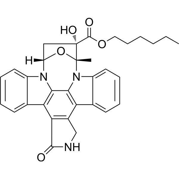
KT5720 is a potent, cell-permeable, specific, reversible and ATP-competitive PKA inhibitor (IC50=3.3 μM). KT5720 is effective in reversing MDR1-mediated multidrug resistance. KT5720 also reduces the excitability of dorsal root ganglion (DRG) neurons by attenuating Hyperpolarization-activated cyclic nucleotide-gated (HCN) channel activity and reducing intracellular Ca2+ concentrations. KT5720 can be used in the study of haematological malignancies as well as HCN and DRG neuron-related diseases.
For research use only. We do not sell to patients.
- Purity : 98.53%
- CAS No.: 108068-98-0
- Formula: C32H31N3O5
- Molecular Weight:537.61
-
Storage:Powder -20°C, 3 years ; In solvent -80°C, 6 months , -20°C, 1 month

To place orders, for customer services and technical support, please contact: MedChemExpress USA
Tel: 609-228-6898 E-mail: [email protected] [email protected]


-
 Biological Activity
-
Chemical Information
- Protocols
- Purity & Documentation
- References
-
 Help & FAQs


-
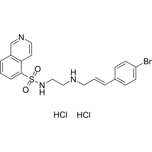
HY-15979A
H-89 dihydrochloridePKA Inhibitor -
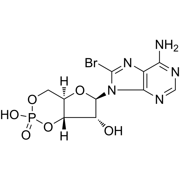
HY-12306A
8-Bromo-cAMPPKA Activator -
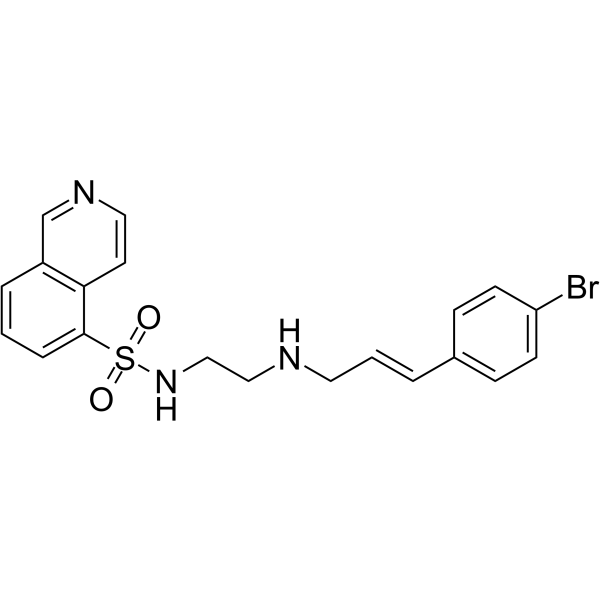
HY-15979
H-89PKA Inhibitor -
HY-N6791
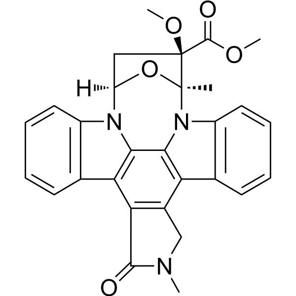
KT5823PKG Inhibitor -
HY-P1291
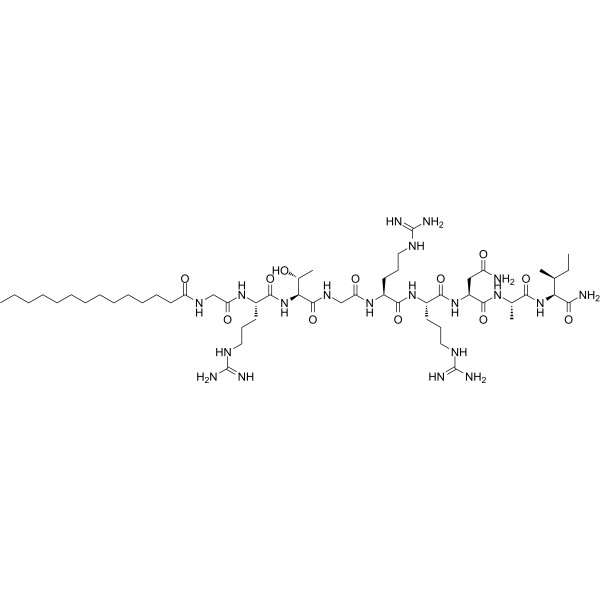
PKI 14-22 amide,myristoylatedPKA Inhibitor -

HY-15371
ForskolinAdenylate Cyclase Activator -
HY-13867
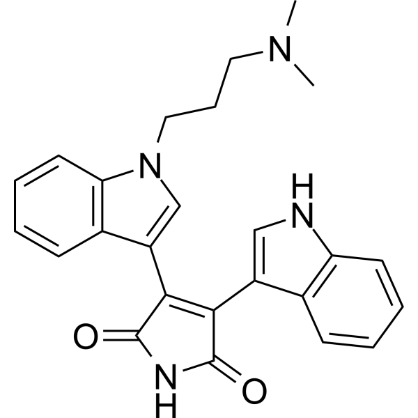
Bisindolylmaleimide IPKC Inhibitor -
HY-100530D
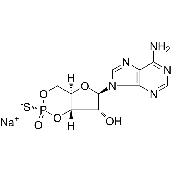
Rp-cAMPS sodium saltPKA I/II Antagonist -
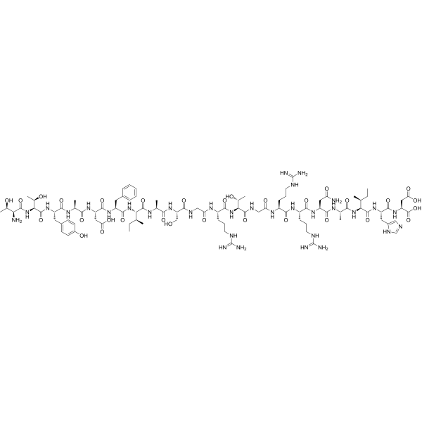
HY-P0222
PKI(5-24)PKA Inhibitor -
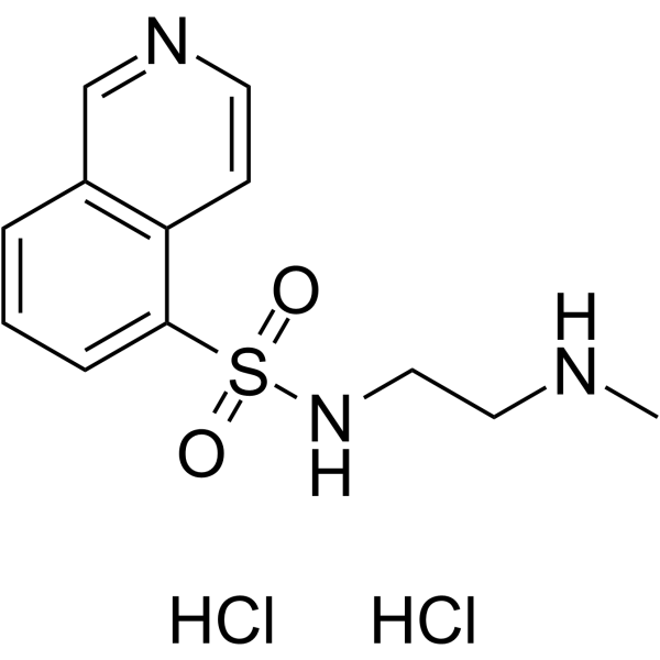
HY-112465
H-8 dihydrochloridecyclic nucleotide-dependent protein kinases Inhibitor -
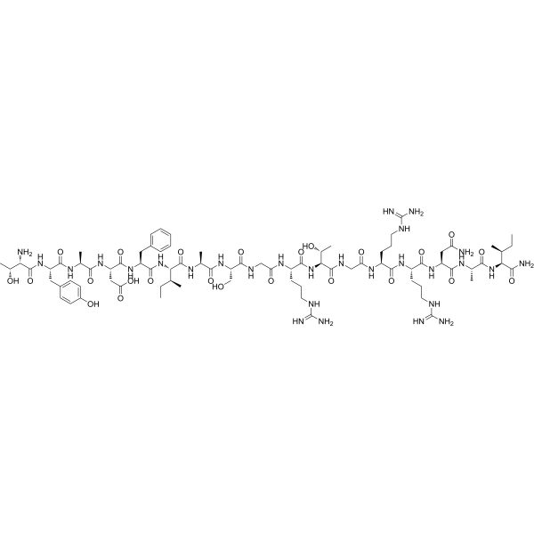
HY-P1290
PKA Inhibitor Fragment (6-22) amidePKA Inhibitor -
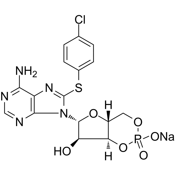
HY-111673
8-CPT-Cyclic AMP sodiumPKA Activator
Publications Citing Use of MedChemExpress (MCE) KT5720
More

Biological Activity
Description
IC50 & Target
IC50: 11 nM (PHK); 300 nM (PDK1); 3.3 µM (PKA)[3].
In Vitro
KT5720 (0-8 μM; 72 h) reverses multidrug resistance in an MDR1 lymphoma cell model[1].
?
KT5720 (3 μM) attenuates Ih in freshly isolated rat DRG neurons and slows down HCN channel activation kinetics[2].
?
KT5720 (3 μM) reduces DRG neurons excitability and reduces DRG neuron intracellular Ca2 + level[2].
MedChemExpress (MCE) has not independently confirmed the accuracy of these methods. They are for reference only. Further protocols information, click here.
-
Cell Line:LM1/MDR cells (carrying and expressing a human MDR1 transgene)
-
Concentration:0-8 µM
-
Incubation Time:72 h
-
Result:Increased sensitivity of LM1/MDR cells to colchicine in a dose-dependent manner.
In Vivo
MedChemExpress (MCE) has not independently confirmed the accuracy of these methods. They are for reference only.
-
Animal Model:MDR1 transgenic mice model[1].
-
Dosage:5 mg/kg
-
Administration:Intraperitoneal injection; single daily for 8 days
-
Result:Exhibited the leukopenia induced by daunorubicin in the MDR mice was equivalent to that measured in non-MDR mice treated with daunorubicin alone.


Chemical Information
-
CAS No. 108068-98-0
-
Appearance Solid
-
Molecular Weight 537.61
-
Formula C32H31N3O5
-
Color White to off-white
-
SMILES
C[C@@]12N3C4=C(C5=CC=CC=C35)C(CNC6=O)=C6C7=C4N(C8=CC=CC=C87)[C@@](C[C@@]2(O)C(OCCCCCC)=O)([H])O1
-
Shipping
Room temperature in continental US; may vary elsewhere.
-
Storage
Powder -20°C 3 years In solvent -80°C 6 months -20°C 1 month


Publications (4)
-
Journal Impact Factor
-
Most Recent
-
Cell Metab
The thermogenic activity of adjacent adipocytes fuels the progression of ccRCC and compromises anti-tumor therapeutic efficacy. [Abstract]2021 Oct 5;33(10):2021-2039.e8. PMID: 34508696 -
Cell Metab
The phytochemical hyperforin triggers thermogenesis in adipose tissue via a Dlat-AMPK signaling axis to curb obesity. [Abstract]2021 Mar 2;33(3):565-580.e7. PMID: 33657393 -
Oncogene
FBXO28 suppresses liver cancer invasion and metastasis by promoting PKA-dependent SNAI2 degradation. [Abstract]2023 Sep;42(39):2878-2891. PMID: 37596321 -
Int J Biochem Cell Biol
Downregulation of Pannexin-1 attenuates sepsis-induced acute lung injury via activation of the cAMP/PKA pathway. [Abstract]2026 Jun-Jul:195-196:106941. PMID: 41850417

Protocols
-
Cell Cytotoxicity Assay
Cytotoxicity assays are usually based on the assessment of cell membrane damage, which can also be indirectly detected by measuring cell viability. Detection methods include MTT assay, CKK-8 assay, LDH assay and ATP assay, etc.
-
Ca2+ Staining Technique
Ca2+ staining is an experimental technique that utilizes specific fluorescent probes (such as Fluo-4 AM, Fura-2, etc.) to qualitatively or quantitatively detect dynamic changes in intracellular Ca2+ concentrations; this is achieved by monitoring the changes in fluorescent signals generated when these probes bind to free intracellular calcium ions. The underlying principle relies primarily on the presence of chelating groups within the probe's molecular structure that possess high affinity for calcium ions.
-
Primary Dorsal Root Ganglion Sensory Neuron Culture
Primary dorsal root ganglion sensory neuron culture isolates DRG neuronal somata from rodent or human ganglia, dissociates tissue enzymatically and mechanically, and maintains post-mitotic sensory neurons in vitro for readouts such as neurite outgrowth, immunocytochemical marker expression, calcium imaging, electrophysiology, RNA/protein analysis, or neuropeptide release assays. The method reflects peripheral sensory neuron biology because DRG neurons are primary sensory neurons whose cell bodies reside in dorsal root ganglia and whose cultured dissociated cells can retain neuronal morphology, sensory-neuron marker expression, and stimulus-responsive properties depending on the downstream assay.


Purity & Documentation
-
Data Sheet (278 KB)
-
SDS (418 KB)
- English - EN (418 KB)
- Français - FR (418 KB)
- Deutsch - DE (418 KB)
- Norwegian - NO (418 KB)
- Español - ES (418 KB)
- Swedish - SV (418 KB)
- Italian - IT (418 KB)
- Korean - KR (418 KB)
- Portuguese - PT (418 KB)
-
Handling Instructions (2659 KB)
References
[1]. Galski H, et al. In vitro and in vivo reversal of MDR1-mediated multidrug resistance by KT-5720: implications on hematological malignancies. Leuk Res. 2006 Sep;30(9):1151-8. [Content Brief]
[2]. Cheng Q, et al. Novel role of KT5720 on regulating hyperpolarization-activated cyclic nucleotide-gated channel activity and dorsal root ganglion neuron excitability. DNA Cell Biol. 2013 Jun;32(6):320-8. [Content Brief]
[3]. Davies SP, et al. Specificity and mechanism of action of some commonly used protein kinase inhibitors. Biochem J. 2000 Oct 1;351(Pt 1):95-105. [Content Brief]

Calculators
Concentration (start) × Volume (start) = Concentration (final) × Volume (final)


KT5720 Related Classifications
HY-N6789 Related Classifications
 Powered by Bioz