



Levamlodipine
Based on 2 publication(s) in Google Scholar
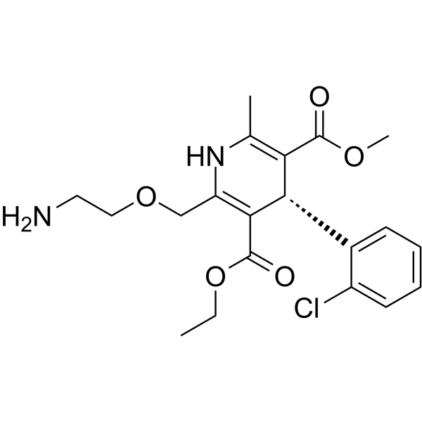
Levamlodipine ((S)-Amlodipine; Levoamlodipin) is an orally active L-type calcium channel blocker and MMP-9 modulator with high permeability and retention properties. Levamlodipine significantly enhances plaque stability and improves lipid profiles by reducing blood pressure, decreasing systolic blood pressure variability, and inhibiting MMP-9 expression in atherosclerotic plaques. Levamlodipine not only alleviates cardiac and aortic hypertrophy and prevents renal atrophy, but also produces synergistic effects in blood pressure reduction and organ protection when combined with bisoprolol (HY-129029). Levamlodipine exerts no significant inhibitory effect on abdominal aortic intimal hyperplasia. When excessively accumulated in the epidermis, Levamlodipine may induce changes in keratin structure, impair the skin barrier and trigger inflammation; long-term use further exacerbates skin irritation caused by local administration. Levamlodipine can be used in research related to hypertension and atherosclerosis.
For research use only. We do not sell to patients.
- Purity : 98.06%
- CAS No.: 103129-82-4
- Formula: C20H25ClN2O5
- Molecular Weight:408.88
-
Storage:Powder -20°C, 3 years , 4°C, 2 years ; In solvent -80°C, 6 months , -20°C, 1 month

To place orders, for customer services and technical support, please contact: MedChemExpress USA
Tel: 609-228-6898 E-mail: [email protected] [email protected]


-
 Biological Activity
-
Chemical Information
-
Solvent & Solubility
- Protocols
- Purity & Documentation
- References
-
 Help & FAQs


-
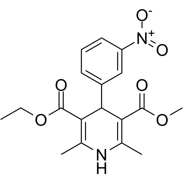
HY-B0424
NitrendipineCalcium Channel Blocker -
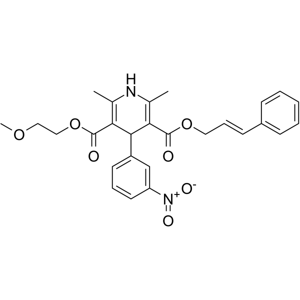
HY-17404
CilnidipineCa Channel Blocker -
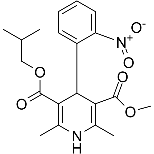
HY-17402
NisoldipineCalcium Channel Blocker -
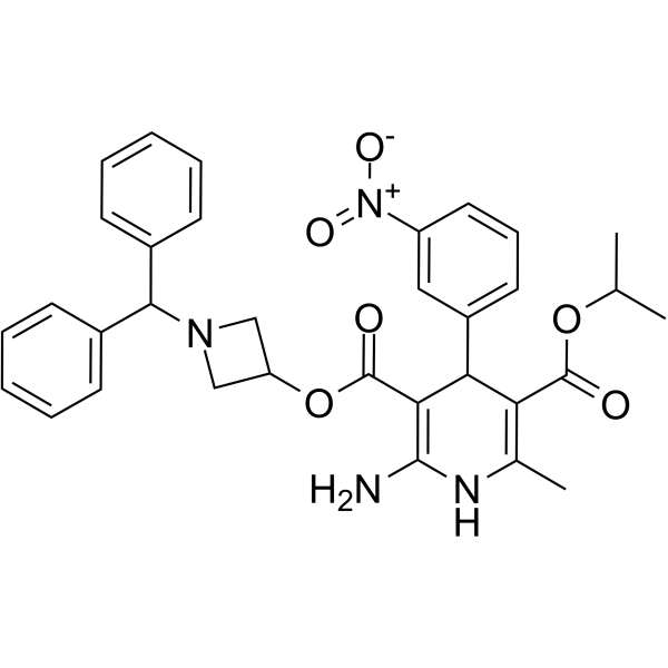
HY-B0023
AzelnidipineCalcium Channel Blocker -
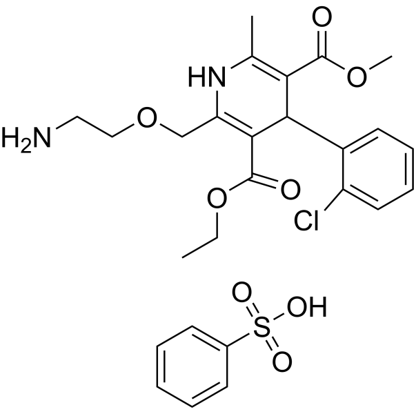
HY-B0317B
Amlodipine besylateCalcium Channel Blocker -
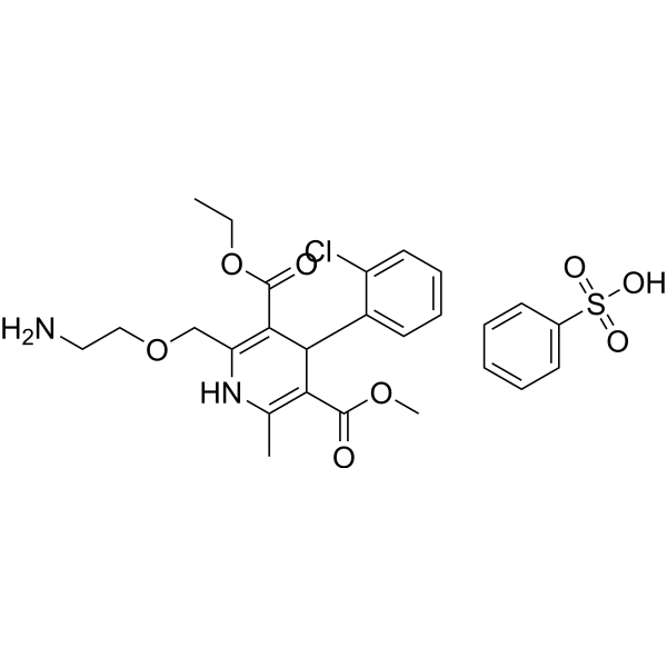
HY-B0317BR
Amlodipine (besylate) (Standard)Calcium Channel Blocker -
HY-B0284
NifedipineCalcium Channel Antagonist -
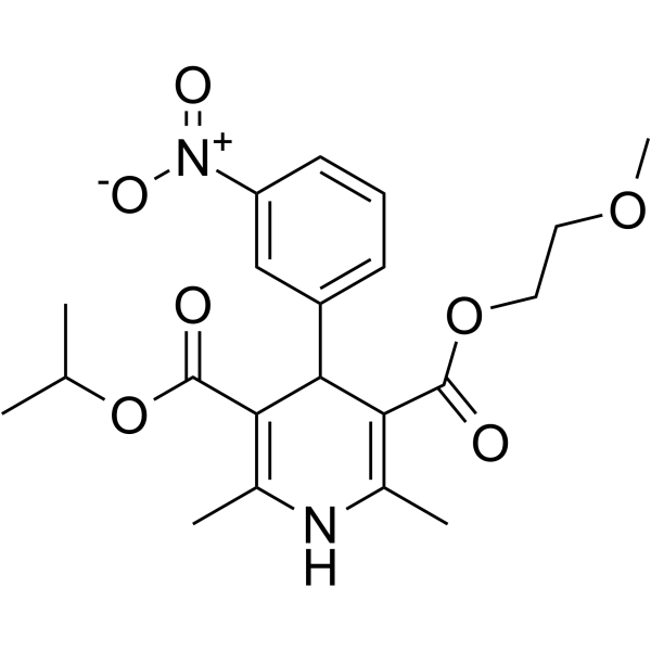
HY-B0265
NimodipineDihydropyridine Calcium Antagonist -
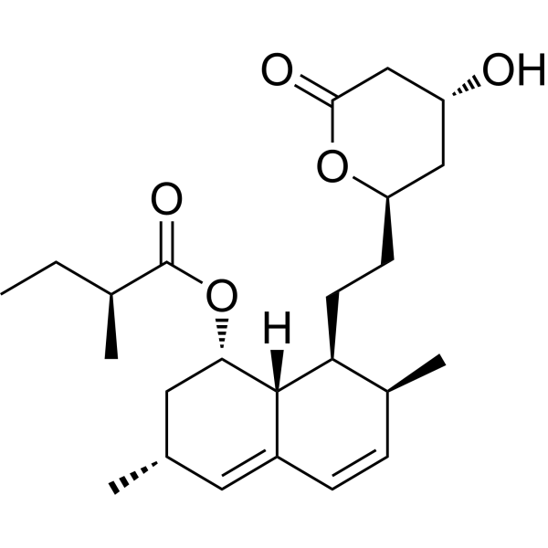
HY-N0504
LovastatinHMG-CoA Inhibitor -
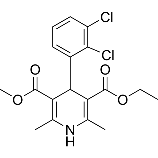
HY-B0309
FelodipineCalcium Channel Antagonist -
HY-B0216
EthinylestradiolEndogenous Metabolite -
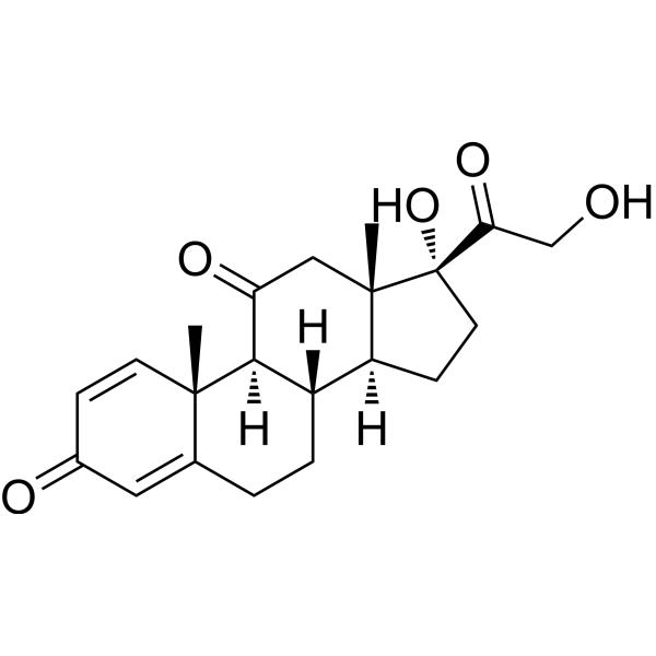
HY-B0214
PrednisoneGlucocorticoid Receptor Agonist
Publications Citing Use of MedChemExpress (MCE) Levamlodipine
MoreAll Calcium Channel Isoforms
More

Biological Activity
Description
IC50 & Target
|
L-type calcium channel |
MMP-9 |
In Vitro
Levamlodipine-Sorbic acid (HY-N0626) ion pair patches (13-15% (w/w) LAM-SA, 10% (w/w) IPM; 72 h) maintain equivalent porcine skin permeability to free levamlodipine patches, and addition of 10% IPM significantly enhances permeability; the optimized 13% LAM-SA, 10% IPM, 70 μm patch achieves high, predictable transdermal delivery over 72 h[3].
MedChemExpress (MCE) has not independently confirmed the accuracy of these methods. They are for reference only.
Parmacokinetics
In Vivo
Levamlodipine (1-4 mg/kg; intragastric administration; single dose; 1 mg/kg/day; p.o.; daily; 16 weeks) reduces blood pressure in male SHR in a dose-dependent manner[2].
The ion-pair transdermal patch of Levamlodipine and sorbic acid (HY-N0626) (12-15% w/w; transdermal administration; single dose; 72 h) significantly reduces skin irritation in rabbits. Compared with non-ion-pair levamlodipine patches, it shows a lower erythema index, milder epidermal thickening, and faster skin recovery[3].
MedChemExpress (MCE) has not independently confirmed the accuracy of these methods. They are for reference only.
-
Animal Model:New Zealand rabbit (5~6 months old, male, 2.0~2.5 kg; induced by high-fat diet feeding + abdominal aortic balloon endothelial injury)[1]
-
Dosage:2.5 mg/d
-
Administration:p.o. (mixed with feed); once daily; 8 weeks
-
Result:Showed a decreasing trend in serum LDL-C level (16.24 mmol/L vs model group 19.41 mmol/L, P=0.08).
Resulted in abdominal aortic plaque thickness of 313.56 μm and intimal hyperplasia index (IHI) of 0.66, with no significant difference compared to model group (405.93 μm, 0.68, P>0.05).
Showed no significant difference in serum hs-CRP, LP-PLA2, PAI-1 levels compared to model group (P>0.05).
Increased serum TNF-α level to 113.11 μg/mL, which was significantly higher than model group's 81.13 μg/mL (P<0.05).
Reduced the positive expression area percentage of MMP-9 in plaques to 52.45%, which was significantly lower than model group's 70.84% (P<0.05).
-
Animal Model:Japanese white rabbits (male, 1.8-2.2 kg)[3]
-
Dosage:12% w/w LAM-SA (6 cm2 skin area); 13.00% w/w LAM-SA (1.13 cm2 skin area); 15% w/w LAM-SA
-
Administration:topical; single exposure; 72 hours
-
Result:Produced post-removal erythema index of 188 at 1 hour, 181 at 24 hours, and 166 at 72 hours for 13% w/w formulation; showed significantly lower ΔEI than non-ion-paired LAM patch at all time points (p < 0.05).
Exhibited only slight epidermal thickening with no inflammatory cell infiltration, and significantly lower epidermal thickness than non-ion-paired LAM patch for 13% w/w formulation.
Produced only mild erythema, with the lowest erythema index among all tested ion-paired formulations for 15% w/w formulation.
-
Animal Model:Wistar rats (male, 180-220 g)[3]
-
Dosage:12% w/w LAM-SA (6 cm2 skin area)
-
Administration:topical; single exposure
-
Result:Achieved Cmax of 182.27 ng/mL at tmax of 36 hours, t1/2 of 23.55 hours, and AUC0-t of 7863.22h·ng/mL.
Reached absolute bioavailability of 81.20%.
Maintained detectable plasma levamlodipine concentrations for at least 4 days, with complete elimination by the fifth day.
- RNA extraction experimental
- Mitochondrial membrane-potential fluorescent assay
- Mitochondrial membrane-potential and mitochondrial mass staining
- Transepithelial/transendothelial electrical resistance assay
- Research Protocol for Cardiovascular Diseases
- Research Protocol for Inflammation-related Diseases
- Dermal Irritation/Dermal Toxicity Study
Clinical Trial
| NCT Number | Sponsor | Condition | Start Date |
Phase

|
|---|---|---|---|---|
| NCT01329991 | 2011-05 | PHASE1 |


Chemical Information
-
CAS No. 103129-82-4
-
Appearance Solid
-
Molecular Weight 408.88
-
Formula C20H25ClN2O5
-
Color White to off-white
-
SMILES
O=C(C1=C(COCCN)NC(C)=C(C(OC)=O)[C@@H]1C2=CC=CC=C2Cl)OCC
-
Synonyms
(S)-Amlodipine; Levoamlodipine
-
Shipping
Room temperature in continental US; may vary elsewhere.
-
Storage
Powder -20°C 3 years 4°C 2 years In solvent -80°C 6 months -20°C 1 month


Publications (2)
-
Journal Impact Factor
-
Most Recent
-
Cell
Structural basis for human Cav1.2 inhibition by multiple drugs and the neurotoxin calciseptine. [Abstract]2023 Nov 22;186(24):5363-5374.e16. PMID: 37972591 -
Proc Natl Acad Sci U S A
Design and structural basis of selective 1,4-dihydropyridine inhibitors of the calcium-activated potassium channel KCa3.1. [Abstract]2025 May 6;122(18):e2425494122. PMID: 40294255


Solvent & Solubility
In Vitro:
DMSO : 100 mg/mL (244.57 mM; Need ultrasonic; Hygroscopic DMSO has a significant impact on the solubility of product, please use newly opened DMSO)
Please refer to the solubility information to select the appropriate solvent. Once prepared, please aliquot and store the solution to prevent product inactivation from repeated freeze-thaw cycles.
Storage method and period of stock solution: -80°C, 6 months; -20°C, 1 month. When stored at -80°C, please use it within 6 months. When stored at -20°C, please use it within 1 month.
Please refer to the solubility information to select the appropriate solvent. Once prepared, please aliquot and store the solution to prevent product inactivation from repeated freeze-thaw cycles.
Storage method and period of stock solution: -80°C, 6 months; -20°C, 1 month. When stored at -80°C, please use it within 6 months. When stored at -20°C, please use it within 1 month.
Concentration (start) × Volume (start) = Concentration (final) × Volume (final)
In Vivo:
Select the appropriate dissolution method based on your experimental animal and administration route.
- For the following dissolution methods, please ensure to first prepare a clear stock solution using an In Vitro approach and then sequentially add co-solvents:
- To ensure reliable experimental results, the clarified stock solution can be appropriately stored based on storage conditions. As for the working solution for In Vivo experiments, it is recommended to prepare freshly and use it on the same day.
- The percentages shown for the solvents indicate their volumetric ratio in the final prepared solution. If precipitation or phase separation occurs during preparation, heat and/or sonication can be used to aid dissolution.
Add each solvent one by one: 10% DMSO 40% PEG300 5% Tween-80 45% Saline
Solubility: ≥ 2.5 mg/mL (6.11 mM); Clear solution
This protocol yields a clear solution of ≥ 2.5 mg/mL (saturation unknown).
Taking 1 mL working solution as an example, add 100 μL DMSO stock solution (25.0 mg/mL) to 400 μL PEG300, and mix evenly; then add 50 μL Tween-80 and mix evenly; then add 450 μL Saline to adjust the volume to 1 mL.
Preparation of Saline: Dissolve 0.9 g sodium chloride in ddH₂O and dilute to 100 mL to obtain a clear Saline solution.
Add each solvent one by one: 10% DMSO 90% (20% SBE-β-CD in Saline)
Solubility: ≥ 2.5 mg/mL (6.11 mM); Clear solution
This protocol yields a clear solution of ≥ 2.5 mg/mL (saturation unknown).
Taking 1 mL working solution as an example, add 100 μL DMSO stock solution (25.0 mg/mL) to 900 μL 20% SBE-β-CD in Saline, and mix evenly.
Preparation of 20% SBE-β-CD in Saline (4°C, storage for one week): 2 g SBE-β-CD powder is dissolved in 10 mL Saline, completely dissolve until clear.
In Vivo Dissolution Calculator
Please enter the basic information of animal experiments:
-
-
-
-
Recommended: Prepare an additional quantity of animals to account for potential losses during experiments.
Please enter your animal formula composition:
-
%DMSO +
Recommended: Keep the proportion of DMSO in working solution below 2% if your animal is weak.
-
%+
-
+%Tween-80 + +
-
%Saline +
The co-solvents required include: DMSO, . All of co-solvents are available by MedChemExpress (MCE). , Tween 80. All of co-solvents are available by MedChemExpress (MCE).
Working solution concentration: 0.22 mg/mL
Method for preparing stock solution: mg drug dissolved in μL DMSO. Stock solution concentration: mg/mL.
1. Take μL DMSO stock solution;
2. Add μL .
μL , mix evenly;
3. Then add μL Tween 80, mix evenly;
4. Then add μL
Please ensure that the stock solution in the first step is dissolved to a clear state, and add co-solvents in sequence. You can use ultrasonic heating (ultrasonic cleaner, recommended frequency 20-40 kHz), vortexing, etc. to assist dissolution.


Purity & Documentation
-
Data Sheet (283 KB)
-
SDS (393 KB)
- English - EN (393 KB)
- Français - FR (393 KB)
- Deutsch - DE (393 KB)
- Norwegian - NO (393 KB)
- Español - ES (393 KB)
- Swedish - SV (393 KB)
- Italian - IT (393 KB)
- Korean - KR (393 KB)
- Portuguese - PT (393 KB)
-
Handling Instructions (2659 KB)
References
[2]. Yang YL, et al. Synergic effects of levamlodipine and bisoprolol on blood pressure reduction and organ protection in spontaneously hypertensive rats. CNS Neurosci Ther. 2012;18(6):471-474. [Content Brief]
[3]. Wu J, et al. Development of levamlodipine long-acting patches based on an ion-pair strategy: Investigation of the mechanism for reducing skin irritation. Int J Pharm. 2024;665:124703. [Content Brief]


Complete Stock Solution Preparation Table
Please refer to the solubility information to select the appropriate solvent. Once prepared, please aliquot and store the solution to prevent product inactivation from repeated freeze-thaw cycles.
Storage method and period of stock solution: -80°C, 6 months; -20°C, 1 month. When stored at -80°C, please use it within 6 months. When stored at -20°C, please use it within 1 month.
| Optional Solvent | Concentration Solvent Mass | 1 mg | 5 mg | 10 mg | 25 mg |
|---|---|---|---|---|---|
| DMSO | 1 mM | 2.4457 mL | 12.2285 mL | 24.4571 mL | 61.1426 mL |
| 5 mM | 0.4891 mL | 2.4457 mL | 4.8914 mL | 12.2285 mL | |
| 10 mM | 0.2446 mL | 1.2229 mL | 2.4457 mL | 6.1143 mL | |
| 15 mM | 0.1630 mL | 0.8152 mL | 1.6305 mL | 4.0762 mL | |
| 20 mM | 0.1223 mL | 0.6114 mL | 1.2229 mL | 3.0571 mL | |
| 25 mM | 0.0978 mL | 0.4891 mL | 0.9783 mL | 2.4457 mL | |
| 30 mM | 0.0815 mL | 0.4076 mL | 0.8152 mL | 2.0381 mL | |
| 40 mM | 0.0611 mL | 0.3057 mL | 0.6114 mL | 1.5286 mL | |
| 50 mM | 0.0489 mL | 0.2446 mL | 0.4891 mL | 1.2229 mL | |
| 60 mM | 0.0408 mL | 0.2038 mL | 0.4076 mL | 1.0190 mL | |
| 80 mM | 0.0306 mL | 0.1529 mL | 0.3057 mL | 0.7643 mL | |
| 100 mM | 0.0245 mL | 0.1223 mL | 0.2446 mL | 0.6114 mL |


Levamlodipine Related Classifications
HY-14744 Related Classifications
 Powered by Bioz