



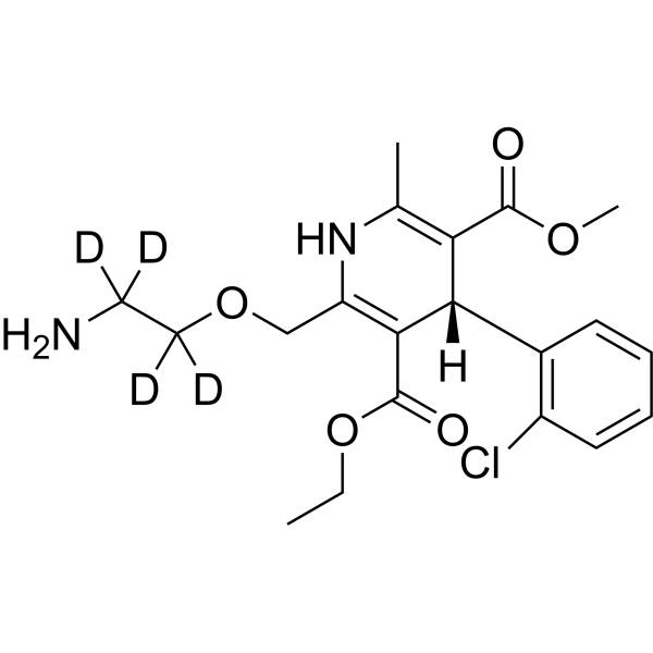
Levamlodipine-d4

Based on 1 Customer Validation
Levamlodipine-d4 is the deuterium labeled Levamlodipine. Levamlodipine ((S)-Amlodipine; Levoamlodipin) is an orally active L-type calcium channel blocker and MMP-9 modulator with high permeability and retention properties. Levamlodipine significantly enhances plaque stability and improves lipid profiles by reducing blood pressure, decreasing systolic blood pressure variability, and inhibiting MMP-9 expression in atherosclerotic plaques. Levamlodipine not only alleviates cardiac and aortic hypertrophy and prevents renal atrophy, but also produces synergistic effects in blood pressure reduction and organ protection when combined with bisoprolol (HY-129029). Levamlodipine exerts no significant inhibitory effect on abdominal aortic intimal hyperplasia. When excessively accumulated in the epidermis, Levamlodipine may induce changes in keratin structure, impair the skin barrier and trigger inflammation; long-term use further exacerbates skin irritation caused by local administration. Levamlodipine can be used in research related to hypertension and atherosclerosis.
商品は「研究用試薬」です。人や動物の医療用・臨床診断用・食品用の製品ではありません。
研究用途以外に使用した場合、当社は一切の責任を負いかねます。
- 純度 : 99.74%
- CAS 番号: 1346617-19-3
- 分子式: C20H21D4ClN2O5
- 分子量:412.90
-
保管条件:
-20°C, protect from light, stored under nitrogen
* In solvent : -80°C, 6 months; -20°C, 1 month (protect from light, stored under nitrogen)

To place orders, for customer services and technical support, please contact: MedChemExpress USA
Tel: 609-228-6898 E-mail: [email protected] [email protected]


-
 生物活性
生物活性
-
化学情報
-
溶剤 & 溶解度
- プロトコル
- 純度とドキュメンテーション
- 参考文献
-
 Help & FAQs


Calcium Channel アイソフォーム固有の製品をすべて表示
More

生物活性
製品説明
体外実験
Stable heavy isotopes of hydrogen, carbon, and other elements have been incorporated into drug molecules, largely as tracers for quantitation during the drug development process. Deuteration has gained attention because of its potential to affect the pharmacokinetic and metabolic profiles of drugs[1].
MedChemExpress (MCE) has not independently confirmed the accuracy of these methods. They are for reference only.
- RNA extraction experimental
- Mitochondrial membrane-potential fluorescent assay
- Mitochondrial membrane-potential and mitochondrial mass staining
- Transepithelial/transendothelial electrical resistance assay
- Research Protocol for Cardiovascular Diseases
- Research Protocol for Inflammation-related Diseases
- Dermal Irritation/Dermal Toxicity Study
アプリケーション
1. This compound can be used as a tracer
2. This compound can be used as an internal standard for quantitative analysis by NMR, GC-MS, or LC-MS.


化学情報
-
CAS 番号 1346617-19-3
-
非標識Cas 103129-82-4
-
性状 Solid
-
分子量 412.90
-
分子式 C20H21D4ClN2O5
-
Color White to off-white
-
SMILES
CCOC(C1=C(COC([2H])([2H])C([2H])([2H])N)NC(C)=C(C(OC)=O)[C@]1([H])C2=C(C=CC=C2)Cl)=O
-
別名
(S)-Amlodipine-d4; Levoamlodipine-d4
-
輸送条件
Room temperature in continental US; may vary elsewhere.
-
保管条件
-20°C, protect from light, stored under nitrogen
* In solvent : -80°C, 6 months; -20°C, 1 month (protect from light, stored under nitrogen)


溶剤 & 溶解度
体外:
DMSO : 100 mg/mL (242.19 mM; Need ultrasonic and warming; Hygroscopic DMSO has a significant impact on the solubility of product, please use newly opened DMSO)
Please refer to the solubility information to select the appropriate solvent. Once prepared, please aliquot and store the solution to prevent product inactivation from repeated freeze-thaw cycles.
Storage method and period of stock solution: -80°C, 6 months; -20°C, 1 month (protect from light, stored under nitrogen). When stored at -80°C, please use it within 6 months. When stored at -20°C, please use it within 1 month.
Please refer to the solubility information to select the appropriate solvent. Once prepared, please aliquot and store the solution to prevent product inactivation from repeated freeze-thaw cycles.
Storage method and period of stock solution: -80°C, 6 months; -20°C, 1 month (protect from light, stored under nitrogen). When stored at -80°C, please use it within 6 months. When stored at -20°C, please use it within 1 month.
濃度 (開始) × 体積 (開始) = 濃度 (終了) × 体積 (終了)


純度とドキュメンテーション
-
データシート (277 KB)
-
SDS (252 KB)
- English - EN (252 KB)
- Français - FR (252 KB)
- Deutsch - DE (252 KB)
- Norwegian - NO (252 KB)
- Español - ES (252 KB)
- Swedish - SV (252 KB)
- Italian - IT (252 KB)
- Korean - KR (252 KB)
- Portuguese - PT (252 KB)
-
取扱説明書 (2659 KB)
参考文献
[1]. Russak EM, et al. Impact of Deuterium Substitution on the Pharmacokinetics of Pharmaceuticals. Ann Pharmacother. 2019 Feb;53(2):211-216. [Content Brief]
[3]. Yang YL, et al. Synergic effects of levamlodipine and bisoprolol on blood pressure reduction and organ protection in spontaneously hypertensive rats. CNS Neurosci Ther. 2012;18(6):471-474. [Content Brief]
[4]. Wu J, et al. Development of levamlodipine long-acting patches based on an ion-pair strategy: Investigation of the mechanism for reducing skin irritation. Int J Pharm. 2024;665:124703. [Content Brief]


Complete Stock Solution Preparation Table
Please refer to the solubility information to select the appropriate solvent. Once prepared, please aliquot and store the solution to prevent product inactivation from repeated freeze-thaw cycles.
Storage method and period of stock solution: -80°C, 6 months; -20°C, 1 month (protect from light, stored under nitrogen). When stored at -80°C, please use it within 6 months. When stored at -20°C, please use it within 1 month.
| Optional Solvent | Concentration Solvent Mass | 1 mg | 5 mg | 10 mg | 25 mg |
|---|---|---|---|---|---|
| DMSO | 1 mM | 2.4219 mL | 12.1095 mL | 24.2189 mL | 60.5473 mL |
| 5 mM | 0.4844 mL | 2.4219 mL | 4.8438 mL | 12.1095 mL | |
| 10 mM | 0.2422 mL | 1.2109 mL | 2.4219 mL | 6.0547 mL | |
| 15 mM | 0.1615 mL | 0.8073 mL | 1.6146 mL | 4.0365 mL | |
| 20 mM | 0.1211 mL | 0.6055 mL | 1.2109 mL | 3.0274 mL | |
| 25 mM | 0.0969 mL | 0.4844 mL | 0.9688 mL | 2.4219 mL | |
| 30 mM | 0.0807 mL | 0.4036 mL | 0.8073 mL | 2.0182 mL | |
| 40 mM | 0.0605 mL | 0.3027 mL | 0.6055 mL | 1.5137 mL | |
| 50 mM | 0.0484 mL | 0.2422 mL | 0.4844 mL | 1.2109 mL | |
| 60 mM | 0.0404 mL | 0.2018 mL | 0.4036 mL | 1.0091 mL | |
| 80 mM | 0.0303 mL | 0.1514 mL | 0.3027 mL | 0.7568 mL | |
| 100 mM | 0.0242 mL | 0.1211 mL | 0.2422 mL | 0.6055 mL |


Levamlodipine-d4 関連分類
HY-14744S 関連分類
 Powered by Bioz