



Liriodenine

Based on 1 Customer Validation
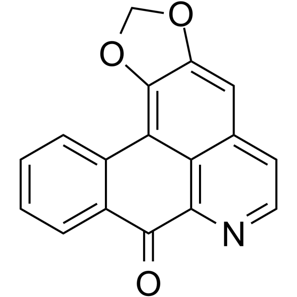
Liriodenine (Spermatheridine; VLT045) is an aporphine alkaloid isolated from the plant?Mitrephora sirikitiae?and has anti-cancer activities. Liriodenine induces cell apoptosis, activates the intrinsic pathway by induction of caspase-3 and caspase-9.
For research use only. We do not sell to patients.
- Purity : 97%
- CAS No.: 475-75-2
- Formula: C17H9NO3
- Molecular Weight:275.26
-
Storage:
-20°C, sealed storage, away from moisture
* In solvent : -80°C, 6 months; -20°C, 1 month (sealed storage, away from moisture)

To place orders, for customer services and technical support, please contact: MedChemExpress USA
Tel: 609-228-6898 E-mail: [email protected] [email protected]


-
 Biological Activity
-
Chemical Information
- Protocols
- Purity & Documentation
- References
-
 Help & FAQs




Biological Activity
Description
Cellular Effect
|
Cell Line

|
Type | Value | Description | References |
|---|---|---|---|---|
| A549 | ED50 |
0.72 μg/mL
Compound: 1
|
Cytotoxicity against human A549 cells
Cytotoxicity against human A549 cells
|
[PMID: 2292689] |
| B16-F10 | IC50 |
>10 μM
Compound: 20
|
Cytotoxicity against mouse B16-F10 cells assessed as reduction in cell viability incubated for 72 hrs by Alamar blue assay
Cytotoxicity against mouse B16-F10 cells assessed as reduction in cell viability incubated for 72 hrs by Alamar blue assay
|
[PMID: 27300257] |
| BV-2 | IC50 |
5 μM
Compound: 13
|
Antineuroinflammatory activity against mouse BV2 cells assessed as inhibition of LPS-induced NO production incubated for 20 hrs by Griess assay
Antineuroinflammatory activity against mouse BV2 cells assessed as inhibition of LPS-induced NO production incubated for 20 hrs by Griess assay
|
[PMID: 33822610] |
| HCT-8 | ED50 |
0.7 μg/mL
Compound: 1
|
Cytotoxicity against human HCT8 cells
Cytotoxicity against human HCT8 cells
|
[PMID: 2292689] |
| HepG2 | IC50 |
8.3 μM
Compound: 20
|
Cytotoxicity against human HepG2 cells assessed as reduction in cell viability incubated for 72 hrs by Alamar blue assay
Cytotoxicity against human HepG2 cells assessed as reduction in cell viability incubated for 72 hrs by Alamar blue assay
|
[PMID: 27300257] |
| HL-60 | IC50 |
5.5 μM
Compound: 20
|
Cytotoxicity against human HL-60 cells assessed as reduction in cell viability incubated for 72 hrs by Alamar blue assay
Cytotoxicity against human HL-60 cells assessed as reduction in cell viability incubated for 72 hrs by Alamar blue assay
|
[PMID: 27300257] |
| HT-29 | IC50 |
>10 μM
Compound: 6
|
Cytotoxicity against human HT-29 cells assessed as inhibition of cell proliferation after 3 days by SRB assay
Cytotoxicity against human HT-29 cells assessed as inhibition of cell proliferation after 3 days by SRB assay
|
[PMID: 23327794] |
| K562 | IC50 |
5 μM
Compound: 20
|
Cytotoxicity against human K562 cells assessed as reduction in cell viability incubated for 72 hrs by Alamar blue assay
Cytotoxicity against human K562 cells assessed as reduction in cell viability incubated for 72 hrs by Alamar blue assay
|
[PMID: 27300257] |
| KB | ED50 |
1 μg/mL
Compound: 1
|
Cytotoxicity against human KB cells
Cytotoxicity against human KB cells
|
[PMID: 2292689] |
| KB | ED50 |
3.1 μg/mL
Compound: liriodenine
|
Cytotoxicity against human KB cells
Cytotoxicity against human KB cells
|
10.1021/np50019a011 |
| KB | IC50 |
5 μM
Compound: 9
|
Cytotoxicity against human KB cells after 3 days by sulforhodamine B assay
Cytotoxicity against human KB cells after 3 days by sulforhodamine B assay
|
[PMID: 19874044] |
| L1210 | ED50 |
2.33 μg/mL
Compound: 1
|
Cytotoxicity against mouse L1210 cells
Cytotoxicity against mouse L1210 cells
|
[PMID: 2292689] |
| MCF7 | IC50 |
5 μM
Compound: 9
|
Cytotoxicity against human MCF7 cells after 3 days by sulforhodamine B assay
Cytotoxicity against human MCF7 cells after 3 days by sulforhodamine B assay
|
[PMID: 19874044] |
| NCI-H460 | IC50 |
5 μM
Compound: 9
|
Cytotoxicity against human NCI-H460 cells after 3 days by sulforhodamine B assay
Cytotoxicity against human NCI-H460 cells after 3 days by sulforhodamine B assay
|
[PMID: 19874044] |
| P388 | ED50 |
0.57 μg/mL
Compound: 1
|
Cytotoxicity against mouse P388 cells
Cytotoxicity against mouse P388 cells
|
[PMID: 2292689] |
| PBMC | IC50 |
34.8 μM
Compound: 20
|
Cytotoxicity against human PBMC cells assessed as reduction in cell viability incubated for 72 hrs by Alamar blue assay
Cytotoxicity against human PBMC cells assessed as reduction in cell viability incubated for 72 hrs by Alamar blue assay
|
[PMID: 27300257] |
| RAW264.7 | IC50 |
1.39 μM
Compound: 129
|
Inhibition of NO production in LPS-stimulated mouse RAW264.7 cells incubated for 24 hrs by Griess reagent-based assay
Inhibition of NO production in LPS-stimulated mouse RAW264.7 cells incubated for 24 hrs by Griess reagent-based assay
|
[PMID: 33454546] |
| RAW264.7 | IC50 |
10.5 μM
Compound: 10
|
Antiinflammatory activity in mouse RAW264.7 assessed as inhibition of LPS-induced NO production preincubated for 30 mins followed by LPS stimulation measured after 24 hrs by Griess reagent based assay
Antiinflammatory activity in mouse RAW264.7 assessed as inhibition of LPS-induced NO production preincubated for 30 mins followed by LPS stimulation measured after 24 hrs by Griess reagent based assay
|
[PMID: 33939429] |
| RAW264.7 | IC50 |
34.7 μM
Compound: 6
|
Inhibition of LPS-stimulated nitric oxide production in mouse RAW264.7 cells incubated for 24 hrs by Griess reagent-based assay
Inhibition of LPS-stimulated nitric oxide production in mouse RAW264.7 cells incubated for 24 hrs by Griess reagent-based assay
|
[PMID: 32247751] |
| SF-268 | IC50 |
5 μM
Compound: 9
|
Cytotoxicity against human SF268 cells after 3 days by sulforhodamine B assay
Cytotoxicity against human SF268 cells after 3 days by sulforhodamine B assay
|
[PMID: 19874044] |
| Vero | CC50 |
48 μM
Compound: 16
|
Cytotoxicity against african green monkey Vero cells
Cytotoxicity against african green monkey Vero cells
|
[PMID: 9584402] |
In Vitro
Liriodenine is against P-388, KB, HT-29, MCF-7, A549, ASK and Hek-293 cells with IC50 values of 9.60 μM, 11.02 μM, 10.62 μM, 9.20 μM and 8.07μM, respectively.
MedChemExpress (MCE) has not independently confirmed the accuracy of these methods. They are for reference only.


Chemical Information
-
CAS No. 475-75-2
-
Appearance Solid
-
Molecular Weight 275.26
-
Formula C17H9NO3
-
Color Light yellow to yellow
-
SMILES
O=C1C2=CC=CC=C2C3=C4OCOC4=CC5=CC=NC1=C53
-
Synonyms
Spermatheridine; VLT045
-
Structure Classification
-
Shipping
Room temperature in continental US; may vary elsewhere.
-
Storage
-20°C, sealed storage, away from moisture
* In solvent : -80°C, 6 months; -20°C, 1 month (sealed storage, away from moisture)


Purity & Documentation
-
Data Sheet (273 KB)
-
SDS (421 KB)
- English - EN (421 KB)
- Français - FR (421 KB)
- Deutsch - DE (421 KB)
- Norwegian - NO (421 KB)
- Español - ES (421 KB)
- Swedish - SV (421 KB)
- Italian - IT (421 KB)
- Korean - KR (421 KB)
- Portuguese - PT (421 KB)
-
Handling Instructions (2659 KB)
References
[1]. Natthinee Anantachoke, et al. Cytotoxic Compounds From the Leaves and Stems of the Endemic Thai Plant Mitrephora sirikitiae. Pharm Biol [Content Brief]
[2]. Noraziah Nordin, et al. Liriodenine, an Aporphine Alkaloid From Enicosanthellum Pulchrum, Inhibits Proliferation of Human Ovarian Cancer Cells Through Induction of Apoptosis via the Mitochondrial Signaling Pathway and Blocking Cell Cycle Progression. Drug Des Devel Ther [Content Brief]

Calculators
Concentration (start) × Volume (start) = Concentration (final) × Volume (final)


Liriodenine Related Classifications
HY-N3376 Related Classifications
 Powered by Bioz