



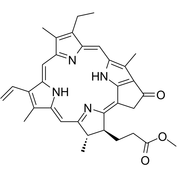
Methyl pyropheophorbide-a
Based on 2 publication(s) in Google Scholar
Methyl pyropheophorbide-a (Pyropheophorbide-a methyl ester), a chlorophyll-a derivative, is a potent photosensitizer that can be used in photodynamic therapy (PDT) of cancer. Methyl pyropheophorbide-a has photodynamic activity and can induce apoptosis and inhibit tumor growth.
For research use only. We do not sell to patients.
- Purity : 99.14%
- CAS No.: 6453-67-4
- Formula: C34H36N4O3
- Molecular Weight:548.67
-
Storage:Powder -20°C, 3 years , 4°C, 2 years ; In solvent -80°C, 6 months , -20°C, 1 month

To place orders, for customer services and technical support, please contact: MedChemExpress USA
Tel: 609-228-6898 E-mail: [email protected] [email protected]


-
 Biological Activity
-
Chemical Information
-
Solvent & Solubility
- Protocols
- Purity & Documentation
- References
-
 Help & FAQs


-
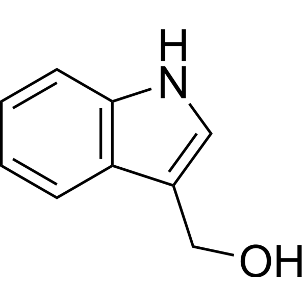
HY-N0170
Indole-3-carbinolEndogenous Metabolite -

HY-N0170R
Indole-3-carbinol (Standard)Endogenous Metabolite -

HY-137473R
Methyl pyropheophorbide-a (Standard)Photosensitizer -
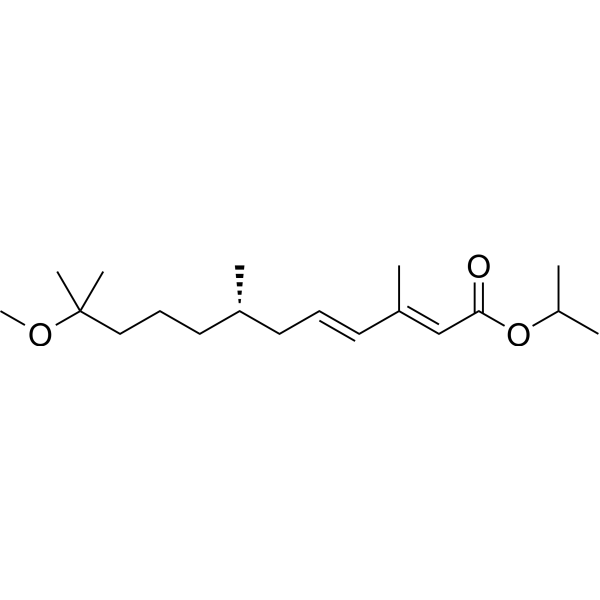
HY-B1161A
S-MethopreneInsecticide -
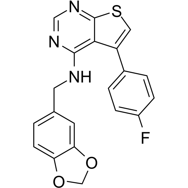
HY-113848
AEM1Keap1-Nrf2 Inhibitor -
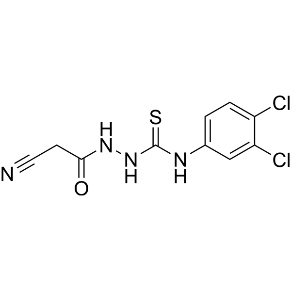
HY-124952
iKIX1Antifungal Agent -

HY-B1161AR
S-Methoprene (Standard)Insecticide -

HY-122566
SMU127
Publications Citing Use of MedChemExpress (MCE) Methyl pyropheophorbide-a
More

Biological Activity
Description
Cellular Effect
|
Cell Line

|
Type | Value | Description | References |
|---|---|---|---|---|
| HeLa | IC50 |
0.28 μM
Compound: 1, MPPa
|
Cytotoxicity against human HeLa cells of light exposed compound after 12 hrs by WST-8 assay
Cytotoxicity against human HeLa cells of light exposed compound after 12 hrs by WST-8 assay
|
[PMID: 22335896] |
In Vitro
Pyropheophorbide-a methyl ester (2 μM; 12 h) inhibits the cell cycle progression from the G0/G1-phases and induces apoptosis at the light dosage of 55.6 kJ/m2 in PC-3M cells[2].
Pyropheophorbide-a methyl ester (0.2-15 μM; 20 h) has no signi?cant dark cytotoxicity in NCI-h446 cells[1].
Pyropheophorbide-a methyl ester (0.1-15 μM; 20 h) mediates a dose-dependent photocytotoxicity in the NCI-h446 cells[1].
Pyropheophorbide-a methyl ester (2-4 μM; 0-24 h) induces apoptosis of NCI-h446 cells in a time-dependent manner[1].
Pyropheophorbide-a methyl ester (0.5-4 μM; 0-20 h) shows a photocytotoxicity in PC-3M cells[2].
MedChemExpress (MCE) has not independently confirmed the accuracy of these methods. They are for reference only.
-
Cell Line:PC-3M cells
-
Concentration:2 μM
-
Incubation Time:12 h
-
Result:Apoptosis rate was about 0.27%, 6.15%, 20.49%, 50.76%, 65.39%, 64.92%, and 64.37% at 1, 3, 6, 12, 18, and 24 h posttreatment, respectively under the conditions of LC75 (2 μM+55.6 kJ/m2).
In Vivo
MedChemExpress (MCE) has not independently confirmed the accuracy of these methods. They are for reference only.
-
Animal Model:Male BALB/c nude mice (6-8 weeks, 16-20 g) bearing PC-3M tumor[2]
-
Dosage:15 mg/kg
-
Administration:Tail vein injection and topical injection
-
Result:The tumor volume and the weight inhibition rate were 46.78%, 41.84% in tail vein injection group and 78.66%, 72.07% in topical injection group, respectively.


Chemical Information
-
CAS No. 6453-67-4
-
Appearance Solid
-
Molecular Weight 548.67
-
Formula C34H36N4O3
-
Color Blue to dark blue
-
SMILES
CC1=C(N2)/C=C(C(CC)=C3C)\N=C3/C=C(C(C=C)=C/4C)\NC4=C/C5=N/C([C@H]([C@@H]5C)CCC(OC)=O)=C(C6)\C2=C1C6=O
-
Synonyms
Pyropheophorbide-a methyl ester
-
Shipping
Room temperature in continental US; may vary elsewhere.
-
Storage
Powder -20°C 3 years 4°C 2 years In solvent -80°C 6 months -20°C 1 month


Publications (2)
-
Journal Impact Factor
-
Most Recent
-
Cell Prolif
2025 Jul 16:e70097. PMID: 40670295 -


Solvent & Solubility
In Vitro:
DMSO : 2.5 mg/mL (4.56 mM; ultrasonic and warming and heat to 60°C; Hygroscopic DMSO has a significant impact on the solubility of product, please use newly opened DMSO)
Please refer to the solubility information to select the appropriate solvent. Once prepared, please aliquot and store the solution to prevent product inactivation from repeated freeze-thaw cycles.
Storage method and period of stock solution: -80°C, 6 months; -20°C, 1 month. When stored at -80°C, please use it within 6 months. When stored at -20°C, please use it within 1 month.
Please refer to the solubility information to select the appropriate solvent. Once prepared, please aliquot and store the solution to prevent product inactivation from repeated freeze-thaw cycles.
Storage method and period of stock solution: -80°C, 6 months; -20°C, 1 month. When stored at -80°C, please use it within 6 months. When stored at -20°C, please use it within 1 month.
Concentration (start) × Volume (start) = Concentration (final) × Volume (final)


Purity & Documentation
-
Data Sheet (281 KB)
-
SDS (252 KB)
- English - EN (252 KB)
- Français - FR (252 KB)
- Deutsch - DE (252 KB)
- Norwegian - NO (252 KB)
- Español - ES (252 KB)
- Swedish - SV (252 KB)
- Italian - IT (252 KB)
- Korean - KR (252 KB)
- Portuguese - PT (252 KB)
-
Handling Instructions (2659 KB)
References
[1]. Sun X, et, al. Photodynamic therapy with pyropheophorbide-a methyl ester in human lung carcinoma cancer cell: efficacy, localization and apoptosis. Photochem Photobiol. 2002 Jun;75(6):644-51. [Content Brief]
[2]. Tian Y, et, al. Cell death induced by MPPa-PDT in prostate carcinoma in vitro and in vivo. Biochem Biophys Res Commun. 2006 Sep 22;348(2):413-20. [Content Brief]


Complete Stock Solution Preparation Table
Please refer to the solubility information to select the appropriate solvent. Once prepared, please aliquot and store the solution to prevent product inactivation from repeated freeze-thaw cycles.
Storage method and period of stock solution: -80°C, 6 months; -20°C, 1 month. When stored at -80°C, please use it within 6 months. When stored at -20°C, please use it within 1 month.
| Optional Solvent | Concentration Solvent Mass | 1 mg | 5 mg | 10 mg | 25 mg |
|---|---|---|---|---|---|
| DMSO | 1 mM | 1.8226 mL | 9.1129 mL | 18.2259 mL | 45.5647 mL |


Methyl pyropheophorbide-a Related Classifications
HY-137473 Related Classifications
 Powered by Bioz