



Obexelimab

Based on 1 Customer Validation
Obexelimab (XmAb5871) is a humanized anti-CD19 antibody. Obexelimab works by inhibiting B cell receptor (BCR) mediated calcium influx and promoting the phosphorylation of Fc γ receptor IIb (FcγRIIb), which reduces B cell activation and function, leading to B cell apoptosis. Obexelimab can be used in research for rheumatoid arthritis and systemic lupus erythematosus.
For research use only. We do not sell to patients.
- Purity : 99.99%
- CAS No.: 1690307-05-1
- Molecular Weight:147.24 kDa
-
Storage:
Please store the product under the recommended conditions in the Certificate of Analysis.


To place orders, for customer services and technical support, please contact: MedChemExpress USA
Tel: 609-228-6898 E-mail: [email protected] [email protected]


-
 Biological Activity
-
Chemical Information
- Protocols
- Purity & Documentation
- References
-
 Help & FAQs


-

HY-P99113
InebilizumabAnti-CD19 mAb -

HY-P9981
TafasitamabCD19 Monoclonal Antibody -

HY-P99285
DenintuzumabHumanized Anti-CD19 mAb -

HY-P9963
BlinatumomabAntineoplastic Agent -
HY-16450
SB 204990ATP Citrate Lyase Inhibitor -
HY-P9913A
Rituximab (anti-CD20)CD20 Inhibitor -

HY-P990006
DuvakitugAnti-TNFSf15/TL1A Antibody -

HY-P99711
LoncastuximabAnti-CD19 mAb -

HY-P991252
XmAb-7195Anti-IGHE mAb


Biological Activity
Description
Isotype
Human IgG1 kappa
Recommend Isotype Controls
Species Reactivity
Human
IC50 & Target
CD19
In Vitro
Obexelimab can induce the expression levels of apoptosis pathway related genes and promote B cell apoptosis[1]. Obexelimab (10 μg/mL, 3 min) inhibits B cell activation and function by suppressing BCR mediated calcium flux and promoting Fc γ RIIb phosphorylation[2].
MedChemExpress (MCE) has not independently confirmed the accuracy of these methods. They are for reference only.
-
Cell Line:B cells isolated from PBMC of healthy and RA donors
-
Concentration:10 μg/mL
-
Incubation Time:3 min
-
Result:Stimulated FcγRIIb phosphorylation in healthy and RA B cells.
In Vivo
MedChemExpress (MCE) has not independently confirmed the accuracy of these methods. They are for reference only.
-
Animal Model:Female C.B-17 SCID mice transplanted with PBMCs[2].
-
Dosage:10 mg/kg
-
Administration:Intraperitoneal injection (i.p.); twice a week
-
Result:Resulted in inhibition of various immune responses in mice, including IgG, IgM, RF, ACPA, and anti-tetanus antibodies.
- Kinase activity and phosphorylation assays
- Western Blot
- Collagen-Induced Arthritis
- Detection of Apoptosis
- TUNEL staining for apoptotic DNA fragmentation
- Annexin V plus membrane-impermeant dye apoptosis staining
- Apoptosis Solutions
- Ca2+ Staining Technique
- Protocol for Kinase activity and phosphorylation assays
Clinical Trial
| NCT Number | Sponsor | Condition | Start Date |
Phase

|
|---|---|---|---|---|
| NCT01329991 | Plexxikon| | 2011-05 | PHASE1 |
Gene ID
Accession
Conjugated
Unconjugated
Reconsititution
The product can be reconstituted/diluted with sterile PBS or saline.
Format
-
Human IgG1 kappa

Application
ELISA, FACS, Functional assay
Verified Bioactivity
-
 Flow Cytometry analysis of Raji cells labelling CD19 (red) with Obexelimab (HY-P99761). Cells were fixed with 4% paraformaldehyde and permeabilised with 90% methanol. Then cells were stained with the primary antibody at 1/200 dilution for an hour at 4℃. Goat Anti-Human IgG H&L (AF488) (HY-P83776) was used as the secondary antibody at 1/1,000 dilution for 30 minutes at 4℃. Human IgG1 kappa (HY-P99001, blue) was used as the isotype control, cells without incubation with primary antibody were used as the unlabeled control (black).
 -
 Immobilized CD19 Protein, Human (HEK293, His, HY-P74330) can bind Obexelimab. The EC50 for this effect is 16.38 ng/mL.
 -
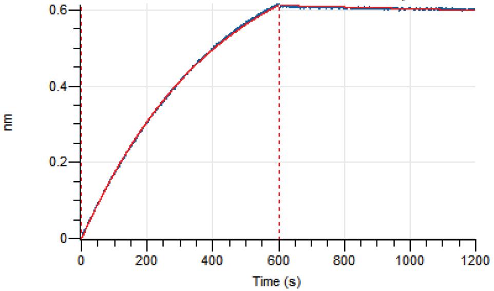 Loaded Obexelimab on AHC2 biosensor, can bind CD19 Protein, Human (HEK293, His, HY-P74330) with an affinity constant of 1.556E-09 M as determined in BLI assay.


Chemical Information
-
CAS No. 1690307-05-1
-
Appearance Liquid
-
Molecular Weight 147.24 kDa
-
Color Colorless to light yellow
-
SMILES
[Obexelimab]
-
Synonyms
XmAb5871
-
Shipping
Shipping with dry ice.
-
Formulation
Please refer to the lot-specific COA for specific buffer information.
-
Storage
Please store the product under the recommended conditions in the Certificate of Analysis.


Purity & Documentation
-
Data Sheet (261 KB)
-
SDS (251 KB)
- English - EN (251 KB)
- Français - FR (251 KB)
- Deutsch - DE (251 KB)
- Norwegian - NO (251 KB)
- Español - ES (251 KB)
- Swedish - SV (251 KB)
- Italian - IT (251 KB)
- Korean - KR (251 KB)
- Portuguese - PT (251 KB)
-
Inhibitory Antibodies User Guide (603 KB)
References
[1]. Boyles JS, et al. A nondepleting anti-CD19 antibody impairs B cell function and inhibits autoimmune diseases. JCI Insight. 2023 Jul 10;8(13):e166137. [Content Brief]
[2]. Chu SY, et al. Suppression of rheumatoid arthritis B cells by XmAb5871, an anti-CD19 antibody that coengages B cell antigen receptor complex and Fcγ receptor IIb inhibitory receptor. Arthritis Rheumatol. 2014 May;66(5):1153-64. [Content Brief]
[3]. Horton HM, et al. Antibody-mediated coengagement of FcγRIIb and B cell receptor complex suppresses humoral immunity in systemic lupus erythematosus. J Immunol. 2011 Apr 1;186(7):4223-33. [Content Brief]

Calculators
Concentration (start) × Volume (start) = Concentration (final) × Volume (final)


Obexelimab Related Classifications
HY-P99761 Related Classifications
 Powered by Bioz