



Osteostatin
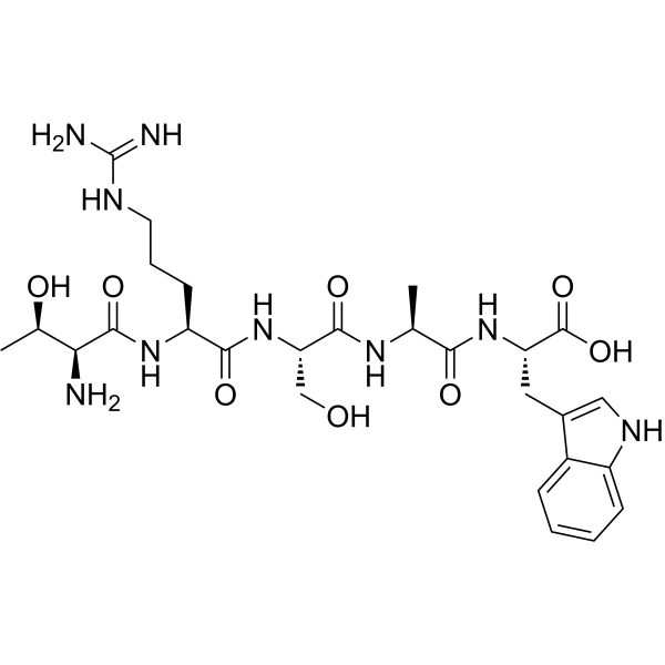
Osteostatin, a fragment of parathyroid hormone-related protein (PTHrP) 107-111, promotes bone repair in animal models of bone defects and prevents bone erosion in inflammatory arthritis, inhibits collagen-induced arthritis and inhibits osteoclastic bone resorption directly. Osteostatin can be used for inflammation and immunology research.
For research use only. We do not sell to patients.
- CAS No.: 138949-73-2
- Formula: C27H41N9O8
- Molecular Weight:619.67
-
Storage:
Please store the product under the recommended conditions in the Certificate of Analysis.

To place orders, for customer services and technical support, please contact: MedChemExpress USA
Tel: 609-228-6898 E-mail: [email protected] [email protected]


-
 Biological Activity
-
Chemical Information
- Protocols
- Purity & Documentation
- References
-
 Help & FAQs




Biological Activity
Description
In Vitro
Osteostatin (100, 250 and 500 nM, 7-9 days) decreases the differentiation of osteoclasts in a concentration-dependent manner[2].
Osteostatin (100, 250 and 500 nM, 7 days) decreases the mRNA levels of cathepsin K, osteoclast associated Ig-like receptor (OSCAR) and NFATc1[2].
Osteostatin (100, 250 and 500 nM, 2 days) inhibits the nuclear translocation of the master transcription factor NFATc1 in osteoclast differentiation [2].
Osteostatin (0.1-100 nM, 2 days) significantly increased cell growth in MC3T3-E1 cells[3].
MedChemExpress (MCE) has not independently confirmed the accuracy of these methods. They are for reference only. Further protocols information, click here.
-
Cell Line:PBMCs are cultured with M-CSF and RANKL
-
Concentration:100, 250 and 500 nM
-
Incubation Time:7-9 days
-
Result:Decreased the differentiation of osteoclasts in a concentration-dependent manner, but it does not modify the resorptive ability of mature osteoclasts.
-
Cell Line:PBMCs are cultured with M-CSF and RANKL
-
Concentration:100, 250 and 500 nM
-
Incubation Time:7 days
-
Result:Decreased the mRNA levels of cathepsin K, osteoclast associated Ig-like receptor (OSCAR) and NFATc1.
-
Cell Line:PBMCs are cultured with M-CSF and RANKL
-
Concentration:100, 250 and 500 nM
-
Incubation Time:2 days
-
Result:Inhibited the nuclear translocation of the master transcription factor in osteoclast differentiation NFATc1.
-
Cell Line:MC3T3-E1 cells
-
Concentration:0-100 nM
-
Incubation Time:2 days
-
Result:Significantly increased cell growth of MC3T3-E1 cells
In Vivo
MedChemExpress (MCE) has not independently confirmed the accuracy of these methods. They are for reference only.
-
Animal Model:Collagen-induced arthritis in male DBA/1 mice[4]
-
Dosage:80 or 120 μg/kg
-
Administration:After the onset of disease s.c. every day for 13 days
-
Result:Reduced serum IgG2a levels as well as T cell activation, with the downregulation of RORγt+CD4+ T cells and upregulation of FoxP3+CD8+ T cells in lymph nodes.
Decreased the levels of key cytokines, such as interleukin (IL)-1β, IL-2, IL-6, IL-17, and tumor necrosis factor-α in mice paws, whereas enhanced IL-10.


Chemical Information
-
CAS No. 138949-73-2
-
Molecular Weight 619.67
-
Formula C27H41N9O8
-
Synonyms
PTHrP (107–111)
-
Sequence
Thr-Arg-Ser-Ala-Trp
-
Sequence Shortening
TRSAW
-
Shipping
Room temperature in continental US; may vary elsewhere.
-
Storage
Please store the product under the recommended conditions in the Certificate of Analysis.

Protocols
-
Collagen-Induced Arthritis
Collagen-induced arthritis (CIA) is an autoimmune murine model of rheumatoid arthritis in which immunization with type II collagen (CII) emulsified in an adjuvant induces a T cell- and autoantibody-driven inflammatory arthritis characterized by synovial hyperplasia, immune cell infiltration, and joint destruction. The model typically relies on genetically susceptible mouse strains (e. g. , DBA/1) and reproduces key features of human rheumatoid arthritis, including anti-collagen immune responses and progressive joint inflammation. Disease onset generally occurs within ~3-4 weeks after immunization, depending on antigen/adjuvant combinations and protocol variation. The immunopathology is driven by adaptive immune activation against CII, leading to systemic and local joint inflammation mediated by pro-inflammatory cytokines and effector immune cells, making CIA a standard preclinical platform for evaluating immunomodulatory and anti-arthritic interventions.
-
Research Protocol for Endocrine Diseases
Endocrine diseases often arise from disrupted hormone production, hormone signaling, or target-tissue responsiveness; for diabetes-focused endocrine disease models, insulin signaling regulates glucose uptake, hepatic glucose output, lipid metabolism, and β-cell compensation. Type 2 diabetes develops through interacting defects in insulin resistance, β-cell dysfunction, adipose inflammation, hepatic glucose overproduction, altered incretin signaling, and ectopic lipid metabolism. A major unresolved question is whether endocrine dysfunction is driven primarily by target-tissue insulin resistance, intrinsic β-cell failure, immune/inflammatory stress, or combined multi-organ failure that differs by disease stage.
-
Research Protocol for Inflammation-related Diseases
The NLRP3 inflammasome is a cytosolic innate immune signaling platform that integrates priming signals and danger-signal activation to promote caspase-1 activation, maturation of IL-1β and IL-18, and gasdermin D-mediated pyroptotic cell death. The core experimental logic is to determine whether inflammatory disease phenotypes are driven by increased NLRP3 expression, ASC-containing inflammasome assembly, caspase-1 cleavage, GSDMD cleavage, and extracellular release of IL-1β/IL-18 rather than by nonspecific cell injury alone. The pathway is strongly linked to inflammation-related disease phenotypes because monosodium urate crystals activate NALP3/NLRP3 inflammasome signaling in gout-like crystal inflammation, cholesterol crystals activate NLRP3 inflammasomes in atherogenesis models, and DSS-induced intestinal inflammation has been reported to involve NLRP3 inflammasome activity. However, experimental colitis studies also show context-dependent protective effects of NLRP3 inflammasome co
-
Osteoclast differentiation from monocyte/macrophage precursors
Osteoclast differentiation is an in vitro induction assay in which monocyte/macrophage-lineage precursors are exposed to macrophage colony-stimulating factor (M-CSF) and receptor activator of NF-κB ligand (RANKL), generating multinucleated osteoclasts that are commonly identified by tartrate-resistant acid phosphatase (TRAP) staining and functionally confirmed by resorption pits on dentin, bone, or mineralized substrates. M-CSF supports survival and expansion of osteoclast precursors, while RANKL binding to RANK drives osteoclast commitment, fusion, maturation, and resorptive function; osteoprotegerin inhibits this pathway by binding RANKL and preventing RANK activation. The main readouts are the number of TRAP-positive multinucleated cells, formation of F-actin rings, and resorbed surface area; TRAP-positive multinucleated cells indicate osteoclast differentiation, whereas pit formation on dentin, bone, or mineralized coating indicates functional bone-resorbing activity.


Purity & Documentation
References
[1]. Fenton AJ, et.al. A potent inhibitor of osteoclastic bone resorption within a highly conserved pentapeptide region of parathyroid hormone-related protein; PTHrP[107-111]. Endocrinology. 1991 Dec;129(6):3424-6. [Content Brief]
[2]. Ibáñez L, et.al. Osteostatin Inhibits M-CSF+RANKL-Induced Human Osteoclast Differentiation by Modulating NFATc1. Int J Mol Sci. 2022 Aug 1;23(15):8551. [Content Brief]
[3]. Lozano D, et.al. Osteostatin-loaded bioceramics stimulate osteoblastic growth and differentiation. Acta Biomater. 2010 Mar;6(3):797-803. [Content Brief]
[4]. Nácher-Juan J, et.al. Osteostatin Inhibits Collagen-Induced Arthritis by Regulation of Immune Activation, Pro-Inflammatory Cytokines, and Osteoclastogenesis. Int J Mol Sci. 2019 Aug 7;20(16):3845. [Content Brief]

Calculators
Concentration (start) × Volume (start) = Concentration (final) × Volume (final)


Osteostatin Related Classifications
HY-P4684 Related Classifications
 Powered by Bioz