



PDD00017272
Based on 2 publication(s) in Google Scholar
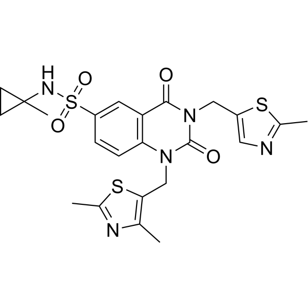
PDD00017272 is an inhibitor of poly(ADP-ribose) glycohydrolase (PARG) (EC50=4.8 nM) and an activator of PARP1/2. PDD00017272 inhibits its activity of hydrolyzing poly(ADP-ribose) (pADPr), resulting in the accumulation of pADPr on chromatin, interfering with DNA damage repair and replication processes, and inducing PARP1/2-dependent cytotoxicity. PDD00017272 can be used in cancer models with DNA repair defects (such as BRCA mutations) or resistance to PARP inhibitors. PDD00017272 has a PARG expression level-correlated inhibitory potency with EC50 of 9.2 nM (PARG cells), the tumor cells with lower PARG expression are more sensitive.
For research use only. We do not sell to patients.
- Purity : 99.01%
- CAS No.: 1945950-20-8
- Formula: C23H25N5O4S3
- Molecular Weight:531.67
-
Storage:Powder -20°C, 3 years , 4°C, 2 years ; In solvent -80°C, 6 months , -20°C, 1 month

To place orders, for customer services and technical support, please contact: MedChemExpress USA
Tel: 609-228-6898 E-mail: [email protected] [email protected]


-
 Biological Activity
-
Chemical Information
-
Solvent & Solubility
- Protocols
- Purity & Documentation
- References
-
 Help & FAQs


-
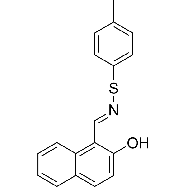
HY-128760
COH34PARG Inhibitor -
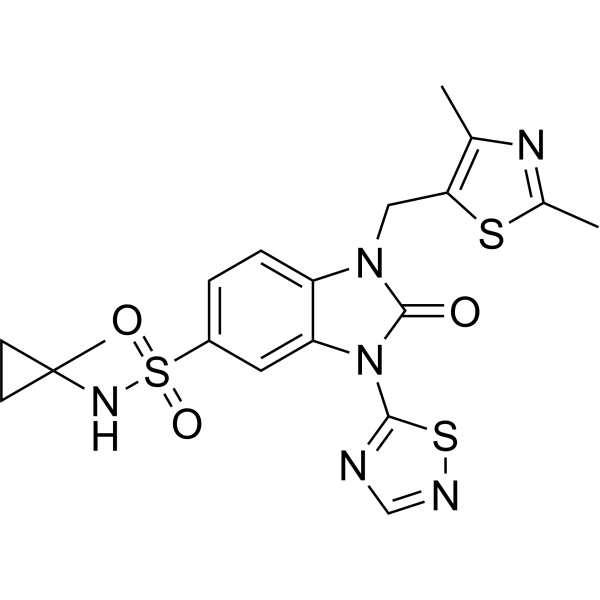
HY-133530
PDD00017238PARG Inhibitor -
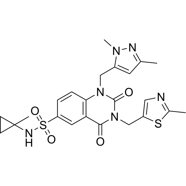
HY-108360
PDD 00017273PARG Inhibitor -
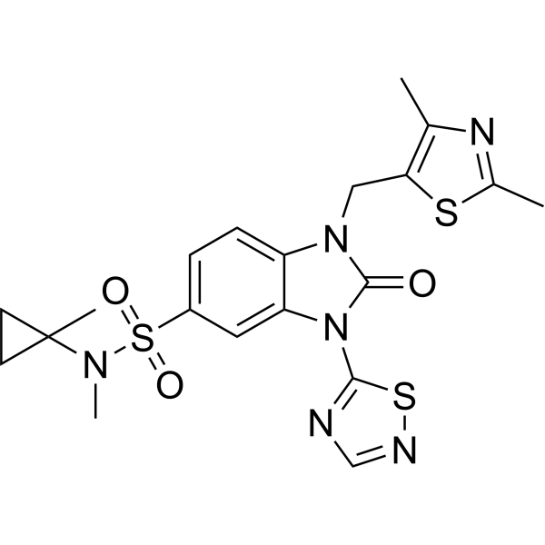
HY-135846
PDD00031705PARG Inhibitor -
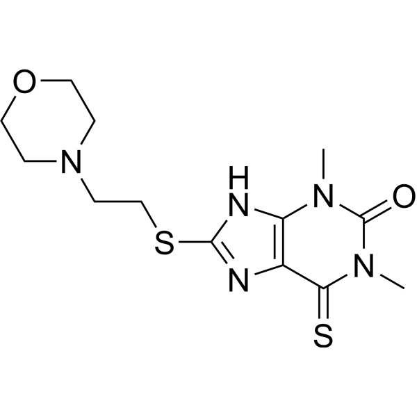
HY-137924
JA2131PARG Inhibitor -
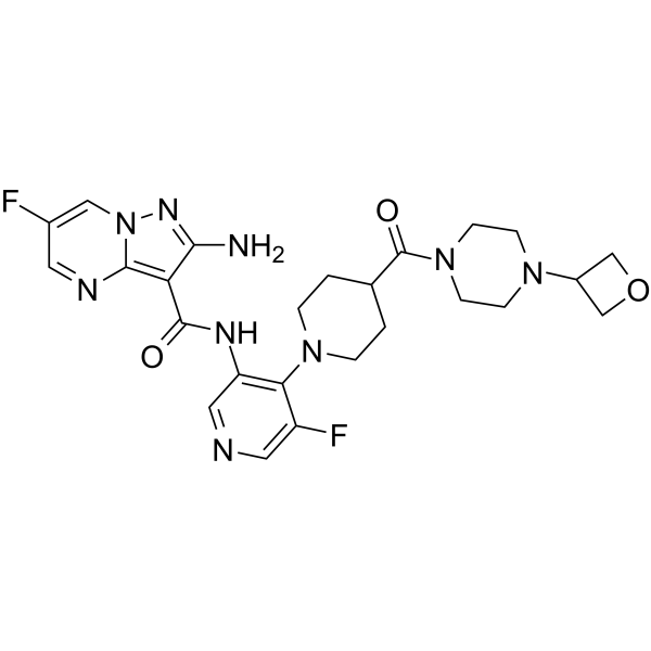
HY-136270
GartisertibATR Inhibitor -
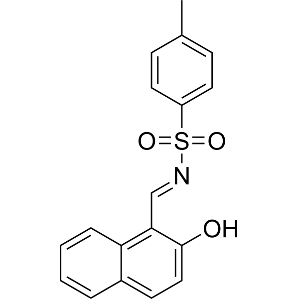
HY-115666
COH34 analog 1PARG Inhibitor -

HY-108360R
PDD 00017273 (Standard)Reference Standard
Publications Citing Use of MedChemExpress (MCE) PDD00017272
More

Biological Activity
Description
In Vitro
PDD00017272 (PARGi) (10 μM; 4 h) induces a significant increase in the level of chromatin-bound pADPr in HEK293A PARG knockout cells, and the signal was mainly concentrated in S phase cells, indicating that inhibition of PARG leads to the accumulation of endogenous pADPr at replication forks[1].
PDD00017272 (compound 34f) (0-100 μM; 72 h) significantly increases the sensitivity of PARG knockout cells to the drug in HEK293A wild-type and PARG knockout cells (IC50=96 μM and 210 nM, respectively), indicating that it has a cytotoxic effect related to PARG activity[1].
MedChemExpress (MCE) has not independently confirmed the accuracy of these methods. They are for reference only. Further protocols information, click here.
-
Cell Line:HEK293A wild-type cells and PARG KO cells
-
Concentration:0.01-100 μM
-
Incubation Time:72 h
-
Result:Showed an IC50 of 210 ± 30 nM against PARG KO cells, whereas exhibited an IC50 of 96 ± 24 μM against wild-type cells.
Reconstitution with wild-type PARG rescued viability, while a catalytically inactive PARG mutant failed to do so, confirming dependency on PARG enzymatic activity.
In Vivo
MedChemExpress (MCE) has not independently confirmed the accuracy of these methods. They are for reference only.


Chemical Information
-
CAS No. 1945950-20-8
-
Appearance Solid
-
Molecular Weight 531.67
-
Formula C23H25N5O4S3
-
Color White to off-white
-
SMILES
O=S(C1=CC2=C(N(CC3=C(C)N=C(C)S3)C(N(CC4=CN=C(C)S4)C2=O)=O)C=C1)(NC5(C)CC5)=O
-
Shipping
Room temperature in continental US; may vary elsewhere.
-
Storage
Powder -20°C 3 years 4°C 2 years In solvent -80°C 6 months -20°C 1 month


Publications (2)
-
Journal Impact Factor
-
Most Recent
-
-
bioRxiv
Mutant p53 Directs PARP to Regulate Replication Stress and Drive Breast Cancer Metastasis. [Abstract]2026 Mar 28:2026.03.26.713220. PMID: 41928956


Solvent & Solubility
In Vitro:
DMSO : ≥ 25 mg/mL (47.02 mM; Hygroscopic DMSO has a significant impact on the solubility of product, please use newly opened DMSO)
* "≥" means soluble, but saturation unknown.
Please refer to the solubility information to select the appropriate solvent. Once prepared, please aliquot and store the solution to prevent product inactivation from repeated freeze-thaw cycles.
Storage method and period of stock solution: -80°C, 6 months; -20°C, 1 month. When stored at -80°C, please use it within 6 months. When stored at -20°C, please use it within 1 month.
Please refer to the solubility information to select the appropriate solvent. Once prepared, please aliquot and store the solution to prevent product inactivation from repeated freeze-thaw cycles.
Storage method and period of stock solution: -80°C, 6 months; -20°C, 1 month. When stored at -80°C, please use it within 6 months. When stored at -20°C, please use it within 1 month.
Concentration (start) × Volume (start) = Concentration (final) × Volume (final)
In Vivo:
Select the appropriate dissolution method based on your experimental animal and administration route.
- For the following dissolution methods, please ensure to first prepare a clear stock solution using an In Vitro approach and then sequentially add co-solvents:
- To ensure reliable experimental results, the clarified stock solution can be appropriately stored based on storage conditions. As for the working solution for In Vivo experiments, it is recommended to prepare freshly and use it on the same day.
- The percentages shown for the solvents indicate their volumetric ratio in the final prepared solution. If precipitation or phase separation occurs during preparation, heat and/or sonication can be used to aid dissolution.
In Vivo Dissolution Calculator
Please enter the basic information of animal experiments:
-
-
-
-
Recommended: Prepare an additional quantity of animals to account for potential losses during experiments.
Please enter your animal formula composition:
-
%DMSO +
Recommended: Keep the proportion of DMSO in working solution below 2% if your animal is weak.
-
%+
-
+%Tween-80 + +
-
%Saline +
The co-solvents required include: DMSO, . All of co-solvents are available by MedChemExpress (MCE). , Tween 80. All of co-solvents are available by MedChemExpress (MCE).
Working solution concentration: 0.22 mg/mL
Method for preparing stock solution: mg drug dissolved in μL DMSO. Stock solution concentration: mg/mL.
1. Take μL DMSO stock solution;
2. Add μL .
μL , mix evenly;
3. Then add μL Tween 80, mix evenly;
4. Then add μL
Please ensure that the stock solution in the first step is dissolved to a clear state, and add co-solvents in sequence. You can use ultrasonic heating (ultrasonic cleaner, recommended frequency 20-40 kHz), vortexing, etc. to assist dissolution.

Protocols
-
RNA extraction experimental
By lysing cells, releasing RNA, and removing impurities such as proteins and DNA, high-purity RNA products are finally obtained. The commonly used traditional method is the guanidine isothiocyanate/phenol/chloroform method (Trizol), which is suitable for a variety of animal materials including animal tissues, microorganisms, cultured cells, etc., and most plant materials.
-
Cell Cytotoxicity Assay
Cytotoxicity assays are usually based on the assessment of cell membrane damage, which can also be indirectly detected by measuring cell viability. Detection methods include MTT assay, CKK-8 assay, LDH assay and ATP assay, etc.
-
Mammalian live/dead viability and cytotoxicity staining
Live/dead viability and cytotoxicity staining assays are based on the simultaneous detection of intracellular esterase activity in metabolically active (viable) cells and membrane integrity loss in non-viable cells. In commonly used dual-staining approaches, membrane-permeant fluorogenic substrates are converted by intracellular esterases into fluorescent products in live cells, while impermeant DNA-binding dyes selectively enter cells with compromised plasma membranes and label nucleic acids in dead or dying cells, enabling discrimination between viable and non-viable populations by fluorescence microscopy or flow cytometry.
-
Genotoxicity/Mutagenicity Study
The bacterial reverse mutation assay detects point mutations that restore amino-acid prototrophy in auxotrophic Salmonella typhimurium or Escherichia coli tester strains; after exposure to a test article, mutagenic activity is read out as an increased number of revertant colonies on minimal agar compared with the vehicle control. The assay uses tester strains with different mutation targets so that base-substitution and frameshift mutagens can be detected, and testing is performed with and without exogenous mammalian metabolic activation because some chemicals require biotransformation to become mutagenic.
-
Cell Viability Determination by MTT Colorimetric Assay
The following protocol uses the MTT colorimetric assay as a classic literature-established method for assessing cell viability/metabolic activity in cultured mammalian cells. MTT[3-(4,5-dimethylthiazol-2-yl)-2,5-diphenyltetrazolium bromide] is reduced by metabolically active cells to a colored formazan product; the amount of formazan is quantified spectrophotometrically and provides an indirect measure of metabolically active viable cells. Importantly, MTT reduction reflects cellular oxidoreductase/metabolic activity rather than an absolute direct count of living cells, so changes in cellular metabolism can alter the signal independently of cell number.


Purity & Documentation
-
Data Sheet (279 KB)
-
SDS (251 KB)
- English - EN (251 KB)
- Français - FR (251 KB)
- Deutsch - DE (251 KB)
- Norwegian - NO (251 KB)
- Español - ES (251 KB)
- Swedish - SV (251 KB)
- Italian - IT (251 KB)
- Korean - KR (251 KB)
- Portuguese - PT (251 KB)
-
Handling Instructions (2659 KB)
References
[1]. Nie L, et al. DePARylation is critical for S phase progression and cell survival. Elife. 2024 Apr 5;12:RP89303. [Content Brief]
[2]. Bohdan Waszkowycz, et al. Cell-Active Small Molecule Inhibitors of the DNA-Damage Repair Enzyme Poly(ADP-ribose) Glycohydrolase (PARG): Discovery and Optimization of Orally Bioavailable Quinazolinedione Sulfonamides. J Med Chem. 2018 Dec 13;61(23):10767-10792. [Content Brief]


Complete Stock Solution Preparation Table
Please refer to the solubility information to select the appropriate solvent. Once prepared, please aliquot and store the solution to prevent product inactivation from repeated freeze-thaw cycles.
Storage method and period of stock solution: -80°C, 6 months; -20°C, 1 month. When stored at -80°C, please use it within 6 months. When stored at -20°C, please use it within 1 month.
| Optional Solvent | Concentration Solvent Mass | 1 mg | 5 mg | 10 mg | 25 mg |
|---|---|---|---|---|---|
| DMSO | 1 mM | 1.8809 mL | 9.4043 mL | 18.8087 mL | 47.0217 mL |
| 5 mM | 0.3762 mL | 1.8809 mL | 3.7617 mL | 9.4043 mL | |
| 10 mM | 0.1881 mL | 0.9404 mL | 1.8809 mL | 4.7022 mL | |
| 15 mM | 0.1254 mL | 0.6270 mL | 1.2539 mL | 3.1348 mL | |
| 20 mM | 0.0940 mL | 0.4702 mL | 0.9404 mL | 2.3511 mL | |
| 25 mM | 0.0752 mL | 0.3762 mL | 0.7523 mL | 1.8809 mL | |
| 30 mM | 0.0627 mL | 0.3135 mL | 0.6270 mL | 1.5674 mL | |
| 40 mM | 0.0470 mL | 0.2351 mL | 0.4702 mL | 1.1755 mL |


PDD00017272 Related Classifications
HY-133531 Related Classifications
 Powered by Bioz