



Ppc-1
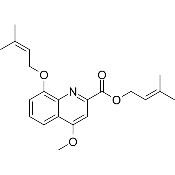
Ppc-1 is a mitochondrial uncoupler. Ppc-1 enhances mitochondrial oxygen consumption without adverse effects on ATP production. Ppc-1 is a cell-permeate interleukin-2 (IL-2) inhibitor. Ppc-1 inhibits the Gram-negative periodontopathogen Porphyromonas gingivalis. Ppc-1 has anti-obesity, antibacterial and anti-inflammatory activities.
Nos produits utilisent uniquement pour la recherche. Nous ne vendons pas aux patients.
- CAS No.: 1245818-17-0
- Formule: C21H25NO4
- Masse moléculaire:355.43
-
Stockage:
Please store the product under the recommended conditions in the Certificate of Analysis.

To place orders, for customer services and technical support, please contact: MedChemExpress USA
Tel: 609-228-6898 E-mail: [email protected] [email protected]


-
 Activité biologique
-
Chemical Information
- Protocole
- Pureté et documentation
- Références
-
 Help & FAQs


-
HY-100410
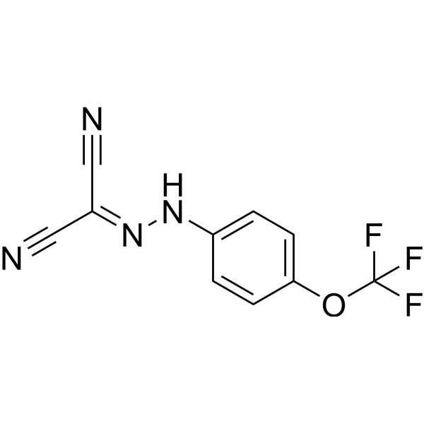
FCCPOXPHOS Uncoupler -
HY-B0158S
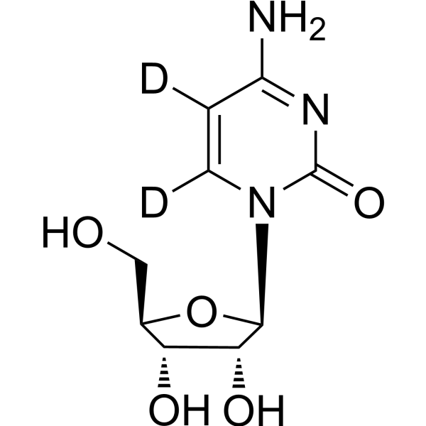
Cytidine-d2Stable Isotope -
HY-129630
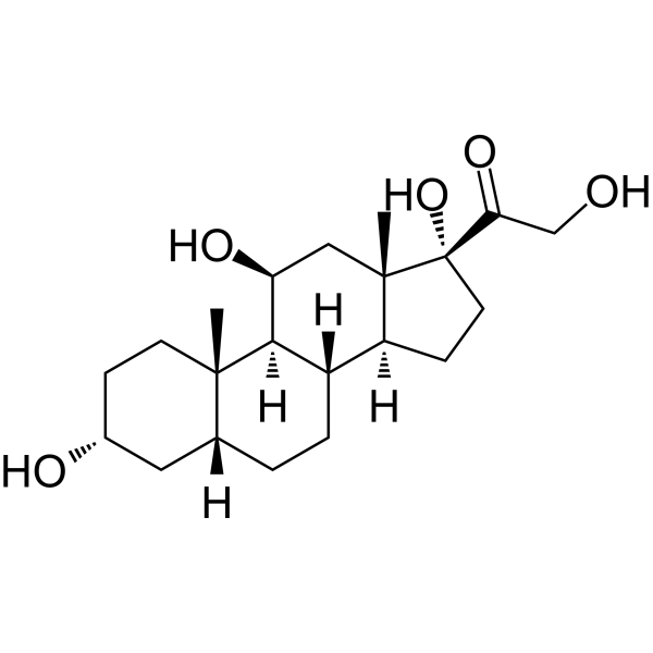
TetrahydrocortisolMetabolite of Hydrocortisone -
HY-N1441
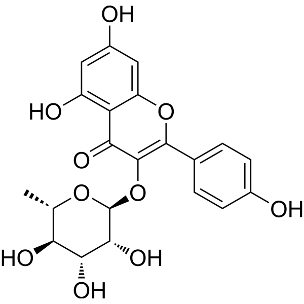
AfzelinFlavonol Glycoside -

HY-W738465
N,N-Dimethylamidino UreaEndogenous metabolite -

HY-100410R
FCCP (Standard)OXPHOS Uncoupler -

HY-P10447
Plipastatin A1Phospholipase A2 Iinhibitor -

HY-N1441R
Afzelin (Standard)Flavonol Glycoside


Activité biologique
Description
IC50 & Target
[1]|
IL-2 |
In Vitro
Ppc-1 treatment (0-10 μM; 24 hours; Jurkat cells) significant inhibits IL-2 production in Jurkat cells with an IC50 of 4 μM[2].
Ppc-1 (compound 6) has antiproliferative activities in K562 human leukemia, Hela cervical carcinoma, and 3T3-L1 mouse embryonic fibroblast cells. Ppc-1 shows about 50% inhibition at 15 μM in all cell lines. Ppc-1 inhibits the growth of K562 cells with an EC50 of 13 μM[3].
Using the U937-3xκB-LUC human monocytic cell line, Ppc-1 dose-dependently inhibits the lipopolysaccharide-induced NF-κB activation[4].
MedChemExpress (MCE) has not independently confirmed the accuracy of these methods. They are for reference only.
In Vivo
Serum fatty acid levels are significantly elevated in mice treated with Ppc-1, while body fat content remained low. After a single administration, Ppc-1 distributes into various tissues of individual animals at low levels. Ppc-1 stimulates adipocytes in culture to release fatty acids, which might explain the elevated serum fatty acids in Ppc-1-treated mice[1].
MedChemExpress (MCE) has not independently confirmed the accuracy of these methods. They are for reference only.
-
Animal Model:Female ICR mice[1]
-
Dosage:0 mg /kg , 0.16 mg /kg, 0.8 mg /kg, 4 mg /kg, and 10 mg /kg
-
Administration:Intraperitoneal injection; once a week; for 8 weeks
-
Result:Suppresseed weight gain in animals.


Chemical Information
-
CAS No. 1245818-17-0
-
Masse moléculaire 355.43
-
Formule C21H25NO4
-
SMILES
O=C(C1=NC2=C(OC/C=C(C)/C)C=CC=C2C(OC)=C1)OC/C=C(C)\C
-
Livraison
Room temperature in continental US; may vary elsewhere.
-
Stockage
Please store the product under the recommended conditions in the Certificate of Analysis.


Pureté et documentation
Références
[1]. Suzuki T, et al. Weight loss by Ppc-1, a novel small molecule mitochondrial uncoupler derived from slime mold. PLoS One. 2015 Feb 10;10(2):e0117088. [Content Brief]
[2]. Ogura M, et al. Prenylated quinolinecarboxylic acid derivative suppresses immune response through inhibition of PAK2. Biochem Pharmacol. 2016 Apr 1;105:55-65. [Content Brief]
[4]. Azelmat J, et al. Antibacterial and Anti-inflammatory Activities of Ppc-1, Active Principle of the Cellular Slime Mold Polysphondylium pseudo-candidum. Med Chem. 2015;11(7):666-9. [Content Brief]

Calculators
Concentration (start) × Volume (start) = Concentration (final) × Volume (final)


Ppc-1 Related Classifications
HY-117843 Related Classifications
 Powered by Bioz