



Coagulation factor VII/F7 Protein, Human (HEK293, His)

Based on 1 Customer Validation
Coagulation factor VII (F7) initiates the extrinsic pathway of blood coagulation. As a circulating zymogen, F7 is activated to F7a through proteolysis induced by factor Xa, factor XIIa, factor IXa, or thrombin. In the presence of tissue factor and calcium ions, F7a catalyzes limited proteolysis of factor X, converting it to factor Xa. F7a can also convert factor IX to factor IXa, emphasizing its pivotal role in coagulation cascades. Coagulation factor VII/F7 Protein, Human (HEK293, His) is the recombinant human-derived Coagulation factor VII/F7, expressed by HEK293, with C-His labeled tag.
- Species: Human
- Source: HEK293
-
Storage:Stored at -20°C for 2 years from date of receipt. After reconstitution, it is stable at 4°C for 1 week or -20°C for longer (with carrier protein). It is recommended to freeze aliquots at -20°C or -80°C for extended storage.


To place orders, for customer services and technical support, please contact: MedChemExpress USA
Tel: 609-228-6898 E-mail: [email protected] [email protected]


-
 Biological Activity
-
Technical Parameters
-
Product Properties
- Documentation
- Help & FAQs




Biological Activity
Description
Coagulation factor VII (F7) initiates the extrinsic pathway of blood coagulation. As a circulating zymogen, F7 is activated to F7a through proteolysis induced by factor Xa, factor XIIa, factor IXa, or thrombin. In the presence of tissue factor and calcium ions, F7a catalyzes limited proteolysis of factor X, converting it to factor Xa. F7a can also convert factor IX to factor IXa, emphasizing its pivotal role in coagulation cascades. Coagulation factor VII/F7 Protein, Human (HEK293, His) is the recombinant human-derived Coagulation factor VII/F7, expressed by HEK293, with C-His labeled tag.
Background
Coagulation factor VII (F7) plays a crucial role in initiating the extrinsic pathway of blood coagulation. As a serine protease circulating in the blood in its zymogen form, factor VII undergoes activation to factor VIIa through minor proteolysis induced by factor Xa, factor XIIa, factor IXa, or thrombin. In the presence of tissue factor and calcium ions, factor VIIa further catalyzes the limited proteolysis of factor X, converting it to factor Xa. Additionally, in the presence of tissue factor and calcium, factor VIIa is capable of converting factor IX to factor IXa, highlighting its pivotal function in the intricate cascade of coagulation processes.
Verified Bioactivity
Measured by its ability to cleave 100 μM fluorogenic peptide substrate Boc-VPR-AMC (HY-P137784) in kinetic mode for 5 minutes. The specific activity is >50 pmol/min/µg. (Activation description: The proenzyme needs to be activated by Tissue Factor (HY-P7787) for an activated form.)
MCE Validation Data
-
Purity - SDS-PAGE
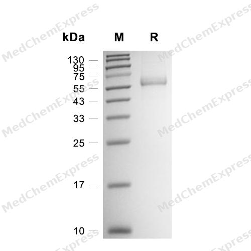
Purity - SDS-PAGE

Assay Procedure
Materials
Assay buffer: 50 mM Tris, 2.5 mM CaCl2, pH 8.5
Coagulation factor VII/F7 Protein, Human (406a.a, HEK293, His) (HY-P78763)
Coagulation factor III/F3 Protein, Human (HEK293, His) (Tissue Factor, HY-P7787)
Substrate: Boc-Val-Pro-Arg-AMC hydrochloride (HY-137784)
Standard: 7-amino, 4-Methyl Coumarin (HY-D0027)
Procedure
1. Dilute 7-amino, 4-Methyl Coumarin to 0, 9.765625, 19.53125, 39.0625, 78.125, 156.25, 312.5, 625, 1250, 2500, 5000, 10000 pmol with assay buffer respectively.
2. Add 100 μL of each concentration of standard to the wells.
3. Read in kinetic mode for 5 minutes with excitation and emission wavelengths of 380 nm and 460 nm (top read).
4. Plot a standard curve with the standard concentration as the abscissa and the measured RFU as the ordinate to obtain the standard curve.
5. Protein activity determination: First reconstitute Coagulation factor VII with sterile water to 100 μg/mL, then dilute Coagulation factor VII to 32 μg/mL with assay buffer.
6. Dilute Tissue Factor to 35.5 μg/mL with assay buffer.
7. Mix equal volumes of Coagulation factor VII and Tissue Factor, and incubate at 37°C for 5 minutes.
8. Dilute the substrate to 200 μM in assay buffer.
9. Transfer 50 μL of activated Coagulation factor VII to the wells of a black well plate.
10. Add 50 μL of 200 μM substrate to each well to start the reaction.
11. Read for 5 minutes in kinetic mode at excitation and emission wavelengths of 380 nm and 460 nm respectively.
12. Calculate the specific activity:
Specific Activity (pmol/min/μg) = | Adjusted Vmax* (RFU/min) x Conversion Factor ** (pmol/RFU) |
| amount of enzyme (μg) |
*Adjusted for Substrate Blank
**Derived using calibration standard
Per Well:
CoagμLation factor VII: 0.8 μg
Substrate: 100 μM


Technical Parameters
-
Species Human
-
Source HEK293
-
Tag C-6*His
-
Accession
P08709-1 (A61-P466)
-
Molecular Construction
-
N-term
-
F7 (A61-P466)
Accession # P08709-1 -
His
-
C-term
-
-
Protein Length
Full Length of Isoform-1
-
Synonyms
F7; FVII Coagulation Protein; Coagulation Factor VII; Proconvertin; Eptacog Alfa; SPCA; Coagulation Factor VII (Serum Prothrombin Conversion Accelerator); Alternative Protein F7; Serum Prothrombin Conversion Accelerator; Factor VII
-
AA Sequence
ANAFLEELRPGSLERECKEEQCSFEEAREIFKDAERTKLFWISYSDGDQCASSPCQNGGSCKDQLQSYICFCLPAFEGRNCETHKDDQLICVNENGGCEQYCSDHTGTKRSCRCHEGYSLLADGVSCTPTVEYPCGKIPILEKRNASKPQGRIVGGKVCPKGECPWQVLLLVNGAQLCGGTLINTIWVVSAAHCFDKIKNWRNLIAVLGEHDLSEHDGDEQSRRVAQVIIPSTYVPGTTNHDIALLRLHQPVVLTDHVVPLCLPERTFSERTLAFVRFSLVSGWGQLLDRGATALELMVLNVPRLMTQDCLQQSRKVGDSPNITEYMFCAGYSDGSKDSCKGDSGGPHATHYRGTWYLTGIVSWGQGCATVGHFGVYTRVSQYIEWLQKLMRSEPRPGVLLRAPFP
-
Predicted Molecular Mass
45.9 kDa
-
Molecular Weight
Approximately 55-65 kDa, based on SDS-PAGE under reducing conditions, due to the glycosylation.
-
Glycosylation
Yes
-
Purity
≥ 95%, as determined by reducing SDS-PAGE.


Product Properties
Lyophilized powder
Lyophilized from a 0.22 μm filtered solution of 50 mM Tris-HCl, 150 mM NaCl, pH 7.5, 8% trehalose.
Note: For SPR assay, please replace the buffer. Primary amine components (e.g., Tris, imidazole) can affect protein-coupled chips.
<1 EU/μg, determined by LAL method.
It is not recommended to reconstitute to a concentration less than 100 μg/mL in ddH2O.
Stored at -20°C for 2 years from date of receipt. After reconstitution, it is stable at 4°C for 1 week or -20°C for longer (with carrier protein). It is recommended to freeze aliquots at -20°C or -80°C for extended storage.
Room temperature in continental US; may vary elsewhere.


Documentation
-
Data Sheet (262 KB)
-
SDS (252 KB)
- English - EN (252 KB)
- Français - FR (252 KB)
- Deutsch - DE (252 KB)
- Norwegian - NO (252 KB)
- Español - ES (252 KB)
- Swedish - SV (252 KB)
- Italian - IT (252 KB)
- Korean - KR (252 KB)
- Portuguese - PT (252 KB)
-
Handling Instructions (2659 KB)

Calculators
Concentration (start) × Volume (start) = Concentration (final) × Volume (final)


 Powered by Bioz

