



Galectin-3/LGALS3 Protein, Human
Based on 4 publication(s) in Google Scholar
Galectin-3/LGALS3 Protein is a type of galactoside-binding lectin that has a high binding affinity for carbohydrates with β-galactoside linkages. Galectin-3/LGALS3 Protein interacts with glycosylated proteins to mediate intercellular interactions and adhesion to the extracellular matrix. Galectin-3/LGALS3 Protein plays various roles, including regulating signaling pathways such as Wnt/β-catenin, affecting cell proliferation and survival, modulating immune functions, and influencing tumor progression. Galectin-3/LGALS3 Protein, Human is a recombinant galectin-3/LGALS3 protein expressed in E. coli.
- Species: Human
- Source: E. coli
-
Storage:Stored at -20°C for 2 years from date of receipt. After reconstitution, it is stable at 4°C for 1 week or -20°C for longer (with carrier protein). It is recommended to freeze aliquots at -20°C or -80°C for extended storage.


To place orders, for customer services and technical support, please contact: MedChemExpress USA
Tel: 609-228-6898 E-mail: [email protected] [email protected]


-
 Biological Activity
-
Technical Parameters
-
Product Properties
- Documentation
- References
- Help & FAQs


-

HY-P70535
Galectin-9/LGALS9 Protein, Human (HEK293, His) -

HY-15141
Staurosporine -
HY-P70543
TGF beta 1/TGFB1 Protein, Human (HEK293) -
HY-P73072
Galectin-1/LGALS1 Protein, Human -
HY-15559
Hoechst 33342 -
HY-12686
5Z-7-Oxozeaenol -
HY-P74131
Galectin-1/LGALS1 Protein, Mouse -
HY-19940
Olitigaltin -
HY-P7085
M-CSF Protein, Mouse -
HY-P70570
HMGB1/HMG-1 Protein, Human -
HY-P77684
Galectin-3/LGALS3 Protein, Mouse (HEK293, His) -
HY-P79079
Galectin-3/LGALS3 Protein, Mouse


Biological Activity
Description
Galectin-3/LGALS3 Protein is a type of galactoside-binding lectin that has a high binding affinity for carbohydrates with β-galactoside linkages. Galectin-3/LGALS3 Protein interacts with glycosylated proteins to mediate intercellular interactions and adhesion to the extracellular matrix. Galectin-3/LGALS3 Protein plays various roles, including regulating signaling pathways such as Wnt/β-catenin, affecting cell proliferation and survival, modulating immune functions, and influencing tumor progression. Galectin-3/LGALS3 Protein, Human is a recombinant galectin-3/LGALS3 protein expressed in E. coli[1][2].
Background
Galectin-3/LGALS3 Protein is widely distributed in most tissues and is extensively expressed in specific tissues, with extracellular, membrane-bound, cytoplasmic, and nuclear localization[1]. The preferred ligand for Galectin-3/LGALS3 Protein is N-acetylglucosamine, which is also the only galectin that contains a conserved N domain and a single carbohydrate recognition domain (CRD). The N domain allows the formation of multimer complexes with proteins that do not bind to carbohydrate targets[1]. Galectin-3/LGALS3 Protein has a variety of cell functions, including regulating environment-sensitive cell adhesion or splitting, modulating clathrin-independent endocytosis, controlling intracellular signaling through interactions with membrane-bound signal complexes, regulating Wnt/β-catenin signaling, affecting the activity of transcription factors like TTF-1 and STAT, influencing mRNA maturation by affecting spliceosome function, repairing DNA damage, inducing late G1 cell cycle arrest, promoting cell proliferation, and enhancing cell survival through anti-apoptotic effects via Bcl2[1]. Galectin-3/LGALS3 Protein is involved in immune function. It enhances innate immunity by cleaning up cellular debris, promoting the production of respiratory burst enzymes, serving as a specific phagocytic signal for MerTK, and acting as a warning signal in the central nervous system; additionally, it can promote adaptive immunity by regulating T cell activation[2]. Galectin-3/LGALS3 Protein is closely related to pathological processes such as tumors, cardiovascular diseases, and acute kidney injury[3][4][5].
In Vitro
High expression of Galectin-3/LGALS3 Protein in MI D1 macrophages can promote neutrophil degranulation and regulate myocardial infarction[3]. Overexpression of Galectin-3/LGALS3 Protein in non-small cell lung cancer may be associated with the occurrence and development of lung cancer[4]. The lack of expression of Galectin-3/LGALS3 Protein in DCT cells of LGALS3 gene knockdown mice can promote AGE-mediated cell apoptosis[5]. The lack of expression of Galectin-3/LGALS3 Protein in metastatic melanoma cells where the LGALS3 gene is silenced can inhibit the expression of the pro-tumor gene autotaxin (ENPP2) and the transcription factor NFAT1[6].
In Vivo
The lack of expression of the Galectin-3/LGALS3 Protein in LGALS3 mutant (LGALS3 -/-) mice leads to a more diverse circadian rhythm pattern in their eating, drinking, and exercise behaviors[1]. The expression level of Galectin-3/LGALS3 Protein is elevated in mice with acute kidney injury caused by rhabdomyolysis (RIAKI), thereby providing protection to the distal nephron from damage mediated by AGEs[5]. Galectin-3/LGALS3 Protein is not expressed in melanoma mice with silenced LGALS3 gene, thereby promoting melanoma growth and metastasis by inhibiting the expression of NFAT1 and autotaxin[6].
Verified Bioactivity
Measured by the ability of the immobilized protein to support the adhesion of MOLT-4 human acute lymphoblastic leukemia cells. The ED50 for this effect is 1-3 μg/mL.


Publications (4)
-
Journal Impact Factor
-
Most Recent
-
Adv Mater
Noninvasive Optogenetics Realized by iPSC-Derived Tentacled Carrier in Alzheimer's Disease Treatment. [Abstract]2025 Aug;37(32):e2419768. PMID: 40434197 -
J Transl Med
Myeloid leukemia-derived galectin-1 downregulates CAR expression to hinder cytotoxicity of CAR T cells. [Abstract]2024 Jan 6;22(1):32. PMID: 38184596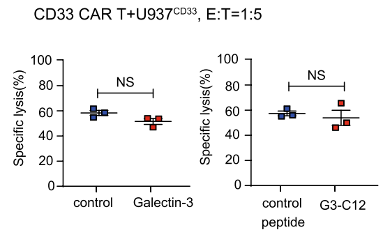
Galectin-3/LGALS3 Protein, Human purchased from MedChemExpress. Usage Cited in: J Transl Med. 2024 Jan 6;22(1):32. [Abstract]
Myeloid leukemia cells use galectin-1 to block CAR T cells function A, The specific lysis after coculture with U937CD33 cells at a 1:5 in the presence of recombinant galectin 3 protein (20 µg/ml) and galectin-3 specific inhibitor G3-C12 (30 µM).
-
Carbohydr Res
A structurally defined galactoglucan from Arisaema erubescens with a multi-target tumor-associated protein binding domain. [Abstract]2025 Nov 29:560:109781. PMID: 41344200 -


Technical Parameters
-
Species Human
-
Source E. coli
-
Tag Tag Free
-
Accession
P17931 (A2-I250)
-
Molecular Construction
-
N-term
-
LGALS3 (A2-I250)
Accession # P17931 -
C-term
-
-
Protein Length
Full Length
-
Synonyms
LGALS3; MAC-2; Prev. LGALS2; MAC2; GALIG; Galactoside-Binding Protein; Epididymis Secretory Sperm Binding Protein; MAC-2 Antigen; Advanced Glycation End-Product Receptor 3; Mac-2 Antigen; Lectin, Galactoside-Binding, Soluble, 3; Galectin; Carbohydrate-Bin
-
AA Sequence
ADNFSLHDALSGSGNPNPQGWPGAWGNQPAGAGGYPGASYPGAYPGQAPPGAYPGQAPPGAYPGAPGAYPGAPAPGVYPGPPSGPGAYPSSGQPSATGAYPATGPYGAPAGPLIVPYNLPLPGGVVPRMLITILGTVKPNANRIALDFQRGNDVAFHFNPRFNENNRRVIVCNTKLDNNWGREERQSVFPFESGKPFKIQVLVEPDHFKVAVNDAHLLQYNHRVKKLNEISKLGISGDIDLTSASYTMI
-
Molecular Weight
Approximately 30 kDa, based on SDS-PAGE under reducing conditions.
-
Purity
≥ 95%, as determined by reducing SDS-PAGE.


Product Properties
Lyophilized powder
1.Lyophilized from a 0.2 μm filtered solution of 50 mM HEPES, 5% Sucrose, 5% Mannitol, 0.06% Tween 80, pH 7.5.
2.Lyophilized from a 0.2 μm filtered solution of PBS, 8% trehalose, pH 7.4.
3.Lyophilized from a 0.2 μm filtered solution of 50 mM HEPES, 150 mMNaCl, 5% Trehalose, pH 7.4.
<1 EU/μg, determined by LAL method.
It is not recommended to reconstitute to a concentration less than 100 μg/mL in ddH2O.
Stored at -20°C for 2 years from date of receipt. After reconstitution, it is stable at 4°C for 1 week or -20°C for longer (with carrier protein). It is recommended to freeze aliquots at -20°C or -80°C for extended storage.
Room temperature in continental US; may vary elsewhere.


Documentation
-
Data Sheet (265 KB)
-
SDS (252 KB)
- English - EN (252 KB)
- Français - FR (252 KB)
- Deutsch - DE (252 KB)
- Norwegian - NO (252 KB)
- Español - ES (252 KB)
- Swedish - SV (252 KB)
- Italian - IT (252 KB)
- Korean - KR (252 KB)
- Portuguese - PT (252 KB)
-
Handling Instructions (2659 KB)


References
[1]. Chaudoin TR, et al. Mice lacking galectin-3 (Lgals3) function have decreased home cage movement. BMC Neurosci. 2018 May 2;19(1):27. [Content Brief]
[2]. Demetriou M, et al. Negative regulation of T-cell activation and autoimmunity by Mgat5 N-glycosylation. Nature. 2001 Feb 8;409(6821):733-9. [Content Brief]
[3]. Chalise U, et al. Macrophages secrete murinoglobulin-1 and galectin-3 to regulate neutrophil degranulation after myocardial infarction. Mol Omics. 2022 Mar 28;18(3):186-195. [Content Brief]
[4]. Yoshimura A, et al. Increased expression of the LGALS3 (galectin 3) gene in human non-small-cell lung cancer. Genes Chromosomes Cancer. 2003 Jun;37(2):159-64. [Content Brief]
[5]. Kulow VA, et al. Galectin-3 protects distal convoluted tubules in rhabdomyolysis-induced kidney injury. Pflugers Arch. 2024 Jul 23. [Content Brief]
[6]. Braeuer RR, et al. Galectin-3 contributes to melanoma growth and metastasis via regulation of NFAT1 and autotaxin. Cancer Res. 2012 Nov 15;72(22):5757-66. [Content Brief]

Calculators
Concentration (start) × Volume (start) = Concentration (final) × Volume (final)


 Powered by Bioz

