



M-CSF Protein, Mouse
Based on 148 publication(s) in Google Scholar
The M-CSF protein is a key orchestrator in regulating the survival, proliferation, and differentiation of hematopoietic precursor cells, particularly mononuclear phagocytes, including macrophages and monocytes. It actively promotes the release of pro-inflammatory chemokines, thereby playing a key role in innate immunity and inflammatory processes. M-CSF Protein, Mouse is the recombinant mouse-derived M-CSF protein, expressed by E. coli.
- Species: Mouse
- Source: E. coli
-
Storage:Stored at -20°C for 2 years from date of receipt. After reconstitution, it is stable at 4°C for 1 week or -20°C for longer (with carrier protein). It is recommended to freeze aliquots at -20°C or -80°C for extended storage.


To place orders, for customer services and technical support, please contact: MedChemExpress USA
Tel: 609-228-6898 E-mail: [email protected] [email protected]


-
 Biological Activity
-
Technical Parameters
-
Product Properties
- Documentation
- References
- Help & FAQs


-

HY-P7425
RANKL/TNFSF11 Protein, Mouse -

HY-18739
Phorbol 12-myristate 13-acetate -
HY-P7361
GM-CSF Protein, Mouse -
HY-P7071
IFN-gamma Protein, Mouse -
HY-P7080
IL-4 Protein, Mouse (CHO) -
HY-D1056
Lipopolysaccharides, from E. coli O55:B5 -
HY-P73388
RANKL/TNFSF11 Protein, Mouse (HEK293, Fc) -
HY-P70460
IL-13 Protein, Mouse -
HY-P70644
IL-4 Protein, Mouse -
HY-P7050
M-CSF Protein, Human -
HY-P7090
TNF-alpha/TNFSF2 Protein, Mouse (156a.a) -

HY-13259
MG-132


Biological Activity
Description
The M-CSF protein is a key orchestrator in regulating the survival, proliferation, and differentiation of hematopoietic precursor cells, particularly mononuclear phagocytes, including macrophages and monocytes. It actively promotes the release of pro-inflammatory chemokines, thereby playing a key role in innate immunity and inflammatory processes. M-CSF Protein, Mouse is the recombinant mouse-derived M-CSF protein, expressed by E. coli.
Background
Macrophage Colony Stimulating Factor (M-CSF) is a pro-inflammatory cytokine, constitutively produced by several cell types, such as fibroblasts, endothelial cells, stromal cells, macrophages, smooth muscle cells and osteoblasts, binds to its receptor CSF1R, and exists in several isoforms- as a secreted glycoprotein, a cell-surface protein and a proteoglycan[1]. M-CSF is involved in the development and proliferation of cells of the monocyte/macrophage lineage and participates in the induction of osteoclasts, which are important in the destruction of bone and cartilage and in the periarticular osteoporotic changes seen in patients with rheumatoid arthritis[2].
In Vitro
M-CSF Protein, Mouse (25 ng/mL, 5 days) can be added into cell medium for differentiation of bone marrow-derived macrophages (BMDMs)[3].
Verified Bioactivity
The ED50 is <3 ng/mL as measured by murine M-NFS-60 cells, corresponding to a specific activity of >3.3 × 10⁵ units/mg.
MCE Validation Data
-
Purity - SDS-PAGE
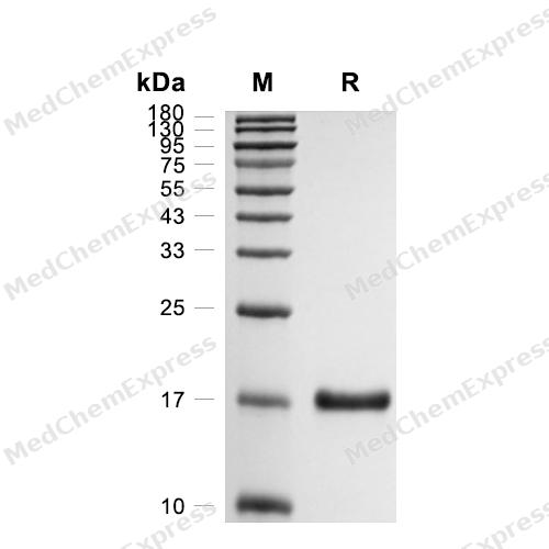
Purity - SDS-PAGE
-
Bioactivity - Cell-Based Assay
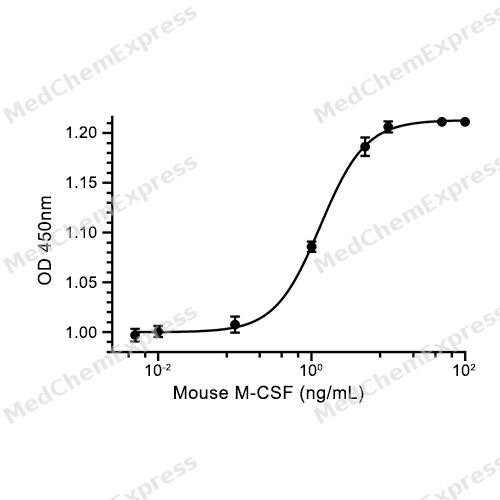
Bioactivity - Cell-Based Assay


Publications (148)
-
Journal Impact Factor
-
Most Recent
-
Science
2026 Feb 26;391(6788):eads4405. PMID: 41747053 -
Nat Nanotechnol
Annexin A1 mRNA-loaded liposomes alleviate acute pancreatitis by suppressing STING pathway and promoting efferocytosis in macrophages. [Abstract]2025 Aug 11. PMID: 40789923 -
Cell Metab
Microbial-derived 3-phenylpropionic acid orchestrates immune-progenitor cell crosstalk to promote beige adipogenesis and energy expenditure. [Abstract]2026 Apr 7;38(4):763-778.e7. PMID: 41742426 -
Cell Metab
Maltol induces diabetic fragility fractures by disrupting the balance of bone remodeling. [Abstract]2026 Mar 27:S1550-4131(26)00095-1. PMID: 41903530 -
Bioact Mater
Designed bone-targeting ROS-responsive nanoplatform for precision glycolysis inhibition in postmenopausal osteoporosis. [Abstract]2025 Nov 29:58:1-18. PMID: 41403870 -
Cell Host Microbe
Engineered probiotics recruit CAR macrophages and establish immune memory to eradicate heterogeneous glioblastoma in mice. [Abstract]2026 Feb 11;34(2):212-229.e7. PMID: 41605215 -
Nat Cell Biol
Psychological stress-induced ALKBH5 deficiency promotes tumour innervation and pancreatic cancer via extracellular vesicle transfer of RNA. [Abstract]2025 Jun;27(6):1035-1047. PMID: 40419796 -
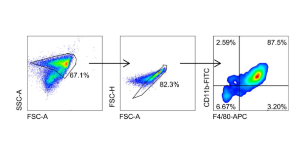
M-CSF Protein, Mouse purchased from MedChemExpress. Usage Cited in: Adv Funct Mater. 2025 Aug 28.
BMDMs were obtained from 6-8 weeks old BALB/c mice. After seven days of in vitro stimulation with macrophage colony-stimulating factor (M-CSF) (20 ng/mL), the cells exhibited a mature macrophage phenotype, with 87.5% of the population being F4/80⁺CD11b⁺, as confirmed by flow cytometry.
-
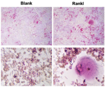
M-CSF Protein, Mouse purchased from MedChemExpress. Usage Cited in: Adv Funct Mater. 2024 May 23.
RAW264.7 cells were cocultured with 30 ng/mL M-CSF (MedChemExpress, USA). Containing 75 ng/mL Rankl (MedChemExpress, USA) were added at 2 days with 30 ng/mL M-CSF. After the appearance of multinucleated giant cells, cells were washed with PBS and fixed with 4% paraformaldehyde. After incubation with TRAP staining solution for 30 min and washed with double steaming, the nuclei were stained with hematoxylin for 2-3 min.
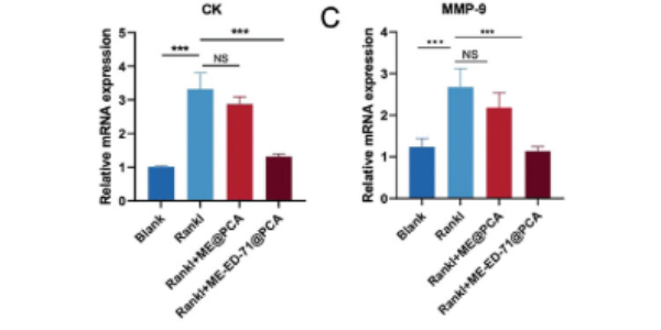
M-CSF Protein, Mouse purchased from MedChemExpress. Usage Cited in: Adv Funct Mater. 2024 May 23.
RAW264.7 cells were cocultured with 30 ng/mL M-CSF (MedChemExpress, USA). Containing 75 ng/mL Rankl (MedChemExpress, USA) were added at 2 days with 30 ng/mL M-CSF. The expression of CK and MMP9 genes after different treatment.
-
Autophagy
Restricting intracellular Salmonella proliferation by coordinating p-TBK1 mediated mitophagy and xenophagy. [Abstract]2026 Mar;22(3):445-467. PMID: 40660474 -
Nat Commun
Identification of splenic IRF7 as a nanotherapy target for tele-conditioning myocardial reperfusion injury. [Abstract]2025 Feb 24;16(1):1909. PMID: 39994192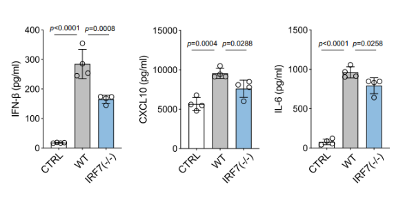
M-CSF Protein, Mouse purchased from MedChemExpress. Usage Cited in: Nat Commun. 2025 Feb 24;16(1):1909. [Abstract]
Levels of pro-inflammatory cytokines in the culture supernatants of BMDMs from wild-type C57BL/6 J mice and IRF7−/− mice after LPS (500 ng/mL) stimulation for 4 h. BMDMs were isolated from male C57BL/6 J mice and IRF7−/− mice. The isolated bone marrow cells were resuspended in BMDM culture medium (DMEM supplemented with 10% fetal bovine serum, 1% penicillin-streptomycin, and 50 ng/mL M-CSF).
-
Nat Commun
Macrophage STING signaling promotes fibrosis in benign airway stenosis via an IL6-STAT3 pathway. [Abstract]2025 Jan 3;16(1):289. PMID: 39753529 -
Nat Commun
Enhanced pericyte-endothelial interactions through NO-boosted extracellular vesicles drive revascularization in a mouse model of ischemic injury. [Abstract]2023 Nov 13;14(1):7334. PMID: 37957174 -
Exp Mol Med
CD300E+ macrophages facilitate liver regeneration after splenectomy in decompensated cirrhotic patients. [Abstract]2025 Feb;57(1):72-85. PMID: 39741181 -
J Adv Res
ADAM8 deficiency in macrophages promotes cardiac repair after myocardial infarction via ANXA2-mTOR-autophagy pathway. [Abstract]2025 Jul:73:483-499. PMID: 39097092 -
Redox Biol
TRPV4 drives macrophage pyroptosis via mitochondrial dysfunction and mtROS-dependent NLRP3 inflammasome activation in acute lung injury. [Abstract]2026 Jun 11:95:104255. PMID: 42314332 -
Redox Biol
Inhibition of NLRP3-mediated crosstalk between hepatocytes and liver macrophages by geniposidic acid alleviates cholestatic liver inflammatory injury. [Abstract]2022 Sep:55:102404. PMID: 35868156 -
-
Acta Pharm Sin B
Icariside Ⅱ, a main compound in Epimedii Folium, induces idiosyncratic hepatotoxicity by enhancing NLRP3 inflammasome activation. [Abstract]2020 Sep;10(9):1619-1633. PMID: 33088683 -
-
Adv Sci (Weinh)
Irgm1 Improves Postinfarction Cardiac Repair by Promoting Neutrophil Clearance and Efferocytosis. [Abstract]2026 Apr;13(21):e14863. PMID: 41738282 -
Adv Sci (Weinh)
Cryoablation Activates the cGAS-STING-CXCL10 Axis in Macrophages to Enhance Anti-Tumor Immunity in NSCLC. [Abstract]2026 Mar 12:e21931. PMID: 41818612 -
Adv Sci (Weinh)
Colorectal Cancer Cell's Weapon: RNF32 Engages SPP1+ Macrophages to Foster Liver Metastasis, Targeted by Indole-3-Acetic Acid. [Abstract]2025 Dec 12:e19735. PMID: 41387204 -
Adv Sci (Weinh)
Carnosic Acid Directly Targets STING C-Terminal Tail to Improve STING-Mediated Inflammatory Diseases. [Abstract]2025 Apr;12(14):e2417686. PMID: 39965124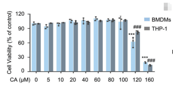
M-CSF Protein, Mouse purchased from MedChemExpress. Usage Cited in: Adv Sci (Weinh). 2025 Apr;12(14):e2417686. [Abstract]
BMDMs were cultured in DMEM containing FBS (10%), penicillin/streptomycin (1%), and M‐CSF (25 ng/mL) for 5 days. Cell viability of BMDMs and THP-1 cells were detected after being administrated with the indicated concentrations of Carnosic acid for 12 h.
-
Adv Sci (Weinh)
Biodegradable Cardiac Occluder with Surface Modification by Gelatin-Peptide Conjugate to Promote Endogenous Tissue Regeneration. [Abstract]2024 Jan;11(2):e2305967. PMID: 37984880 -
Adv Sci (Weinh)
Retinal Microenvironment-Protected Rhein-GFFYE Nanofibers Attenuate Retinal Ischemia-Reperfusion Injury via Inhibiting Oxidative Stress and Regulating Microglial/Macrophage M1/M2 Polarization. [Abstract]2023 Oct;10(30):e2302909. PMID: 37653617 -
Adv Sci (Weinh)
Extracellular Matrix/Glycopeptide Hybrid Hydrogel as an Immunomodulatory Niche for Endogenous Cardiac Repair after Myocardial Infarction. [Abstract]2023 Aug;10(23):e2301244. PMID: 37318159 -
Cell Rep Med
Targeted extracellular vesicle-photoimmunotherapy remodels stromal-immune microenvironment to boost chemo-immunotherapy in preclinical models. [Abstract]2026 Jun 3:102843. PMID: 42235518 -
Cell Death Differ
2026 Mar 17. PMID: 41844895 -
J Control Release
"Cytokine-microfactories" recruit DCs and deliver tumor antigens via gap junctions for immunotherapy. [Abstract]2021 Sep 10:337:417-430. PMID: 34324896 -
Cell Death Dis
Metformin alleviates inflammation through suppressing FASN-dependent palmitoylation of Akt. [Abstract]2021 Oct 12;12(10):934. PMID: 34642298 -
Cancer Lett
Targeting C/EBPβ/SCD1/C16:1 loop-driven macrophage M2 polarization by Bruceine D improves anti-PD-1 immunotherapy in colorectal cancer. [Abstract]2026 Sep 28:656:218655. PMID: 42250753 -
Cancer Lett
Sonrotoclax (BGB-11417) synergistically amplifies the radiotherapy-elicited anti-tumor immune response. [Abstract]2025 Aug 10:625:217759. PMID: 40311913 -
Small
ROS Responsive Cerium Oxide Biomimetic Nanoparticles Alleviates Calcium Oxalate Crystals Induced Kidney Injury via Suppressing Oxidative Stress and M1 Macrophage Polarization. [Abstract]2025 Jan;21(3):e2405417. PMID: 39629501 -
Small
A Bone-Penetrating Precise Controllable Drug Release System Enables Localized Treatment of Osteoporotic Fracture Prevention via Modulating Osteoblast-Osteoclast Communication. [Abstract]2023 Jun;19(26):e2207195. PMID: 36971278 -
J Immunother Cancer
Loss of Annexin A1 in macrophages restrains efferocytosis and remodels immune microenvironment in pancreatic cancer by activating the cGAS/STING pathway. [Abstract]2024 Sep 4;12(9):e009318. PMID: 39237260 -
Cell Commun Signal
Intestinal epithelial PIEZO1 regulates macrophage-to-myofibroblast transition via epithelial-derived TGF-α signaling in Crohn's disease. [Abstract]2026 May 28. PMID: 42210253 -
Cell Commun Signal
c-Ski is a novel repressor of NF-κB through interaction with p65 and HDAC1 in U937 cells. [Abstract]2025 Apr 2;23(1):165. PMID: 40176138 -
Cell Commun Signal
2024 Jun 7;22(1):315. PMID: 38849890 -
Cell Commun Signal
Tricyclic antidepressants induce liver inflammation by targeting NLRP3 inflammasome activation. [Abstract]2023 May 25;21(1):123. PMID: 37231437 -
NPJ Biofilms Microbiomes
Parabacteroides goldsteinii and its metabolite 7-KLCA attenuate endometriosis via TGR5 to reprogram macrophages by modulating the PPARγ/GPR132 axis. [Abstract]2026 May 22. PMID: 42173876 -
Mol Ther
Inhalable lipid nanoparticles for macrophage-specific STING gene editing to ameliorate pulmonary fibrosis. [Abstract]2026 Mar 6:S1525-0016(26)00188-7. PMID: 41795185 -
-
Phytomedicine
Oxymatrine attenuates melanoma progression and metastasis through SORBS2-mediated suppression of M2 macrophage polarization. [Abstract]2026 Jun:155:158148. PMID: 41956023 -
Phytomedicine
Xiao-Qing-Long-Tang alleviates allergic rhinitis by inhibiting M1 macrophage polarization and modulating the TRPV1 channel. [Abstract]2026 Jan:150:157732. PMID: 41455386 -
Phytomedicine
Disruption of cholesterol homeostasis triggers NLRP3-cGAS-STING axis-dependent hepatic fibrosis and honokiol intervention effects. [Abstract]2025 Jul 25:143:156904. PMID: 40449452 -
Phytomedicine
Panax notoginseng saponins ameliorate LPS-induced acute lung injury by promoting STAT6-mediated M2-like macrophage polarization. [Abstract]2025 Apr:139:156513. PMID: 40010033 -
Phytomedicine
Tryptanthrin suppresses multiple inflammasome activation to regulate NASH progression by targeting ASC protein. [Abstract]2024 Aug:131:155758. PMID: 38843643 -
Chin Herb Med
Amplifying protection against acute lung injury: Targeting both inflammasome and cGAS-STING pathway by Lonicerae Japonicae Flos-Forsythiae Fructus drug pair. [Abstract]2024 Apr 30;16(3):422-434. PMID: 39072201 -
Adv Healthc Mater
Ion-Chelating Hydrogel Regulates Porous Zinc Scaffold Degradation for Osteoimmune Bone Repair. [Abstract]2026 Mar 26:e05864. PMID: 41885000 -
Acta Biomater
Anti-inflammatory itaconate-loaded, cell-adhesive peptide-conjugated artificial small diameter vascular grafts for blood vessel regeneration. [Abstract]2025 Jun 19:S1742-7061(25)00453-2. PMID: 40543793 -
Acta Pharmacol Sin
2022 Apr;43(4):992-1000. PMID: 34341510 -
J Orthop Translat
Mitochondrial metabolism restoration via Tramiprosate suppresses mitochondrial ROS-driven foamy macrophage senescence post spinal cord injury. [Abstract]2026 Mar 2:57:101049. PMID: 41808881 -
J Transl Med
Erbin accelerates TFEB-mediated lysosome biogenesis and autophagy and alleviates sepsis-induced inflammatory responses and organ injuries. [Abstract]2023 Dec 17;21(1):916. PMID: 38105228 -
-
Int J Biol Macromol
Functional characterization and engineering of the sugar donor specificity of a 2″-O-xylosyltransferase from Panax notoginseng for rare saponins biosynthesis. [Abstract]2026 May:362:152095. PMID: 42002181 -
Regen Biomater
Triamcinolone acetonide-loaded chitosan-polyethylene glycol hydrogel for preventing esophageal stricture post-endoscopic submucosal dissection. [Abstract]2026 Jan 14:13:rbag002. PMID: 41716490 -
Regen Biomater
In situ injectable chemico-biological cascade-driven antioxidant nanoparticles for periodontitis treatment. [Abstract]2025 Dec 8:12:rbaf125. PMID: 41480412 -
Int J Biol Macromol
Prediction and validation based on scRNA-seq: ETS2 targets CEBPB to mediate osteoclast differentiation in osteoarthritis progression. [Abstract]2025 Aug;319(Pt 4):145639. PMID: 40602581 -
Mol Med
Extinguishing the flames of inflammation: retardant effect of chlorquinaldol on NLRP3-driven diseases. [Abstract]2024 Dec 19;30(1):245. PMID: 39701924 -
Antioxidants (Basel)
Ferroptosis in Recurrent Vulvovaginal Candidiasis Through Integrated Bioinformatics and Experimental Validation. [Abstract]2026 Mar 24;15(4):407. PMID: 42072050 -
-
Free Radic Biol Med
Tartrate-Resistant Acid Phosphatase 5 (TRAP/ACP5) aggravates atherosclerosis by regulating macrophage polarization and promoting ferroptosis. [Abstract]2026 Apr:247:486-501. PMID: 41692316 -
Free Radic Biol Med
Fyn deficiency drives pro-inflammatory macrophage activation to exacerbate septic acute lung injury. [Abstract]2026 Mar 16:246:80-92. PMID: 41490764 -
Free Radic Biol Med
Gut microbial metabolite alleviates osteoporosis by attenuating AKT-NFATc1 signaling pathway and ROS production. [Abstract]2025 Nov 23:243:351-366. PMID: 41290102 -
Free Radic Biol Med
Nepetin limits NLRP3 inflammasome activation and alleviates NLRP3-driven inflammatory diseases via PINK1-dependent mitophagy. [Abstract]2025 Feb 1:227:420-433. PMID: 39653129 -
Free Radic Biol Med
Retinal cell-targeted liposomal ginsenoside Rg3 attenuates retinal ischemia-reperfusion injury via alleviating oxidative stress and promoting microglia/macrophage M2 polarization. [Abstract]2023 Sep:206:162-179. PMID: 37380044 -
Clin Transl Med
2026 Feb;16(2):e70616. PMID: 41674095 -
ACS Appl Mater Interfaces
Ex Vivo High-Dose Ionizing Irradiation-Conditioned Nanovesicles for Enhancing the Immune Effects of Tumor Radiotherapy. [Abstract]2025 Aug 20;17(33):46705-46719. PMID: 40773763 -
Stem Cell Res Ther
Exosomes from adipose-derived stem cells accelerate wound healing by increasing the release of IL-33 from macrophages. [Abstract]2025 Feb 21;16(1):80. PMID: 39984984 -
Cell Rep
Adoptive macrophages suppress glioblastoma growth by reversing immunosuppressive microenvironment through programmed phenotype repolarization. [Abstract]2025 Sep 26;44(10):116350. PMID: 41014558 -
Cell Rep
Depletion of CUL4B in macrophages ameliorates diabetic kidney disease via miR-194-5p/ITGA9 axis. [Abstract]2023 May 23;42(6):112550. PMID: 37224018 -
Chin Med
Multi-omics and network pharmacology reveal Huayu-Tongbi decoction reduced arthritis-related bone erosion. [Abstract]2025 Jul 2;20(1):100. PMID: 40598199 -
-
J Med Chem
Design and Synthesis of FR-β Targeting Chimeric Molecules for Reprogramming Tumor-Associated Macrophages Using 6-Substituted Pyrrolo[2,3- d]pyrimidines as Targeting Ligands. [Abstract]2025 Apr 12. PMID: 40219974 -
-
J Ethnopharmacol
2022 Nov 15:298:115593. PMID: 35973629 -
J Agric Food Chem
Natural Dietary Flavonoid Apigenin Mitigates Ulcerative Colitis via Modulating the AMPK/NF-κB/NLRP3 Signaling Axis. [Abstract]2025 Dec 22. PMID: 41428381 -
Cell Mol Life Sci
m6A-modified MIR670HG suppresses tumor liver metastasis through enhancing Kupffer cell phagocytosis. [Abstract]2025 Apr 28;82(1):185. PMID: 40293529 -
Biochem Pharmacol
The deubiquitinase OTUD1 deubiquitinates TIPE2 and plays a protective role in sepsis-induced lung injury by targeting TAK1-mediated MAPK and NF-κB signaling. [Abstract]2024 Sep:227:116418. PMID: 38996928 -
Life Sci
Esaxerenone inhibits renal macrophage proliferation through MR/TGF-β1 pathway in db/db mice. [Abstract]2025 Dec 1:382:124008. PMID: 41067301 -
Life Sci
Fibroblast to macrophage-like cell transition in renal inflammatory injury through the MR/CSF1 pathway induced by aldosterone. [Abstract]2025 Jul 1:372:123627. PMID: 40216224 -
Inflamm Res
Aldosterone induces renal lymphangiogenesis through macrophage-lymphatic endothelial cell transformation and Inhibition by esaxerenone. [Abstract]2025 May 24;74(1):85. PMID: 40413281 -
-
Commun Biol
2025 Nov 24;8(1):1639. PMID: 41286500 -
-
Mar Drugs
Structural and Functional Insights into a Novel Aspergillus ochraceus Polysaccharide from the Weddell Sea: Implications for Melanoma Immunotherapy In Vitro. [Abstract]2025 Jun 10;23(6):246. PMID: 40559655 -
Int Immunopharmacol
The TGFB1-Wnt/β-catenin axis programs a neuroprotective IGF1+ microglial state during epileptogenesis. [Abstract]2026 Jun 1:178:116579. PMID: 41911647 -
Int Immunopharmacol
GPR132 drives macrophage M1 polarization and aggravates inflammation-associated liver injury. [Abstract]2026 Apr 1:174:116349. PMID: 41679180 -
-
Int Immunopharmacol
Targeting periodontal inflammatory microenvironment to ameliorate periodontitis: A nitrogen ions implantation technique. [Abstract]2026 Feb 1:170:116055. PMID: 41448003 -
Int Immunopharmacol
Targeting RXRα inhibits foamy macrophage formation and neuroinflammation by promoting cholesterol efflux channels post traumatic spinal cord injury. [Abstract]2026 Jan 1;168(Pt 2):115945. PMID: 41317538 -
Int Immunopharmacol
Periprosthetic osteolysis is mediated by the m6A-dependent regulation of CEMIP via YTHDF2. [Abstract]2026 Jan 1;168(Pt 2):115895. PMID: 41308363 -
Int Immunopharmacol
Calreticulin-driven immunogenic cell death promotes osteoclast differentiation and osteoarthritis progression via the LRP1/Rac1 signaling. [Abstract]2025 Oct 10:163:115277. PMID: 40714468 -
Int Immunopharmacol
Intratumoral microbiota-driven macrophage reprogramming in pancreatic cancer via Blautia metabolite 6-hydroxyhexanoic acid. [Abstract]2025 Oct 30:164:115421. PMID: 40865404 -
Int J Mol Sci
Aldosterone-Induced Transformation of Vascular Smooth Muscle Cells into Macrophage-like Cells Participates in Inflammatory Vascular Lesions. [Abstract]2025 Apr 3;26(7):3345. PMID: 40244230 -
Int Immunopharmacol
VCPIP1 negatively regulates NF-κB signaling pathways by deubiquitinating and stabilizing Erbin in MDP-stimulated macrophages. [Abstract]2024 Nov 16;143(Pt 3):113622. PMID: 39550842 -
Int Immunopharmacol
Monotropein inhibits colitis associated cancer through VDR/JAK1/STAT1 regulation of macrophage polarization. [Abstract]2023 Aug 24;124(Pt A):110838. PMID: 37633235 -
Int Immunopharmacol
AKBA alleviates experimental pancreatitis by inhibiting oxidative stress in Macrophages through the Nrf2/HO-1 pathway. [Abstract]2023 Aug:121:110501. PMID: 37364326 -
Int Immunopharmacol
Psoralidin, a major component of Psoraleae Fructus, induces inflammasome activation and idiosyncratic liver injury. [Abstract]2021 Mar:92:107352. PMID: 33422760 -
Inflammation
Activation of β2-Adrenergic Receptor Alleviates Viral Myocarditis by Regulating Energy Metabolism in Monocyte-derived Macrophages via the AMPK Pathway. [Abstract]2026 Jan 15;49(1):48. PMID: 41537900 -
Arthritis Res Ther
Trim21 deficiency alleviates osteoporosis by inhibiting osteoclast differentiation through regulating Txnip. [Abstract]2025 Aug 1;27(1):163. PMID: 40751213 -
Front Cell Dev Biol
Tcirg1 deficiency delays osteoarthritis progression by impairing lysosome acidification and peripheral accumulation in osteoclasts. [Abstract]2025 Sep 9:13:1621648. PMID: 40995561 -
Int Dent J
Nitrogen Species Modulate Macrophage ER Stress to Preserve Alveolar Bone: A Translational Adjunct for Periodontal Care. [Abstract]2026 Feb 3;76(2):109400. PMID: 41637951 -
Chem Biol Interact
2023 Oct 1:384:110696. PMID: 37689331 -
Molecules
Constituents from Dolichos lablab L. Flowers and Their Anti-Inflammatory Effects via Inhibition of IL-1β Release. [Abstract]2024 Aug 7;29(16):3751. PMID: 39202831 -
Biochim Biophys Acta Mol Basis Dis
Neutrophil-derived thrombospondin-1 (THBS1) drives type 2 diabetes-induced osteoporosis via CD36-PPARγ-POU2F2 signaling. [Abstract]2025 Dec 15;1872(3):168139. PMID: 41407214 -
Mol Pharm
Curcumin-Loaded Liposomes (hPLipo/Cur) with Liver-Targeting Properties for Efficient NAFLD Treatment by Alleviating Mitochondrial ROS-Mediated Ferroptosis via NRF2 Pathway. [Abstract]2026 Apr 6;23(4):2398-2411. PMID: 41725216 -
PLoS Pathog
Schistosoma japonicum leishmanolysin SjLLPi1 facilitates the invasion of cercariae into the host skin. [Abstract]2025 Aug 26;21(8):e1013446. PMID: 40857348 -
Mediators Inflamm
Artesunate Restrains Maturation of Dendritic Cells and Ameliorates Heart Transplantation-Induced Acute Rejection in Mice through the PERK/ATF4/CHOP Signaling Pathway. [Abstract]2021 Aug 21:2021:2481907. PMID: 34462628 -
Mediators Inflamm
The Proresolving Lipid Mediator Maresin1 Alleviates Experimental Pancreatitis via Switching Macrophage Polarization. [Abstract]2021 Mar 9:2021:6680456. PMID: 33776575 -
Mol Cell Biochem
Upregulation of YY1 in M2 macrophages promotes secretion of exosomes containing hsa-circ-0000326 via super-enhancers to facilitate prostate cancer progression. [Abstract]2025 Jun;480(6):3873-3888. PMID: 39960585 -
-
-
FASEB J
Macrophage WTAP Deficiency Protects Atherosclerosis by Improving Macrophage Apoptosis in an m6A-Independent Manner. [Abstract]2025 Nov 15;39(21):e71182. PMID: 41148185 -
FASEB J
Blocking the ADAM9/ITGAV Pathway Ameliorates Sepsis-Induced Acute Lung Injury by Promoting Macrophage Efferocytosis. [Abstract]2025 Aug 15;39(15):e70845. PMID: 40736047 -
FASEB J
2019 Sep;33(9):9775-9784. PMID: 31166814 -
Clin Transl Allergy
2024 Dec;14(12):e70014. PMID: 39644500 -
J Biol Chem
Hepatitis B virus promotes hepatocellular carcinogenesis by activating IL-6-dependent tumor-macrophage crosstalk and M2-like macrophage polarization. [Abstract]2026 Jun 22:113283. PMID: 42331105 -
J Cell Physiol
Conditional knockout of the PDK-1 gene in osteoblasts affects osteoblast differentiation and bone formation. [Abstract]2021 Jul;236(7):5432-5445. PMID: 33377210 -
-
-
Vet Res
Comprehensive multiomic analysis of extracellular vesicles from Mycoplasma bovis-infected bovine mammary epithelial cells identifies proteins and miRNAs that induce inflammatory responses in macrophages. [Abstract]2025 Sep 25;56(1):185. PMID: 40999451 -
J Orthop Surg Res
Epimedium Brevicornu and Curculigo orchioides inhibit osteoclast autophagy by degrading the level of miRNA-199 to regulate the mTOR signaling pathway. [Abstract]2025 Jul 9;20(1):631. PMID: 40635043 -
J Orthop Surg Res
Licochalcone D inhibits osteoclast differentiation and postmenopausal osteoporosis by inactivating the NF-κB signaling pathway. [Abstract]2025 Jul 28;20(1):713. PMID: 40722101 -
Mol Immunol
Hypericin impedes M2 macrophage polarization and protects against Hepatocellular carcinoma. [Abstract]2025 Mar 27:181:160-168. PMID: 40153953 -
J Pharm Pharmacol
Anoectochilus roxburghii polysaccharide reduces D-GalN/LPS-induced acute liver injury by regulating the activation of multiple inflammasomes. [Abstract]2024 Sep 3;76(9):1212-1224. PMID: 38985664 -
Mol Immunol
Brevilin A inhibits NLRP3 inflammasome activation in vivo and in vitro by acting on the upstream of NLRP3-induced ASC oligomerization. [Abstract]2021 Jul:135:116-126. PMID: 33892379 -
J Biochem Mol Toxicol
Proanthocyanidins suppress NLRP3 inflammasome and M1 macrophage polarization to alleviate severe acute pancreatitis in mice. [Abstract]2022 Oct 13;e23242. PMID: 36229953 -
Immun Inflamm Dis
Formoterol Reduces the Pro-Inflammatory Phenotype by Enhancing the Activity of Glutaminase in Monocyte-Derived Macrophages in the CVB3-Induced Viral Myocarditis. [Abstract]2024 Nov;12(11):e70073. PMID: 39601476 -
Chem Biodivers
Chemical Constituents of the Deep-Sea-Derived Penicillium limosum ZEN48 and Their Osteoclasts' Inhibitory Activity. [Abstract]2026 Mar;23(3):e03812. PMID: 41838563 -
Genes Nutr
Protective effects of Nogo-B deficiency in NAFLD mice and its multiomics analysis of gut microbiology and metabolism. [Abstract]2024 Aug 24;19(1):17. PMID: 39182019 -
FEBS Open Bio
Anti-inflammatory properties of ophioglonin derived from the fern Ophioglossum vulgatum L. via inactivating NF-κB and MAPK signaling pathways. [Abstract]2025 Jan;15(1):122-139. PMID: 39455284 -
-
-
-
Cancer Chemother Pharmacol
Sulforaphane inhibits multiple myeloma cell-induced osteoclast differentiation and macrophage proliferation by elevating ferroportin1. [Abstract]2024 Dec 11;95(1):3. PMID: 39661165 -
-
-
-
-
-
-
Front Med
Yinqiao powder effectively alleviates acute lung injury via regulating cGAS-STING signaling pathway. [Abstract]2025 Dec 27. PMID: 41454071 -
Heliyon
New compatible pair of TCM: Paeoniae Radix Alba effectively alleviate Psoraleae Fructus-induced liver injury by suppressing NLRP3 inflammasome activation. [Abstract]2024 Jul 14;10(14):e34591. PMID: 39130485 -
-
-
Heliyon
Long noncoding RNA HOXC-AS3 remodels lipid metabolism and promotes the proliferation of transformed macrophages in the glioma stem cell microenvironment by regulating the hnRNPA1/CaM axis. [Abstract]2023 Aug 9;9(8):e19034. PMID: 37609424


Technical Parameters
-
Species Mouse
-
Source E. coli
-
Tag Tag Free
-
Accession
P07141-1 (K33-P187)
-
Molecular Construction
-
N-term
-
M-CSF (K33-P187)
Accession # P07141-1 -
C-term
-
-
Protein Length
Partial Extracellular Domain
-
Synonyms
CSF1; Macrophage Colony Stimulating Factor 1; Colony Stimulating Factor 1; Lanimostim; MCSF; MGC31930; Proteoglycan Macrophage Colony-Stimulating Factor; PG-M-CSF; Macrophage Colony-Stimulating Factor 1; CSF-1; M-CSF; CSF1-HERV-LTR71B Fusion Protein; Colo
-
AA Sequence
KEVSEHCSHMIGNGHLKVLQQLIDSQMETSCQIAFEFVDQEQLDDPVCYLKKAFFLVQDIIDETMRFKDNTPNANATERLQELSNNLNSCFTKDYEEQNKACVRTFHETPLQLLEKIKNFFNETKNLLEKDWNIFTKNCNNSFAKCSSRDVVTKP
-
Predicted Molecular Mass
18 kDa
-
Molecular Weight
Approximately 17-20 kDa, based on SDS-PAGE under non reducing (N) conditions.
-
Structure/Form
Disulfide-linked homodimer
-
Purity
≥ 95%, as determined by reducing SDS-PAGE.


Product Properties
Lyophilized powder
1.Lyophilized from a 0.22 μm filtered solution of 50 mM Tris, 150 mM NaCl, pH 8.0.
2.Lyophilized from a 0.22 μm filtered solution of 50 mM Tris-HCl, 150 mM NaCl, pH 8.0.
3.Lyophilized from a 0.22 μm filtered solution of PBS, pH 7.4, 8% trehalose.
Please refer to the lot-specific COA for specific buffer information.
Note: For SPR assay, please replace the buffer. Primary amine components (e.g., Tris, imidazole) can affect protein-coupled chips.
<0.2 EU/μg, determined by LAL method.
It is not recommended to reconstitute to a concentration less than 100 μg/mL in ddH2O. For long term storage it is recommended to add a carrier protein (0.1% BSA, 5% HSA, 10% FBS or 5% Trehalose).
Stored at -20°C for 2 years from date of receipt. After reconstitution, it is stable at 4°C for 1 week or -20°C for longer (with carrier protein). It is recommended to freeze aliquots at -20°C or -80°C for extended storage.
Room temperature in continental US; may vary elsewhere.


Documentation
-
Data Sheet (263 KB)
-
SDS (252 KB)
- English - EN (252 KB)
- Français - FR (252 KB)
- Deutsch - DE (252 KB)
- Norwegian - NO (252 KB)
- Español - ES (252 KB)
- Swedish - SV (252 KB)
- Italian - IT (252 KB)
- Korean - KR (252 KB)
- Portuguese - PT (252 KB)
-
Handling Instructions (2659 KB)


References
[1]. Hamilton JA. Colony-stimulating factors in inflammation and autoimmunity. Nat Rev Immunol. 2008 Jul;8(7):533-44. [Content Brief]
[2]. Rioja I, et al. Potential novel biomarkers of disease activity in rheumatoid arthritis patients: CXCL13, CCL23, transforming growth factor alpha, tumor necrosis factor receptor superfamily member 9, and macrophage colony-stimulating factor. Arthritis Rheum. 2008 Aug;58(8):2257-67. [Content Brief]
[3]. Mu W, et al., Carnosic Acid Directly Targets STING C-Terminal Tail to Improve STING-Mediated Inflammatory Diseases. Adv Sci (Weinh). 2025 Apr;12(14):e2417686. [Content Brief]

Calculators
Concentration (start) × Volume (start) = Concentration (final) × Volume (final)


 Powered by Bioz

