



M-CSF Protein, Human
Based on 23 publication(s) in Google Scholar
M-CSF (CSF-1) protein is a homodimeric hematopoietic growth factor and proinflammatory cytokine, belonging to the colony stimulating factor. M-CSF can act specifically on the monocyte-macrophage lineage, drive monocyte proliferation and differentiation into macrophages/osteoclasts, and induce M1 macrophages to enhance ADCC anti-tumor activity and M2 macrophages to promote VEGF angiogenesis. M-CSF Protein, Human is a recombinant human M-CSF protein expressed in E. coli with tag-free.
- Species: Human
- Source: E. coli
-
Storage:Stored at -20°C for 2 years from date of receipt. After reconstitution, it is stable at 4°C for 1 week or -20°C for longer (with carrier protein). It is recommended to freeze aliquots at -20°C or -80°C for extended storage.


To place orders, for customer services and technical support, please contact: MedChemExpress USA
Tel: 609-228-6898 E-mail: [email protected] [email protected]


-
 Biological Activity
-
Technical Parameters
-
Product Properties
- Documentation
- References
- Help & FAQs


-

HY-18739
Phorbol 12-myristate 13-acetate -

HY-P70445
IL-4 Protein, Human -
HY-P7016A
GM-CSF Protein, Human -
HY-P7025
IFN-gamma Protein, Human -
HY-P7085
M-CSF Protein, Mouse -
HY-P7044
IL-6 Protein, Human -
HY-P70781
SCF Protein, Human -
HY-P7007
BMP-4 Protein, Human -
HY-P7424
RANKL/TNFSF11 Protein, Human -

HY-D1056
Lipopolysaccharides, from E. coli O55:B5 -

HY-10431
SB-431542 -
HY-P70568
IL-13 Protein, Human (HEK293, His)


Biological Activity
Description
M-CSF (CSF-1) protein is a homodimeric hematopoietic growth factor and proinflammatory cytokine, belonging to the colony stimulating factor. M-CSF can act specifically on the monocyte-macrophage lineage, drive monocyte proliferation and differentiation into macrophages/osteoclasts, and induce M1 macrophages to enhance ADCC anti-tumor activity and M2 macrophages to promote VEGF angiogenesis[1][2][3][4]. M-CSF Protein, Human is a recombinant human M-CSF protein expressed in E. coli with tag-free.
Background
1. M-CSF Protein Protein Characteristics[1][2][3]
M-CSF, or macrophage colony stimulating factor, CSF-1, is a disulfide-linked homodimer belonging to the colony stimulating factor (CSF) family. M-CSF is a hematopoietic growth factor and proinflammatory cytokine with multiple glycosylation sites, belonging to the hematopoietic growth factor family along with GM-CSF and G-CSF. M-CSF affects the survival and function of tissue macrophages and has anti-tumor activity[1]. M-CSF is expressed in a variety of cells, such as fibroblasts, endothelial cells, stromal cells, macrophages, smooth muscle cells and osteoblasts, and has multiple subtypes. M-CSF can be a secreted glycoprotein, a cell surface protein and a proteoglycan, binding to its receptor CSF1R[2]. M-CSF can specifically act on the monocyte-macrophage lineage, participate in the development and proliferation of monocyte/macrophage lineage cells, and participate in the induction of osteoclasts. Osteoclasts are very important for the destruction of bone and cartilage and the changes in periarticular osteoporosis in patients with rheumatoid arthritis[3].
2. M-CSF Protein Functional Characteristics[1][4]
M-CSF is regulated by LPS and IFN-γ upstream, and activates NF-κB and STAT3 downstream, regulating inflammatory factors such as IL-12 and TNF-α. M-CSF induces macrophage polarization to M1 type, and enhances antibody-dependent cellular cytotoxicity (ADCC) by upregulating surface molecules such as CD80 and CD16, exerting anti-tumor effects. M-CSF may induce macrophage polarization to M2 type, promoting VEGF secretion and angiogenesis.
3. Application of M-CSF Protein[1][4][5][6]
Oncology: Clinical trials have shown that M-CSF combined with monoclonal antibodies can enhance tumor-targeted killing, but its pro-angiogenic effect may promote metastasis.
Infection and Immunity: Enhance the antifungal (such as Candida) and anti-tuberculosis activity of macrophages, and use it to treat infections in immunocompromised patients.
Hematopoietic Repair: Restore monocyte counts after chemotherapy/transplantation and shorten the neutrophil deficiency period.
Bone Disease: Regulate osteoclasts, used in osteoporosis and rheumatoid arthritis research.
Verified Bioactivity
The ED50 is 0.4-3.0 ng/mL as measured by M-NFS-60 cells, corresponding to a specific activity of 3.3 × 10⁵-2.5 × 10⁶ units/mg.
MCE Validation Data
-
Purity - SDS-PAGE
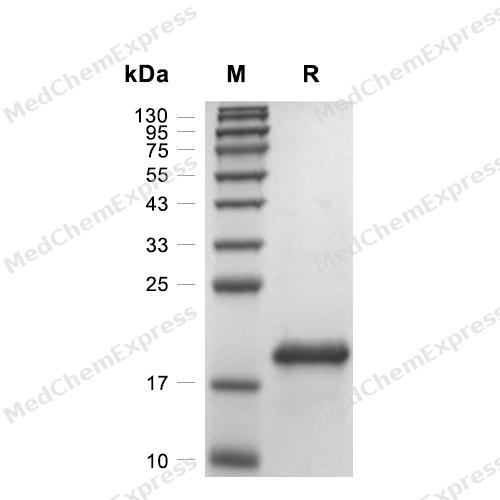
Purity - SDS-PAGE
-
Bioactivity - Cell-Based Assay
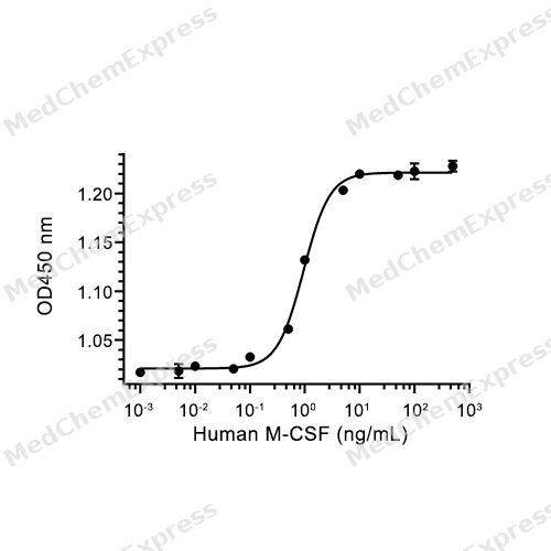
Bioactivity - Cell-Based Assay


Publications (23)
-
Journal Impact Factor
-
Most Recent
-
Cell
De novo assembly of nuclear stress bodies rearranges and enhances NFIL3 to restrain acute inflammatory responses. [Abstract]2025 Aug 21;188(17):4586-4603.e31. PMID: 40436014 -
Redox Biol
Stromal cell-derived itaconate promotes endometriosis via macrophage NRF2 and lysosomal pH modulation. [Abstract]2026 May:92:104101. PMID: 41780193 -
Adv Sci (Weinh)
Colorectal Cancer Cell's Weapon: RNF32 Engages SPP1+ Macrophages to Foster Liver Metastasis, Targeted by Indole-3-Acetic Acid. [Abstract]2025 Dec 12:e19735. PMID: 41387204 -
Adv Sci (Weinh)
Deciphering the Role and Mechanism of Decidual Monocyte-Derived Macrophage Infiltration in Obstetric Antiphospholipid Syndrome at Single-Cell Resolution. [Abstract]2025 Aug 7:e03480. PMID: 40776470 -
Adv Sci (Weinh)
4-Octyl Itaconate Alleviates Myocardial Ischemia-Reperfusion Injury Through Promoting Angiogenesis via ERK Signaling Activation. [Abstract]2025 Mar;12(10):e2411554. PMID: 39836624 -
Cell Rep Med
Microneedle delivery of CAR-M-like engineered macrophages alleviates intervertebral disc degeneration through enhanced efferocytosis capacity. [Abstract]2025 Apr 15;6(4):102079. PMID: 40199328 -
Cell Death Differ
Macrophage junctional adhesion molecule-like (JAML) protein promotes NLRP3 inflammasome activation in the development of atherosclerosis. [Abstract]2025 Mar 28. PMID: 40148467 -
Cell Death Differ
TREM2 macrophage promotes cardiac repair in myocardial infarction by reprogramming metabolism via SLC25A53. [Abstract]2024 Feb;31(2):239-253. PMID: 38182899 -
Cancer Lett
Targeting C/EBPβ/SCD1/C16:1 loop-driven macrophage M2 polarization by Bruceine D improves anti-PD-1 immunotherapy in colorectal cancer. [Abstract]2026 Sep 28:656:218655. PMID: 42250753 -
Phytomedicine
Direct inhibition of macrophage sting signaling by curcumol protects against myocardial infarction via attenuating the inflammatory response. [Abstract]2025 Mar:138:156403. PMID: 39889491 -
BMC Med
Neutrophil extracellular trap-induced intermediate monocytes trigger macrophage activation syndrome in adult-onset Still's disease. [Abstract]2023 Dec 20;21(1):507. PMID: 38124139 -
Cell Rep
IFNα-induced BST2+ tumor-associated macrophages facilitate immunosuppression and tumor growth in pancreatic cancer by ERK-CXCL7 signaling. [Abstract]2024 Apr 23;43(4):114088. PMID: 38602878 -
Clin Sci
IL-33 promotes rheumatoid arthritis progression by enhancing pro-inflammatory macrophage development in the synovial microenvironment. [Abstract]2026 Mar 11;140(3):359-376. PMID: 41615676 -
Food Funct
Fucoxanthin prevents septic cardiomyopathy by targeting BRD2 in M1-polarized macrophages. [Abstract]2026 Jan 5. PMID: 41489867 -
Cancer Biol Ther
CircATP5C1 promotes triple-negative breast cancer progression by binding IGF2BP2 to modulate CSF-1 secretion. [Abstract]2025 Dec;26(1):2479926. PMID: 40176374 -
-
Invest Ophthalmol Vis Sci
Adalimumab Reprograms M1 Macrophages to Attenuate Th1/Th17 Responses in Behçet's Uveitis and Vogt-Koyanagi-Harada Syndrome. [Abstract]2026 Jun 1;67(6):4. PMID: 42227805 -
J Biol Chem
Hepatitis B virus promotes hepatocellular carcinogenesis by activating IL-6-dependent tumor-macrophage crosstalk and M2-like macrophage polarization. [Abstract]2026 Jun 22:113283. PMID: 42331105 -
Regen Ther
Denosumab ameliorates osteoarthritis by protecting cartilage against degradation and modulating subchondral bone remodeling. [Abstract]2024 Mar 26:27:181-190. PMID: 38840731 -
Cytokine
The activation of CaN/NFAT signaling pathway in macrophages aggravated Lactobacillus casei cell wall extract-induced Kawasaki disease vasculitis. [Abstract]2023 Sep:169:156304. PMID: 37487381 -
Tissue Cell
Exosomes secreted from M2-polarized macrophages inhibit osteoclast differentiation via CYLD. [Abstract]2025 Apr:93:102645. PMID: 39671756 -
-


Technical Parameters
-
Species Human
-
Source E. coli
-
Tag Tag Free
-
Accession
P09603-3 (E33-S190)
-
Molecular Construction
-
N-term
-
M-CSF (E33-S190)
Accession # P09603-3 -
C-term
-
-
Protein Length
Partial
-
Synonyms
CSF1; Macrophage Colony Stimulating Factor 1; Colony Stimulating Factor 1; Lanimostim; MCSF; MGC31930; Proteoglycan Macrophage Colony-Stimulating Factor; PG-M-CSF; Macrophage Colony-Stimulating Factor 1; CSF-1; M-CSF; CSF1-HERV-LTR71B Fusion Protein; Colo
-
AA Sequence
EEVSEYCSHMIGSGHLQSLQRLIDSQMETSCQITFEFVDQEQLKDPVCYLKKAFLLVQDIMEDTMRFRDNTPNAIAIVQLQELSLRLKSCFTKDYEEHDKACVRTFYETPLQLLEKVKNVFNETKNLLDKDWNIFSKNCNNSFAECSSQGHERQSEGS
-
Molecular Weight
Approximately 18 kDa under reduced (R) conditions, 28 kDa under non reducing (N) conditions, based on SDS-PAGE, due to the glycosylation.
-
Structure/Form
Disulfide-linked homodimer
-
Purity
≥ 95%, as determined by reducing SDS-PAGE.


Product Properties
Lyophilized powder
1.Lyophilized from a 0.22 μm filtered solution of 50 mM Tris-HCl, 300 mM NaCl, pH 8.0.
2.Lyophilized from a 0.22 μm filtered solution of 50 mM Tris-HCl, 300 mM NaCl, pH 8.0, 5% trehalose, 5% mannitol, 0.01% Tween 80.
Please refer to the lot-specific COA for specific buffer information.
Note: For SPR assay, please replace the buffer. Primary amine components (e.g., Tris, imidazole) can affect protein-coupled chips.
<1 EU/μg, determined by LAL method.
It is not recommended to reconstitute to a concentration less than 100 μg/mL in ddH2O or PBS or Tris-HCl, pH 8.0. For long term storage it is recommended to add a carrier protein (0.1% BSA, 5% HSA, 10% FBS or 5% Trehalose).
Stored at -20°C for 2 years from date of receipt. After reconstitution, it is stable at 4°C for 1 week or -20°C for longer (with carrier protein). It is recommended to freeze aliquots at -20°C or -80°C for extended storage.
Room temperature in continental US; may vary elsewhere.


Documentation
-
Data Sheet (265 KB)
-
SDS (252 KB)
- English - EN (252 KB)
- Français - FR (252 KB)
- Deutsch - DE (252 KB)
- Norwegian - NO (252 KB)
- Español - ES (252 KB)
- Swedish - SV (252 KB)
- Italian - IT (252 KB)
- Korean - KR (252 KB)
- Portuguese - PT (252 KB)
-
Handling Instructions (2659 KB)


References
[1]. Garnick MB, et al. Preclinical and clinical evaluation of recombinant human macrophage colony-stimulating factor (rhM-CSF). Int J Cell Cloning. 1990 Jan;8 Suppl 1:356-71. [Content Brief]
[2]. Hamilton JA. Colony-stimulating factors in inflammation and autoimmunity. Nat Rev Immunol. 2008 Jul;8(7):533-44. [Content Brief]
[3]. Rioja I, et al. Potential novel biomarkers of disease activity in rheumatoid arthritis patients: CXCL13, CCL23, transforming growth factor alpha, tumor necrosis factor receptor superfamily member 9, and macrophage colony-stimulating factor. Arthritis Rheum. 2008 Aug;58(8):2257-67. [Content Brief]
[4]. Eubank TD, et al. M-CSF induces vascular endothelial growth factor production and angiogenic activity from human monocytes. J Immunol. 2003 Sep 1;171(5):2637-43. [Content Brief]
[5]. Jaguin M, et al. Polarization profiles of human M-CSF-generated macrophages and comparison of M1-markers in classically activated macrophages from GM-CSF and M-CSF origin. Cell Immunol. 2013 Jan;281(1):51-61. [Content Brief]
[6]. Sakurai T, et al. Effect of macrophage colony-stimulating factor (M-CSF) on mouse immune responses in vivo. Immunopharmacol Immunotoxicol. 1998 Feb;20(1):79-102. [Content Brief]

Calculators
Concentration (start) × Volume (start) = Concentration (final) × Volume (final)


 Powered by Bioz

