



HGF Protein, Human (HEK293, His)
Based on 2 publication(s) in Google Scholar
HGF Protein is a multifunctional glycoprotein. By binding to the transmembrane tyrosine kinase receptor Met, HGF Protein plays a key role in cell proliferation, migration, differentiation and tissue repair. Abnormalities of HGF Protein are closely associated with tumor growth, metastasis and organ fibrosis. HGF Protein, Human (HEK293, His) is a recombinant HGF protein expressed by HEK293 and tagged with C-His.
- Species: Human
- Source: HEK293
-
Storage:Stored at -20°C for 2 years from date of receipt. After reconstitution, it is stable at 4°C for 1 week or -20°C for longer (with carrier protein). It is recommended to freeze aliquots at -20°C or -80°C for extended storage.


To place orders, for customer services and technical support, please contact: MedChemExpress USA
Tel: 609-228-6898 E-mail: [email protected] [email protected]


-
 Biological Activity
-
Technical Parameters
-
Product Properties
- Documentation
- References
- Help & FAQs


-

HY-P7109
EGF Protein, Human -
HY-10432
A 83-01 -

HY-15371
Forskolin -

HY-14648
Dexamethasone -
HY-P7007
BMP-4 Protein, Human -
HY-P70311
Activin A Protein, Human/Mouse/Rat (HEK293) -
HY-P7114
RSPO1/R-spondin-1 Protein, Human (CHO, His) -
HY-P7048
KGF-2/FGF-10 Protein, Human (169a.a) -
HY-P7110A
VEGF165 Protein, Human (HEK293) -

HY-10182
Laduviglusib -
HY-P74662
OSM Protein, Human (HEK293) -

HY-18739
Phorbol 12-myristate 13-acetate


Biological Activity
Description
HGF Protein is a multifunctional glycoprotein. By binding to the transmembrane tyrosine kinase receptor Met, HGF Protein plays a key role in cell proliferation, migration, differentiation and tissue repair. Abnormalities of HGF Protein are closely associated with tumor growth, metastasis and organ fibrosis. HGF Protein, Human (HEK293, His) is a recombinant HGF protein expressed by HEK293 and tagged with C-His[1][2][3].
Background
HGF is a multifunctional cytokine mainly secreted by mesenchymal cells. HGF activates downstream signaling pathways by binding to the transmembrane tyrosine kinase receptor Met. HGF is indispensable for embryonic development, and knockout of related genes leads to embryonic lethality. HGF has multiple biological functions, including inducing cell proliferation, migration, morphogenesis, and angiogenesis. HGF is involved in the regeneration and repair of multiple organs such as the liver, kidney, and lung, and can improve organ damage through anti-apoptotic, anti-fibrotic, and anti-inflammatory effects. Meanwhile, abnormal activation of the HGF/Met signaling pathway is closely associated with tumor invasion and metastasis. HGF has dual functions: it can promote tissue repair and inhibit tumor progression through antagonistic mechanisms[1][2][3].
In Vitro
HGF Protein (Human; 0-10 ng/mL; 72 h) promotes DNA synthesis in primary rat and human hepatocytes[4].
In Vivo
HGF Protein (Human; 500 μg; intra-arterial injection; 2 times or 3 mg; intravenous injection; 5 times) induces angiogenesis in a rabbit hindlimb ischemia model, significantly promoting collateral vessel formation and improving angiographic scores[5].
Verified Bioactivity
1.The ability to induce IL-11 secretion by Saos-2 human osteosarcoma cells has an ED50 value of <2 ng/mL.
2.Immobilized Human HGF, His Tag at 1 μg/mL (100 μl/Well) on the plate. Dose response curve for Human HGF R, hFc Tag with the EC50 of ≤28.2 ng/mL determined by ELISA.
3. Measured by its binding ability in a functional ELISA. Immobilized Human HGF at 2 μg/mL (100 μL/well) can bind Human HGFR. The ED50 for this effect is 14.09-20 ng/mL.
MCE Validation Data
-
Purity - SDS-PAGE
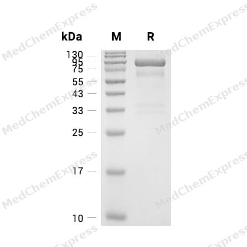
Purity - SDS-PAGE
-
Bioactivity - ELISA
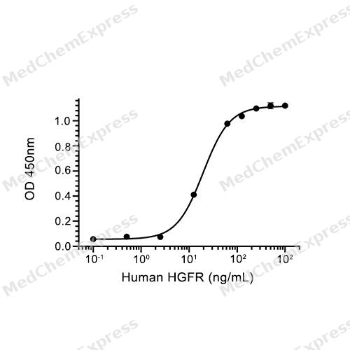
Bioactivity - ELISA
-
Bioactivity - Cell-Based Assay
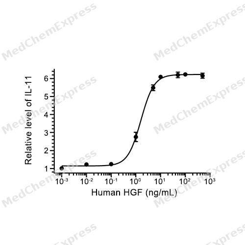
Bioactivity - Cell-Based Assay


Publications (2)
-
Journal Impact Factor
-
Most Recent
-
Adv Sci (Weinh)
ETV4 Promotes Colorectal Cancer Progression by Reprogramming Asparagine Metabolism to Remodel the Stromal Microenvironment. [Abstract]2026 Mar 20:e16557. PMID: 41861091 -
Sci Adv
GPAM mediates mitochondrial dysfunction and the progression of alcoholic liver disease through lipid remodeling. [Abstract]2026 Jun 26;12(26):eaef1896. PMID: 42341117


Technical Parameters
-
Species Human
-
Source HEK293
-
Tag C-His
-
Accession
P14210-1 (Q32-S728)
-
Molecular Construction
-
N-term
-
HGF (Q32-S728)
Accession # P14210 -
His
-
C-term
-
-
Protein Length
Full Length of Isoform-1
-
Synonyms
HGF; Hepatocyte Growth Factor (Hepapoietin A; Scatter Factor); Prev. DFNB39; Lung Fibroblast-Derived Mitogen; HPTA; Hepatopoietin-A; SF; Scatter Factor; F-TCF; Deafness, Autosomal Recessive 39; HGFB; Hepatopoietin A; Hepatocyte Growth Factor; Fibroblast-Derived Tumor Cytotoxic Factor
-
AA Sequence
QRKRRNTIHEFKKSAKTTLIKIDPALKIKTKKVNTADQCANRCTRNKGLPFTCKAFVFDKARKQCLWFPFNSMSSGVKKEFGHEFDLYENKDYIRNCIIGKGRSYKGTVSITKSGIKCQPWSSMIPHEHSFLPSSYRGKDLQENYCRNPRGEEGGPWCFTSNPEVRYEVCDIPQCSEVECMTCNGESYRGLMDHTESGKICQRWDHQTPHRHKFLPERYPDKGFDDNYCRNPDGQPRPWCYTLDPHTRWEYCAIKTCADNTMNDTDVPLETTECIQGQGEGYRGTVNTIWNGIPCQRWDSQYPHEHDMTPENFKCKDLRENYCRNPDGSESPWCFTTDPNIRVGYCSQIPNCDMSHGQDCYRGNGKNYMGNLSQTRSGLTCSMWDKNMEDLHRHIFWEPDASKLNENYCRNPDDDAHGPWCYTGNPLIPWDYCPISRCEGDTTPTIVNLDHPVISCAKTKQLRVVNGIPTRTNIGWMVSLRYRNKHICGGSLIKESWVLTARQCFPSRDLKDYEAWLGIHDVHGRGDEKCKQVLNVSQLVYGPEGSDLVLMKLARPAVLDDFVSTIDLPNYGCTIPEKTSCSVYGWGYTGLINYDGLLRVAHLYIMGNEKCSQHHRGKVTLNESEICAGAEKIGSGPCEGDYGGPLVCEQHKMRMVLGVIVPGRGCAIPNRPGIFVRVAYYAKWIHKIILTYKVPQS
-
Predicted Molecular Mass
80.5 kDa
-
Molecular Weight
Approximately 30-40 kDa (β subunit) & 50-65 kDa (α subunit) & 80-100 kDa (α subunit+β subunit),based on SDS-PAGE under reducing conditions, due to the glycosylation.
-
Glycosylation
Yes
-
Structure/Form
disulfide-linked heterodimer
-
Purity
≥ 95%, as determined by reducing SDS-PAGE.


Product Properties
Lyophilized powder
1.Lyophilized from a 0.22 μm filtered solution of 20 mM Tris-HCl, 500 mM NaCl, pH 8.0.
2.Lyophilized from a 0.22 μm filtered solution of PBS, 200mM L-arginine, pH 7.4.
3.Lyophilized from a 0.22 μm filtered solution of PBS, pH 7.4, 5% trehalose, 5% mannitol, 0.01% Tween 80.
4.Lyophilized from a 0.22 μm filtered solution of PBS, pH 6.5, 8% trehalose.
Please refer to the lot-specific COA for specific buffer information.
Note: For SPR assay, please replace the buffer. Primary amine components (e.g., Tris, imidazole) can affect protein-coupled chips.
<1 EU/μg, determined by LAL method.
It is not recommended to reconstitute to a concentration less than 100 μg/mL in ddH2O.
Stored at -20°C for 2 years from date of receipt. After reconstitution, it is stable at 4°C for 1 week or -20°C for longer (with carrier protein). It is recommended to freeze aliquots at -20°C or -80°C for extended storage.
Room temperature in continental US; may vary elsewhere.


Documentation
-
Data Sheet (268 KB)
-
SDS (254 KB)
- English - EN (254 KB)
- Français - FR (254 KB)
- Deutsch - DE (254 KB)
- Norwegian - NO (254 KB)
- Español - ES (254 KB)
- Swedish - SV (254 KB)
- Italian - IT (254 KB)
- Korean - KR (254 KB)
- Portuguese - PT (254 KB)
-
Handling Instructions (2659 KB)


References
[1]. Cecchi F, et al. Targeting the HGF/Met signalling pathway in cancer. Eur J Cancer. 2010 May;46(7):1260-70. [Content Brief]
[2]. Stella MC, et al. HGF: a multifunctional growth factor controlling cell scattering. Int J Biochem Cell Biol. 1999 Dec;31(12):1357-62. [Content Brief]
[3]. Nakamura T, et al. The discovery of hepatocyte growth factor (HGF) and its significance for cell biology, life sciences and clinical medicine. Proc Jpn Acad Ser B Phys Biol Sci. 2010;86(6):588-610. [Content Brief]
[4]. Strain AJ, et al. Native and recombinant human hepatocyte growth factors are highly potent promoters of DNA synthesis in both human and rat hepatocytes. J Clin Invest. 1991 May;87(5):1853-7. [Content Brief]
[5]. Morishita R, et al. Therapeutic angiogenesis induced by human recombinant hepatocyte growth factor in rabbit hind limb ischemia model as cytokine supplement therapy. Hypertension. 1999 Jun;33(6):1379-84. [Content Brief]

Calculators
Concentration (start) × Volume (start) = Concentration (final) × Volume (final)


 Powered by Bioz

