



MEGF10 Protein, Human (HEK293, His)

Based on 1 Customer Validation
The MEGF10 protein is a membrane receptor that plays a key role in phagocytosis by macrophages and astrocytes, specifically the clearance of apoptotic cells through C1q binding to phosphatidylserine. MEGF10 cooperates with ABCA1 to contribute to the formation of large vacuoles and may facilitate amyloid-beta peptide uptake. MEGF10 Protein, Human (HEK293, His) is the recombinant human-derived MEGF10 protein, expressed by HEK293 , with C-His labeled tag.
- Species: Human
- Source: HEK293
-
Storage:Stored at -20°C for 2 years from date of receipt. After reconstitution, it is stable at 4°C for 1 week or -20°C for longer (with carrier protein). It is recommended to freeze aliquots at -20°C or -80°C for extended storage.


To place orders, for customer services and technical support, please contact: MedChemExpress USA
Tel: 609-228-6898 E-mail: [email protected] [email protected]


-
 Biological Activity
-
Technical Parameters
-
Product Properties
- Documentation
- Help & FAQs




Biological Activity
Description
The MEGF10 protein is a membrane receptor that plays a key role in phagocytosis by macrophages and astrocytes, specifically the clearance of apoptotic cells through C1q binding to phosphatidylserine. MEGF10 cooperates with ABCA1 to contribute to the formation of large vacuoles and may facilitate amyloid-beta peptide uptake. MEGF10 Protein, Human (HEK293, His) is the recombinant human-derived MEGF10 protein, expressed by HEK293 , with C-His labeled tag.
Background
MEGF10 Protein, a membrane receptor, plays a pivotal role in phagocytosis by macrophages and astrocytes, particularly in the clearance of apoptotic cells. As a receptor for C1q, an eat-me signal, MEGF10 binds to phosphatidylserine on the surface of apoptotic cells, facilitating their engulfment. The protein collaborates with ABCA1 during this process and contributes to the formation of large intracellular vacuoles, potentially involved in the uptake of amyloid-beta peptides. In addition to its role in apoptotic cell clearance, MEGF10 is essential for astrocyte-mediated clearance of apoptotic neurons in the developing cerebellum. Furthermore, MEGF10 influences muscle cell proliferation, adhesion, and motility, playing a crucial role in the regulation of myogenesis and the balance between skeletal muscle satellite cell proliferation and differentiation through the notch signaling pathway. Additionally, MEGF10 may contribute to the mosaic spacing of specific neuron subtypes in the retina, ensuring an even distribution across the retina for comprehensive visual field processing. The protein forms homomers and interacts with GULP1, ABCA1, AP2M1, and NOTCH1, highlighting its multifaceted roles in various cellular processes.
Verified Bioactivity
Measured in a cell proliferation assay using C2C12 cells. The ED50 for this effect is 32.22 ng/mL, corresponding to a specific activity is 3.10×10⁴ units/mg.
MCE Validation Data
-
Purity - SDS-PAGE
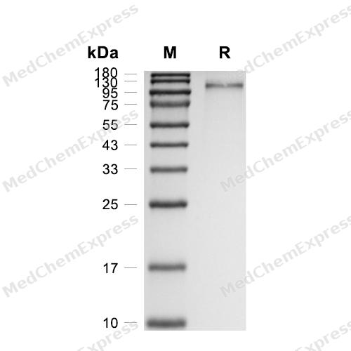
Purity - SDS-PAGE
-
Bioactivity - Cell-Based Assay
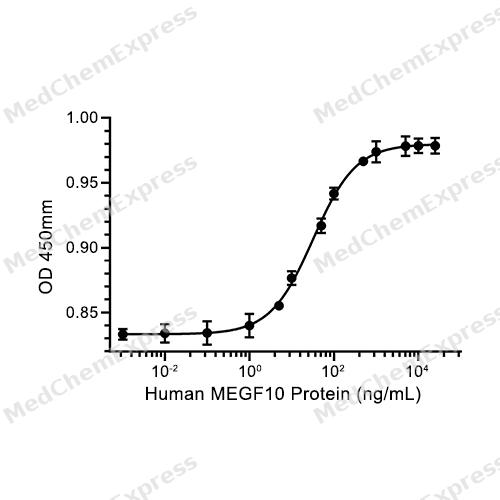
Bioactivity - Cell-Based Assay

Assay Procedure
Materials
MEGF10 Protein, Human (HEK293, His) (HY-P76491)
Cell Line for Activity Assay: C2C12 Cells
Cell medium: DMEM +10% FBS+1% P/S
Penicillin-Streptomycin (100×), Sterile (HY-K1006)
Cell Counting Kit-8 (CCK8 Solution, HY-K0301)
Procedure
1. Using DMEM complete medium, seed cells into a 96-well plate at a density of 1500 cells/well, with a volume of 100 μL per well (Do not add cells to the peripheral wells of the 96-well plate; instead, add an equal volume of PBS solution).
2. After the cells adhere to the plate, discard the medium and subject the cells to starvation treatment overnight using DMEM medium containing 1% FBS.
3. Discard the medium in the wells, add 100 μL of DMEM medium (with 1% FBS) containing MEGF10 protein at different concentrations to the 96-well plate, making the final concentrations of PHPT1 protein in each well 0, 0.001, 0.01, 0.1, 1, 5, 10, 50, 100, 500, 2500, 5000, 10000, 25000 ng/mL.
4. Incubate the cells in a 37°C, 5% CO2 incubator for 48 h.
5. After the incubation period, under dark conditions, add 10 μL of CCK8 solution to each well and incubate in a 37°C, 5% CO2 incubator for 2 h.
6. Measure the OD value at 450 nm using a microplate reader.
7. Use GraphPad Prism software to plot a curve with the OD value as the ordinate and protein concentration as the abscissa, and calculate the ED50 value.


Technical Parameters
-
Species Human
-
Source HEK293
-
Tag C-10*His
-
Accession
Q96KG7-1 (L26-G857)
-
Molecular Construction
-
N-term
-
MEGF10 (L26-G857)
Accession # Q96KG7-1 -
His
-
C-term
-
-
Protein Length
Extracellular Domain
-
Synonyms
MEGF10; Multiple EGF-Like-Domains 10; Multiple EGF Like Domains 10; CMYO10A; KIAA1780; CMYO10B; SR-F3; CMYP10A; Multiple Epidermal Growth Factor-Like Domains Protein 10; CMYP10B; Multiple EGF-Like Domains Protein 10; EMARDD
-
AA Sequence
LNLEDPNVCSHWESYSVTVQESYPHPFDQIYYTSCTDILNWFKCTRHRVSYRTAYRHGEKTMYRRKSQCCPGFYESGEMCVPHCADKCVHGRCIAPNTCQCEPGWGGTNCSSACDGDHWGPHCTSRCQCKNGALCNPITGACHCAAGFRGWRCEDRCEQGTYGNDCHQRCQCQNGATCDHVTGECRCPPGYTGAFCEDLCPPGKHGPQCEQRCPCQNGGVCHHVTGECSCPSGWMGTVCGQPCPEGRFGKNCSQECQCHNGGTCDAATGQCHCSPGYTGERCQDECPVGTYGVLCAETCQCVNGGKCYHVSGACLCEAGFAGERCEARLCPEGLYGIKCDKRCPCHLENTHSCHPMSGECACKPGWSGLYCNETCSPGFYGEACQQICSCQNGADCDSVTGKCTCAPGFKGIDCSTPCPLGTYGINCSSRCGCKNDAVCSPVDGSCTCKAGWHGVDCSIRCPSGTWGFGCNLTCQCLNGGACNTLDGTCTCAPGWRGEKCELPCQDGTYGLNCAERCDCSHADGCHPTTGHCRCLPGWSGVHCDSVCAEGRWGPNCSLPCYCKNGASCSPDDGICECAPGFRGTTCQRICSPGFYGHRCSQTCPQCVHSSGPCHHITGLCDCLPGFTGALCNEVCPSGRFGKNCAGICTCTNNGTCNPIDRSCQCYPGWIGSDCSQPCPPAHWGPNCIHTCNCHNGAFCSAYDGECKCTPGWTGLYCTQRCPLGFYGKDCALICQCQNGADCDHISGQCTCRTGFMGRHCEQKCPSGTYGYGCRQICDCLNNSTCDHITGTCYCSPGWKGARCDQAGVIIVGNLNSLSRTSTALPADSYQIG
-
Molecular Weight
Approximately 95-120 kDa, based on SDS-PAGE under reducing conditions, due to the glycosylation.
-
Glycosylation
Yes
-
Purity
≥ 95%, as determined by reducing SDS-PAGE.


Product Properties
Lyophilized powder.
Lyophilized from a 0.22 μm filtered solution of PBS, pH 7.4.
<1 EU/μg, determined by LAL method.
It is not recommended to reconstitute to a concentration less than 100 μg/mL in ddH2O. For long term storage it is recommended to add a carrier protein (0.1% BSA, 5% HSA, 10% FBS or 5% Trehalose).
Stored at -20°C for 2 years from date of receipt. After reconstitution, it is stable at 4°C for 1 week or -20°C for longer (with carrier protein). It is recommended to freeze aliquots at -20°C or -80°C for extended storage.
Room temperature in continental US; may vary elsewhere.


Documentation
-
Data Sheet (241 KB)
-
SDS (251 KB)
- English - EN (251 KB)
- Français - FR (251 KB)
- Deutsch - DE (251 KB)
- Norwegian - NO (251 KB)
- Español - ES (251 KB)
- Swedish - SV (251 KB)
- Italian - IT (251 KB)
- Korean - KR (251 KB)
- Portuguese - PT (251 KB)
-
Handling Instructions (2659 KB)

Calculators
Concentration (start) × Volume (start) = Concentration (final) × Volume (final)


 Powered by Bioz

