



PDGF-AA Protein, Human
Based on 2 publication(s) in Google Scholar
PDGF-A protein is a key growth factor that regulates embryonic development, cell proliferation, migration, survival, and chemotaxis, and exerts potent mitogenic effects on mesenchymal cells. It is essential for alveolar septal formation, gastrointestinal tract development, interstitial cell maturation, spermatogenesis, and oligodendrocyte development, and contributes to spinal cord and cerebellar myelination. PDGF-AA Protein, Human is the recombinant human-derived PDGF-AA protein, expressed by E. coli , with tag free.
- Species: Human
- Source: E. coli
-
Storage:Stored at -20°C for 2 years from date of receipt. After reconstitution, it is stable at 4°C for 1 week or -20°C for longer (with carrier protein). It is recommended to freeze aliquots at -20°C or -80°C for extended storage.


To place orders, for customer services and technical support, please contact: MedChemExpress USA
Tel: 609-228-6898 E-mail: [email protected] [email protected]


-
 Biological Activity
-
Technical Parameters
-
Product Properties
- Documentation
- Help & FAQs


-

HY-P7055
PDGF-BB Protein, Human -
HY-P7109
EGF Protein, Human -
HY-P7331
FGF basic/bFGF Protein, Human (154a.a) -
HY-P7004
FGF basic/bFGF Protein, Human (146a.a) -
HY-A0070A
Liothyronine -

HY-15371
Forskolin -
HY-P7018
IGF-I/IGF-1 Protein, Human (70a.a) -
HY-P70456
Neurotrophin-3 Protein, Human -
HY-P7121
HGF Protein, Human (CHO) -

HY-P0035
Insulin (human) -
HY-B0511
Biotin -
HY-12848
SAG


Biological Activity
Description
PDGF-A protein is a key growth factor that regulates embryonic development, cell proliferation, migration, survival, and chemotaxis, and exerts potent mitogenic effects on mesenchymal cells. It is essential for alveolar septal formation, gastrointestinal tract development, interstitial cell maturation, spermatogenesis, and oligodendrocyte development, and contributes to spinal cord and cerebellar myelination. PDGF-AA Protein, Human is the recombinant human-derived PDGF-AA protein, expressed by E. coli , with tag free.
Background
PDGF-A Protein takes center stage as a pivotal growth factor regulating embryonic development, cell proliferation, migration, survival, and chemotaxis. Acknowledged for its potent mitogenic effects on mesenchymal cells, PDGF-A is indispensable for various developmental processes, including the formation of lung alveolar septa, gastrointestinal tract development, Leydig cell maturation, spermatogenesis, and the normal development of oligodendrocytes, contributing to myelination in the spinal cord and cerebellum. Furthermore, PDGF-A assumes a crucial role in wound healing dynamics. The intricacies of its signaling pathway involve modulation through the formation of heterodimers with PDGFB, demonstrating its regulatory versatility. Structurally, PDGF-A adopts a homodimeric configuration with an antiparallel disulfide-linked dimer, and it forms heterodimers with PDGFB, engaging in interactions with PDGFRA homodimers and heterodimers formed by PDGFRA and PDGFRB. Additionally, PDGF-A exhibits an interaction with CSPG4, adding another layer to its multifaceted involvement in cellular processes.
Verified Bioactivity
Measured in a cell proliferation assay using Balb/c 3T3 mouse fibroblast cells. The ED50 for this effect is <20 ng/mL.
MCE Validation Data
-
Purity - SDS-PAGE
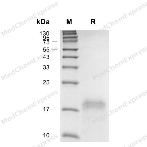
Purity - SDS-PAGE
-
Bioactivity - Cell-Based Assay
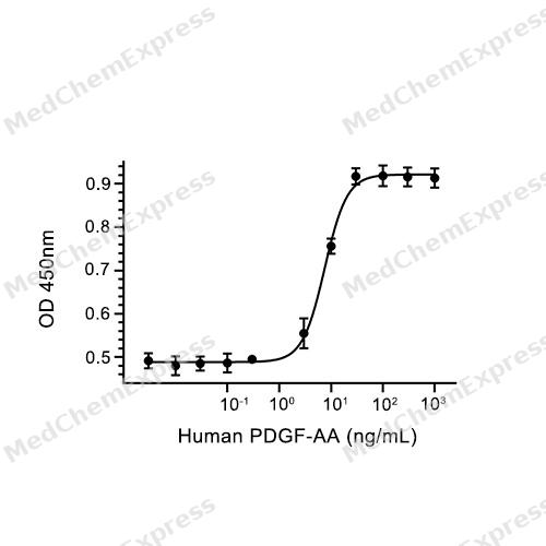
Bioactivity - Cell-Based Assay


Publications (2)
-
Journal Impact Factor
-
Most Recent
-
Int J Dent
The Effect of Differentiation of Human Dental Pulp Stem Cells to Glial Cells on the Sensory Nerves of the Dental Pulp. [Abstract]2024 Apr 5:2024:3746794. PMID: 38715867 -
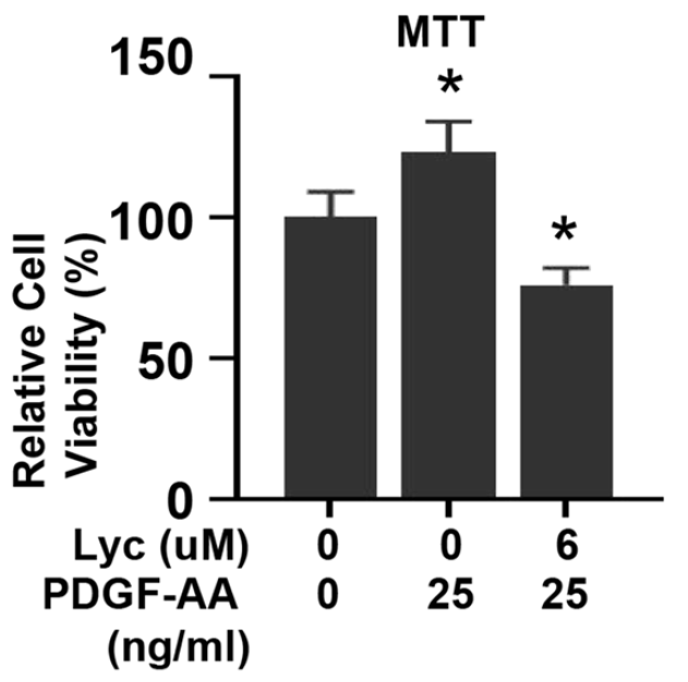
PDGF-AA Protein, Human purchased from MedChemExpress. Usage Cited in: Research Square Preprint. 2022 Feb.
Pretreatment with PDGF-AA Protein, Human (25 ng/ml) stimulated growth of HUVECs.
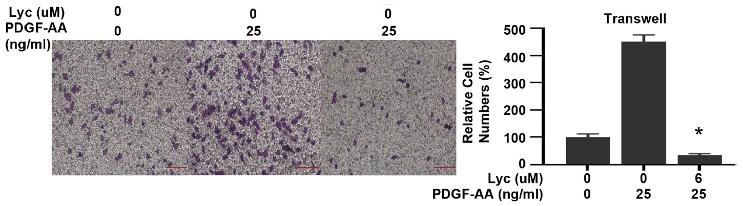
PDGF-AA Protein, Human purchased from MedChemExpress. Usage Cited in: Research Square Preprint. 2022 Feb.
PDGF-AA Protein, Human (25 ng/ml) stimulation increased HUVEC migration to the lower chamber.
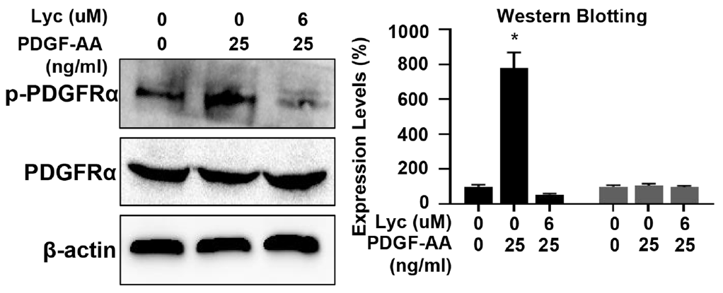
PDGF-AA Protein, Human purchased from MedChemExpress. Usage Cited in: Research Square Preprint. 2022 Feb.
PDGF-AA Protein, Human (25 ng/ml) induced activation of PDGFRα in HUVECs.
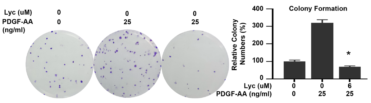
PDGF-AA Protein, Human purchased from MedChemExpress. Usage Cited in: Research Square Preprint. 2022 Feb.
PDGF-AA Protein, Human (25 ng/ml) induced colony formation of HUVECs.
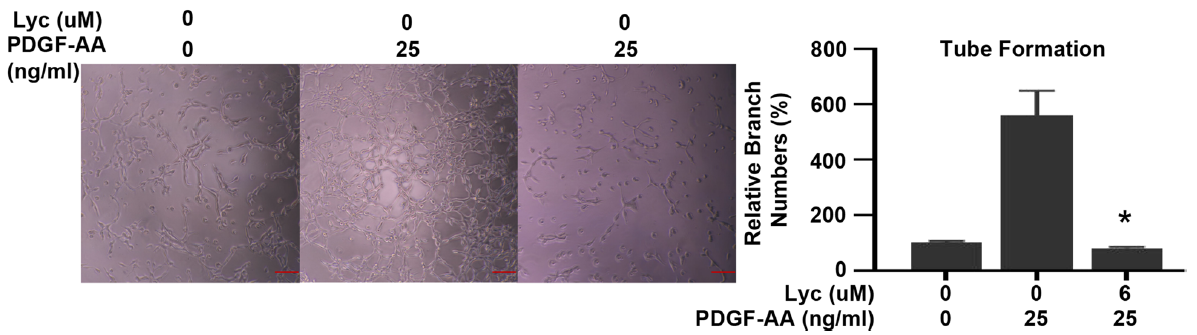
PDGF-AA Protein, Human purchased from MedChemExpress. Usage Cited in: Research Square Preprint. 2022 Feb.
PDGF-AA Protein, Human (25 ng/ml) increased tube formation in HUVECs.


Technical Parameters
-
Species Human
-
Source E. coli
-
Tag Tag Free
-
Accession
P04085-1 (S87-T211)
-
Molecular Construction
-
N-term
-
PDGF-AA (S87-T211)
Accession # P04085-1 -
C-term
-
-
Protein Length
Full Length of Isoform-1 Mature Protein
-
Synonyms
PDGFA; Platelet-Derived Growth Factor Alpha Chain; Platelet Derived Growth Factor Subunit A; Platelet-Derived Growth Factor A Chain; Platelet-Derived Growth Factor Alpha Polypeptide; PDGF Subunit A; PDGF1; PDGF A-Chain; Platelet-Derived Growth Factor Subu
-
AA Sequence
SIEEAVPAVCKTRTVIYEIPRSQVDPTSANFLIWPPCVEVKRCTGCCNTSSVKCQPSRVHHRSVKVAKVEYVRKKPKLKEVQVRLEEHLECACATTSLNPDYREEDTGRPRESGKKRKRKRLKPT
-
Predicted Molecular Mass
14.3 kDa
-
Molecular Weight
Approximately 16 kDa under reduced (R) conditions, 27-33 kDa under non reducing (N) conditions, based on SDS-PAGE, due to the glycosylation.
-
Purity
≥ 95%, as determined by reducing SDS-PAGE.


Product Properties
Lyophilized powder
1.Lyophilized from a 0.22 μm filtered solution of 20 mM Glycine-HCl, 6% sucrose, 4% mannitol, 0.02% Tween 80, pH 3.0.
2.Lyophilized from a 0.22 μm filtered solution of 50 mM Tris-HCl, 300 mM NaCl, 200 mM arginine, pH 8.0.
3.Lyophilized from a 0.22 μm filtered solution of 0.1% TFA, 20% Acetonitrile, pH 2.5.
Please refer to the lot-specific COA for specific buffer information.
Note: For SPR assay, please replace the buffer. Primary amine components (e.g., Tris, imidazole) can affect protein-coupled chips.
<1 EU/μg, determined by LAL method.
It is not recommended to reconstitute to a concentration less than 100 μg/mL in ddH2O. For long term storage it is recommended to add a carrier protein (0.1% BSA, 5% HSA, 10% FBS or 5% Trehalose).
Stored at -20°C for 2 years from date of receipt. After reconstitution, it is stable at 4°C for 1 week or -20°C for longer (with carrier protein). It is recommended to freeze aliquots at -20°C or -80°C for extended storage.
Room temperature in continental US; may vary elsewhere.


Documentation
-
Data Sheet (236 KB)
-
SDS (252 KB)
- English - EN (252 KB)
- Français - FR (252 KB)
- Deutsch - DE (252 KB)
- Norwegian - NO (252 KB)
- Español - ES (252 KB)
- Swedish - SV (252 KB)
- Italian - IT (252 KB)
- Korean - KR (252 KB)
- Portuguese - PT (252 KB)
-
Handling Instructions (2659 KB)

Calculators
Concentration (start) × Volume (start) = Concentration (final) × Volume (final)


 Powered by Bioz

