



Resolvin D1
Based on 1 publication(s) in Google Scholar
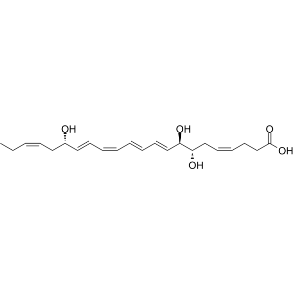
Resolvin D1 (RvD1), an endogenous pro-resolving mediator of inflammation, is derived from omega-3 docosahexaenoic acid during the resolution phase of acute inflammation. Resolvin D1 blocks proinflammatory neutrophil migration by regulating actin polymerization, reduces TNF-α–mediated inflammation in macrophages, and enhances phagocytosis of apoptotic cells by macrophages.
For research use only. We do not sell to patients.
- Purity : 99.6%
- CAS No.: 872993-05-0
- Formula: C22H32O5
- Molecular Weight:376.49
-
Storage:
Solution, -20°C, 2 years

To place orders, for customer services and technical support, please contact: MedChemExpress USA
Tel: 609-228-6898 E-mail: [email protected] [email protected]


-
 Biological Activity
-
Chemical Information
- Protocols
- Purity & Documentation
- References
-
 Help & FAQs


Publications Citing Use of MedChemExpress (MCE) Resolvin D1
MoreAll Endogenous Metabolite Isoforms
More

Biological Activity
Description
IC50 & Target
[1]|
Human Endogenous Metabolite |
In Vitro
Resolvin D1 (RvD1) (1-100 nM; 15 minutes) induces dose-dependent short-term functional changes in primary human macrophages. RvD1 triggers intracellular Ca2+ release, blocks chemotactic migration, and stimulates phagocytosis of microbial particles with maximal efficiency at 10 nM[1].? Resolvin D1 (0-500?nM; 72 hours) shows a strong inhibition of LPS-induced TRAP and cathepsin K expression by RvD1 at different concentrations in RAW264.7 macrophages[2].
MedChemExpress (MCE) has not independently confirmed the accuracy of these methods. They are for reference only. Further protocols information, click here.
In Vivo
MedChemExpress (MCE) has not independently confirmed the accuracy of these methods. They are for reference only.
-
Animal Model:18-20 g thirty 8-week-old female DBA/1J mice (collagen antibody-induced arthritis (CAIA))[3]
-
Dosage:100, 500, and 1000 ng (or 1000 ng, daily on days 4-10)
-
Administration:i.p. injection; daily on days 10
-
Result:Exhibit a reduced arthritic score.


Chemical Information
-
CAS No. 872993-05-0
-
Appearance Liquid
-
Molecular Weight 376.49
-
Formula C22H32O5
-
Color Colorless to light yellow
-
SMILES
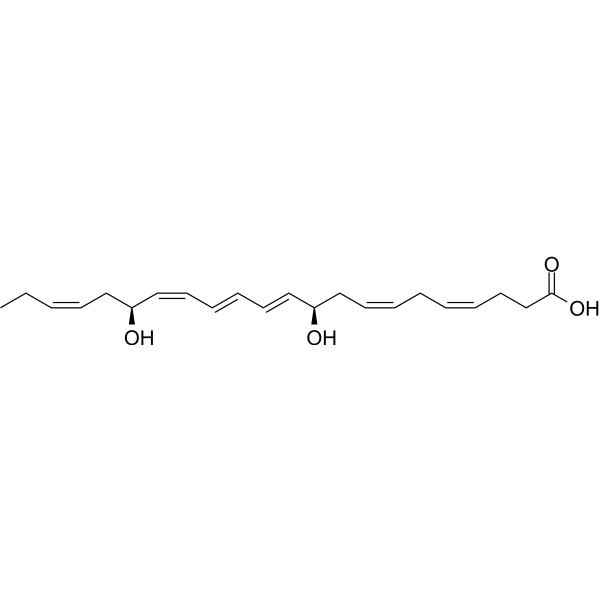
CC/C=C\C[C@@H](/C=C/C=C\C=C\C=C\[C@H]([C@H](C/C=C\CCC(O)=O)O)O)O
-
Synonyms
RvD1
-
Structure Classification
-
Initial Source
-
Shipping
Room temperature in continental US; may vary elsewhere.
-
Storage
Solution, -20°C, 2 years


Publications (1)
-
Journal Impact Factor
-
Most Recent

Protocols
-
Phagocytosis Functional Assay
A phagocytosis functional assay measures the ability of phagocytic cells, such as neutrophils, macrophages, monocytes, or microglia/macrophages, to bind and internalize particulate targets including bacteria, yeast particles, beads, or myelin particles. Fluorescent flow-cytometry assays detect target uptake as fluorescence associated with gated phagocytes, while pH-sensitive dyes such as pHrodo increase signal in acidic phagosomal compartments and therefore preferentially report internalized particles rather than particles remaining outside the cell. Microscopy or high-content imaging can be used to confirm intracellular localization and, in some protocols, to follow uptake kinetics.
-
Apoptosis
Apoptosis, also called programmed cell death, is generally characterized by distinct morphological characteristics.
-
TUNEL staining for apoptotic DNA fragmentation
TUNEL staining detects DNA strand breaks by using terminal deoxynucleotidyl transferase to add labeled nucleotides to exposed 3′-OH DNA termini, generating either microscopic staining in fixed cells or tissue sections, or fluorescence/cytometric signal in cell suspensions. TUNEL positivity reflects DNA fragmentation but should not be interpreted alone as definitive apoptosis, because TUNEL can also label necrotic, autolytic, mechanically damaged, or DNA-repair-associated DNA breaks.
-
Annexin V plus membrane-impermeant dye apoptosis staining
Annexin V-based apoptosis assays rely on the detection of phosphatidylserine (PS) externalization from the inner leaflet of the plasma membrane to the outer leaflet, an early biochemical hallmark of apoptosis. Fluorescently labeled Annexin V binds PS in a calcium-dependent manner, enabling identification of early apoptotic cells by flow cytometry or fluorescence microscopy. When combined with a membrane-impermeant DNA-binding dye (e. g. , propidium iodide), this approach allows discrimination between viable (Annexin V−/dye−), early apoptotic (Annexin V+/dye−), and late apoptotic or necrotic (Annexin V+/dye+) cell populations by assessing membrane integrity and PS exposure.
-
Cell migration
Cell migration is a method that plays an important role in wound healing, cell differentiation, embryonic development, etc.
-
Carrageenan-Induced Paw Edema
Carrageenan-induced paw edema is an acute inflammation model in which intraplantar injection of carrageenan induces localized inflammatory swelling characterized by vascular permeability, leukocyte infiltration, and production of inflammatory mediators such as prostaglandins and cytokines, making it widely used to evaluate anti-inflammatory agents in vivo. The resulting paw volume or thickness increase is quantified over time as a direct readout of inflammatory intensity and drug efficacy, typically reflecting cyclooxygenase-mediated prostaglandin-driven edema formation and immune cell recruitment in peripheral tissue[20].
-
Zymosan-Induced Peritonitis
Zymosan-induced peritonitis is a sterile acute-inflammation model produced by intraperitoneal injection of zymosan, a yeast cell-wall particle preparation, followed by quantification of leukocyte recruitment and soluble inflammatory mediators in peritoneal lavage fluid. Low-dose zymosan peritonitis is commonly used as a self-resolving acute inflammation model in which neutrophil recruitment occurs early and monocyte/macrophage accumulation follows later. The assay readouts include total peritoneal leukocyte number, differential neutrophil and monocyte/macrophage counts, peritoneal cytokines and chemokines, plasma or peritoneal exudation, and optional lipidomic or metabolomic changes during inflammation and resolution. Early neutrophil recruitment after zymosan depends strongly on complement and mast-cell C5a receptor signaling, whereas later monocyte recruitment is linked to MCP-1/CCL2 production.
-
Apoptosis Solutions
Apoptosis is a regulated, generally non-lytic cell-death pathway that removes unwanted, damaged, infected, or abnormal cells through coordinated morphological changes, caspase activation, DNA fragmentation, and membrane remodeling. The intrinsic apoptosis pathway is controlled mainly by mitochondrial outer membrane permeabilization, BCL-2 family proteins, cytochrome c release, apoptosome formation, caspase-9 activation, and downstream executioner caspase-3/7 activation. The extrinsic apoptosis pathway is initiated by death receptors such as Fas, TNFR, and TRAIL receptors, which recruit adaptor proteins and activate caspase-8 before engaging executioner caspases or mitochondrial amplification through BID cleavage. Apoptosis is linked to many phenotypes, including cancer cell killing, tissue homeostasis, immune regulation, neurodegeneration, infection response, and treatment-induced cytotoxicity; unresolved questions include how apoptosis interacts with necroptosis, pyroptosis, ferroptos
-
Research Protocol for Inflammation-related Diseases
The NLRP3 inflammasome is a cytosolic innate immune signaling platform that integrates priming signals and danger-signal activation to promote caspase-1 activation, maturation of IL-1β and IL-18, and gasdermin D-mediated pyroptotic cell death. The core experimental logic is to determine whether inflammatory disease phenotypes are driven by increased NLRP3 expression, ASC-containing inflammasome assembly, caspase-1 cleavage, GSDMD cleavage, and extracellular release of IL-1β/IL-18 rather than by nonspecific cell injury alone. The pathway is strongly linked to inflammation-related disease phenotypes because monosodium urate crystals activate NALP3/NLRP3 inflammasome signaling in gout-like crystal inflammation, cholesterol crystals activate NLRP3 inflammasomes in atherogenesis models, and DSS-induced intestinal inflammation has been reported to involve NLRP3 inflammasome activity. However, experimental colitis studies also show context-dependent protective effects of NLRP3 inflammasome co
-
Phalloidin F-actin cytoskeleton staining
Phalloidin F-actin staining detects polymerized filamentous actin in fixed and permeabilized specimens by using fluorescent phalloidin or phalloidin-derived phallotoxins that bind actin filaments and generate a fluorescence microscopy readout corresponding to F-actin organization, including stress fibers, cortical actin, filament bundles, and tissue-specific actin networks. Phalloidin stabilizes F-actin by reducing actin subunit dissociation from filament ends, and fluorescent phallotoxins were established as tools for visualizing actin-containing structures in eukaryotic cells.


Purity & Documentation
-
Data Sheet (268 KB)
-
SDS (393 KB)
- English - EN (393 KB)
- Français - FR (393 KB)
- Deutsch - DE (393 KB)
- Norwegian - NO (393 KB)
- Español - ES (393 KB)
- Swedish - SV (393 KB)
- Italian - IT (393 KB)
- Korean - KR (393 KB)
- Portuguese - PT (393 KB)
-
Handling Instructions (2659 KB)
References
[1]. Schmid M, et al. Resolvin D1 Polarizes Primary Human Macrophages toward a Proresolution Phenotype through GPR32. J Immunol. 2016 Apr 15;196(8):3429-37. [Content Brief]
[2]. Gao Y, et al. Resolvin D1 Improves the Resolution of Inflammation via Activating NF-κB p50/p50-Mediated Cyclooxygenase-2 Expression in Acute Respiratory Distress Syndrome. J Immunol. 2017 Aug 9. pii: ji1700315. [Content Brief]
[3]. Benabdoun HA, et al. In vitro and in vivo assessment of the proresolutive and antiresorptive actions of resolvin D1: relevance to arthritis. Arthritis Res Ther. 2019 Mar 12;21(1):72. [Content Brief]

Calculators
Concentration (start) × Volume (start) = Concentration (final) × Volume (final)


Resolvin D1 Related Classifications
HY-125527 Related Classifications
 Powered by Bioz