



Acid Red 94 sodium
Based on 1 publication(s) in Google Scholar
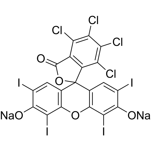
Acid Red 94 sodium, a synthetic fluorescein derivative, is a deep red dye primarily composed of 4,5,6,7-tetrachloro-2,4,5,7-tetraiodo fluorescein. It is widely used as an ophthalmic diagnostic agent to detect dry or damaged cells on the ocular surface. Acid Red 94 sodium exhibits antitumor activity and can inhibit Friend Leukemia Virus (FLV) infection through photodynamic action. Additionally, Acid Red 94 sodium can inhibit Aβ aggregation through light irradiation. Acid Red 94 sodium holds potential for use in cancer, viral infections, and neurodegenerative disease research.
For research use only. We do not sell to patients.
- Purity : 99.49%
- CAS No.: 632-69-9
- Formula: C20H2Cl4I4Na2O5
- Molecular Weight:1017.64
-
Storage:
RT, sealed storage, away from moisture and light.
In solvent -80°C, 1 year , -20°C, 6 months

To place orders, for customer services and technical support, please contact: MedChemExpress USA
Tel: 609-228-6898 E-mail: [email protected] [email protected]


-
 Biological Activity
-
Chemical Information
-
Solvent & Solubility
- Protocols
- Purity & Documentation
- References
-
 Help & FAQs


-

HY-D0214R
Acid Red 94 sodium (Standard)Biological Dye -
HY-D0505
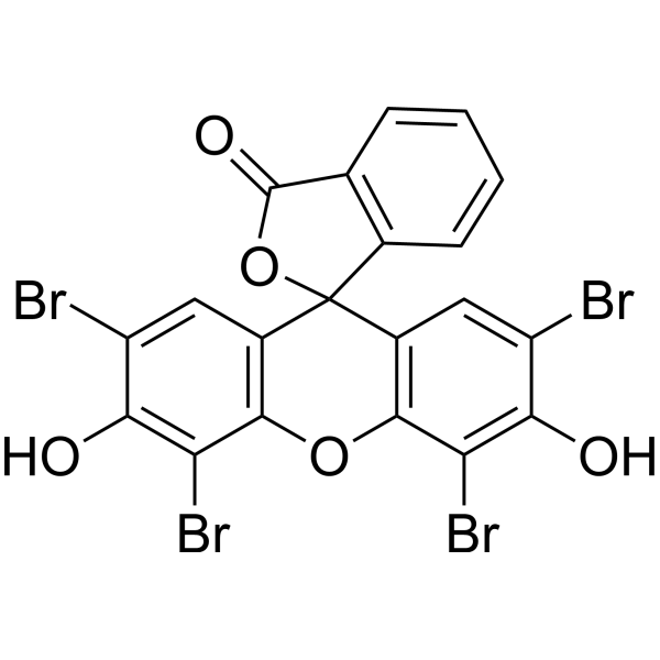
Eosin YDye Molecule -
HY-13594
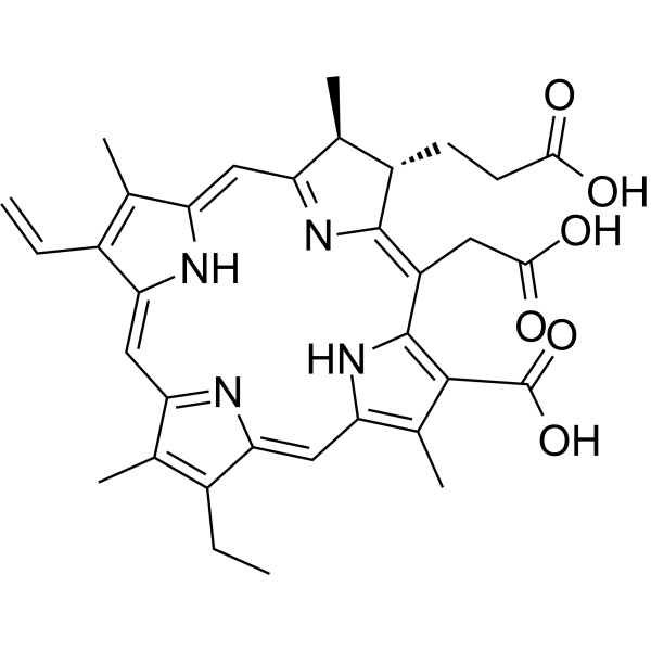
Chlorin e6Photosensitizer -
HY-D0309
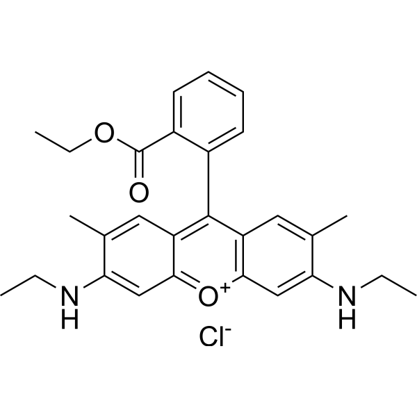
Rhodamine 6GLaser Dye -
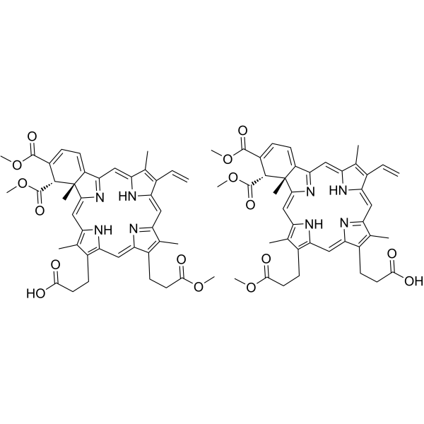
HY-B0146
VerteporfinYAP Inhibitor -
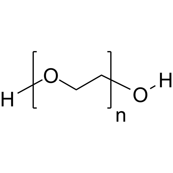
HY-Y0873
PEG300Neutral Polymer -
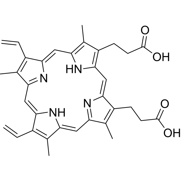
HY-B1247
Protoporphyrin IXEndogenous Metabolite,Cancer Inhibitor -
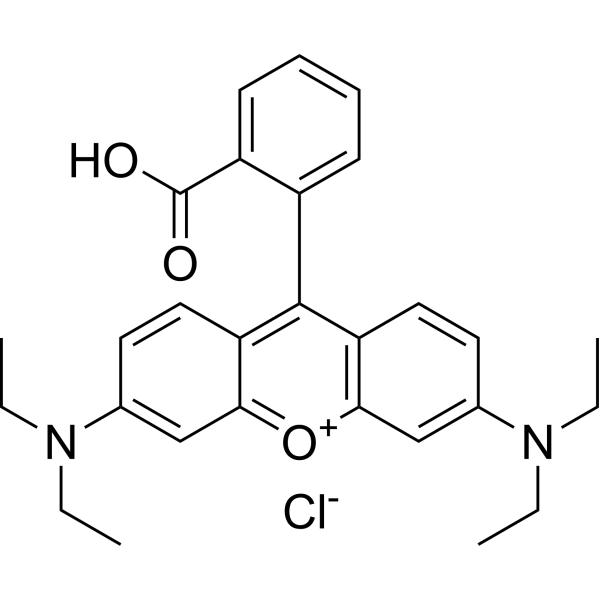
HY-Y0016
Rhodamine BFluorescent Dye -
HY-D0259
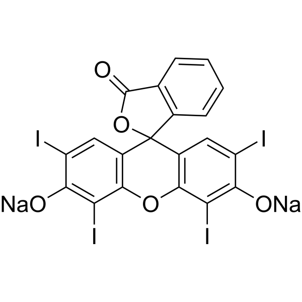
Erythrosine BFluorescent Dyes -

HY-D0214E
Acid Red 94 sodium (80%)Photosensitizer -
HY-101878
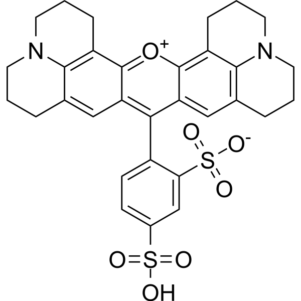
Texas RedRed Fluorescent Dye -
HY-B1102
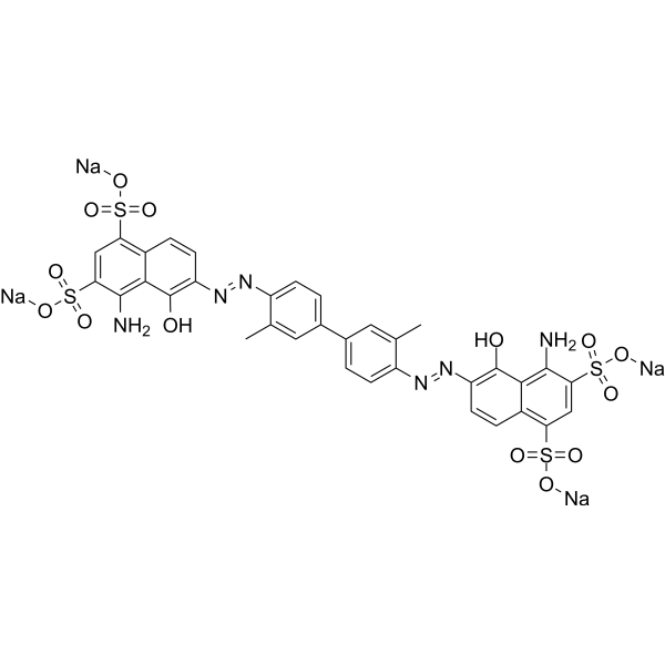
Evans BlueL-glutamate Uptake Inhibitor
Publications Citing Use of MedChemExpress (MCE) Acid Red 94 sodium
More

Biological Activity
Description
Cellular Effect
|
Cell Line

|
Type | Value | Description | References |
|---|---|---|---|---|
| B16-F10 | IC50 |
>5 μM
Compound: 1; RBNa
|
Photodynamic anticancer activity in mouse B16F10 cells preincubated for 2 hrs followed by light exposure at 27 J/cm2 for 30 mins and subsequent incubation for 24 hrs by MTT assay
Photodynamic anticancer activity in mouse B16F10 cells preincubated for 2 hrs followed by light exposure at 27 J/cm2 for 30 mins and subsequent incubation for 24 hrs by MTT assay
|
[PMID: 29324346] |
| HepG2 | IC50 |
>5 μM
Compound: 1; RBNa
|
Photodynamic anticancer activity in human HepG2 cells preincubated for 2 hrs followed by light exposure at 27 J/cm2 for 30 mins and subsequent incubation for 24 hrs by MTT assay
Photodynamic anticancer activity in human HepG2 cells preincubated for 2 hrs followed by light exposure at 27 J/cm2 for 30 mins and subsequent incubation for 24 hrs by MTT assay
|
[PMID: 29324346] |
| MCF7 | IC50 |
>5 μM
Compound: 1; RBNa
|
Photodynamic anticancer activity in human MCF7 cells preincubated for 2 hrs followed by light exposure at 27 J/cm2 for 30 mins and subsequent incubation for 24 hrs by MTT assay
Photodynamic anticancer activity in human MCF7 cells preincubated for 2 hrs followed by light exposure at 27 J/cm2 for 30 mins and subsequent incubation for 24 hrs by MTT assay
|
[PMID: 29324346] |
In Vitro
Acid Red 94 (300 µM, 48 h) exerts toxic effects on MCF-7 cells by inducing apoptosis[3]. Acid Red 94 (5 µM, under light irradiation, 24 h) inhibits Aβ aggregation, likely by binding to Ab42 and preventing its conformational transition[4].
MedChemExpress (MCE) has not independently confirmed the accuracy of these methods. They are for reference only. Further protocols information, click here.
-
Cell Line:MCF-7 (Breast cancer)
-
Concentration:100 µM, 200 µM, 300 µM
-
Incubation Time:24 h, 48 h, 72 h
-
Result:Decreased MCF-7 cell viability in a concentration- and time-dependent manner.
-
Cell Line:MCF-7 (Breast cancer)
-
Concentration:300 µM
-
Incubation Time:48 h
-
Result:Significantly increased Bax protein expression.
In Vivo
MedChemExpress (MCE) has not independently confirmed the accuracy of these methods. They are for reference only.
-
Animal Model:BALB/c mice bearing MT-901 breast cancer model[5]
-
Dosage:50 µL (10% Rose Bengal solution, PV-10, prepared by dissolving 10% Rose Bengal disodium salt in 0.9% NaCl (saline solution))
-
Administration:Intralesional injection (IL) injected into the tumor lesion, once a week, total duration 14 days
-
Result:Significantly reduced the volume of both the injected tumors and the lung metastases.
-
Animal Model:Friend Leukemia Virus (FLV) infection in BALB/c mice model[6]
-
Dosage:7 µg, 22 µg, 67 µg, 200 µg
-
Administration:Intraperitoneal injection (i.p.), mixed with FLV inoculum, illuminated for 1 hour (photodynamic action)
-
Result:Significantly inhibited FLV infection upon illumination at low doses.


Chemical Information
-
CAS No. 632-69-9
-
Appearance Solid
-
Molecular Weight 1017.64
-
Formula C20H2Cl4I4Na2O5
-
Color Brown to reddish brown
-
SMILES
O=C1OC2(C3=C(OC4=C2C=C(I)C(O[Na])=C4I)C(I)=C(O[Na])C(I)=C3)C5=C1C(Cl)=C(Cl)C(Cl)=C5Cl
-
Shipping
Room temperature in continental US; may vary elsewhere.
-
Storage
RT, sealed storage, away from moisture and light
In solvent -80°C 1 year -20°C 6 months


Publications (1)
-
Journal Impact Factor
-
Most Recent


Solvent & Solubility
In Vitro:
H2O : 20 mg/mL (19.65 mM; ultrasonic and warming and heat to 60°C)
Please refer to the solubility information to select the appropriate solvent. Once prepared, please aliquot and store the solution to prevent product inactivation from repeated freeze-thaw cycles.
Storage method and period of stock solution: -80°C, 1 year; -20°C, 6 months. When stored at -80°C, please use it within 1 year. When stored at -20°C, please use it within 6 months.
* Note: If you choose water as the stock solution, please dilute it to the working solution, then filter and sterilize it with a 0.22 μm filter before use.
Please refer to the solubility information to select the appropriate solvent. Once prepared, please aliquot and store the solution to prevent product inactivation from repeated freeze-thaw cycles.
Storage method and period of stock solution: -80°C, 1 year; -20°C, 6 months. When stored at -80°C, please use it within 1 year. When stored at -20°C, please use it within 6 months.
* Note: If you choose water as the stock solution, please dilute it to the working solution, then filter and sterilize it with a 0.22 μm filter before use.
Concentration (start) × Volume (start) = Concentration (final) × Volume (final)

Protocols
-
Research Protocol for Infectious Diseases
Infectious-disease experiments test how pathogens interact with host barriers, innate immune receptors, inflammatory signaling, pathogen replication, and tissue injury; pattern-recognition receptors such as TLRs, RIG-I-like receptors, NOD-like receptors, and inflammasomes detect microbial molecules and activate NF-κB, interferon, and cytokine responses. The central hypothesis is that infection severity reflects the balance between pathogen burden and host response: protective inflammation restricts pathogen growth, whereas excessive or mislocalized inflammation contributes to tissue damage and disease phenotype. Unresolved questions include which host pathways are protective versus pathogenic, why some infection models fail to translate to human disease, and which combined readouts best predict clinically relevant infection outcomes.
-
Subcutaneous Cell-Line-Derived Xenograft
Subcutaneous cell-line-derived xenograft (CDX) models are established by implanting cultured human cancer cell lines into immunodeficient mice, where the injected cells form localized tumors that can be monitored in vivo as a measure of tumorigenic potential, growth kinetics, and treatment response. These models are widely used in oncology research because they allow reproducible tumor formation and enable comparative assessment of tumor growth between different cell lines or genetic manipulations in a controlled in vivo microenvironment. Subcutaneous implantation of cancer cells in immunodeficient mice is a standard approach for evaluating tumor growth behavior and therapeutic response across multiple cancer types, including prostate, esophageal, pancreatic, and colon cancer models.
-
Research Protocol for Neurological Diseases
PINK1/Parkin-mediated mitophagy pathway is a mitochondrial quality-control signaling axis in which mitochondrial depolarization stabilizes PINK1 on damaged mitochondria, activates Parkin recruitment and E3 ubiquitin ligase activity, promotes ubiquitination of outer mitochondrial membrane proteins, recruits selective autophagy adaptors, and drives lysosomal degradation of damaged mitochondria. In neurological disease research, this pathway is experimentally important because neurons, especially dopaminergic neurons, are highly dependent on mitochondrial integrity, and defective mitochondrial turnover can lead to mitochondrial dysfunction, oxidative stress, impaired neuronal survival, α-synuclein accumulation, and neuroinflammatory damage-associated signals. The genetic disease link is strongest in Parkinson’s disease because mutations in PRKN/parkin cause autosomal recessive juvenile parkinsonism, mutations in PINK1 cause hereditary early-onset Parkinson’s disease, and Drosophila studie
-
Alzheimer’s Disease Modeling
Alzheimer’s Disease (AD) is a neurodegenerative disorder characterized by a progressive decline in cognitive functions and loss of specific types of neurons and synapses. Alzheimer's symptoms can be simulated in mice by injecting drugs (such as Aβ) or genetically modified.


Purity & Documentation
-
Data Sheet (287 KB)
-
SDS (396 KB)
- English - EN (396 KB)
- Français - FR (396 KB)
- Deutsch - DE (396 KB)
- Norwegian - NO (396 KB)
- Español - ES (396 KB)
- Swedish - SV (396 KB)
- Italian - IT (396 KB)
- Korean - KR (396 KB)
- Portuguese - PT (396 KB)
-
Handling Instructions (2659 KB)
References
[1]. Doughty MJ, et, al. Fluorescence characteristics of sodium fluorescein-rose bengal ophthalmic solution mixtures. Cont Lens Anterior Eye. 2014 Oct; 37(5): 358-62. [Content Brief]
[2]. Roat MI, et, al. The antiviral effects of rose bengal and fluorescein. Arch Ophthalmol. 1987 Oct; 105(10): 1415-7. [Content Brief]
[3]. Mousavi SH, et al. Direct toxicity of Rose Bengal in MCF-7 cell line: role of apoptosis. Food Chem Toxicol. 2009 Apr;47(4):855-9. [Content Brief]
[4]. Lee JS, et al. Photo-induced inhibition of Alzheimer's β-amyloid aggregation in vitro by rose bengal. Biomaterials. 2015 Jan;38:43-9. [Content Brief]
[5]. Toomey P, et al. Intralesional injection of rose bengal induces a systemic tumor-specific immune response in murine models of melanoma and breast cancer. PLoS One. 2013 Jul 17;8(7):e68561. [Content Brief]
[6]. Stevenson NR, Lenard J. Antiretroviral activities of hypericin and rose bengal: photodynamic effects on Friend leukemia virus infection of mice. Antiviral Res. 1993 Jun;21(2):119-27. [Content Brief]


Complete Stock Solution Preparation Table
Please refer to the solubility information to select the appropriate solvent. Once prepared, please aliquot and store the solution to prevent product inactivation from repeated freeze-thaw cycles.
Storage method and period of stock solution: -80°C, 1 year; -20°C, 6 months. When stored at -80°C, please use it within 1 year. When stored at -20°C, please use it within 6 months.
| Optional Solvent | Concentration Solvent Mass | 1 mg | 5 mg | 10 mg | 25 mg |
|---|---|---|---|---|---|
| H2O | 1 mM | 0.9827 mL | 4.9133 mL | 9.8267 mL | 24.5666 mL |
| 5 mM | 0.1965 mL | 0.9827 mL | 1.9653 mL | 4.9133 mL | |
| 10 mM | 0.0983 mL | 0.4913 mL | 0.9827 mL | 2.4567 mL | |
| 15 mM | 0.0655 mL | 0.3276 mL | 0.6551 mL | 1.6378 mL |
* Note: If you choose water as the stock solution, please dilute it to the working solution, then filter and sterilize it with a 0.22 μm filter before use.


Acid Red 94 sodium Related Classifications
HY-D0214 Related Classifications
 Powered by Bioz