



Salicylcurcumin
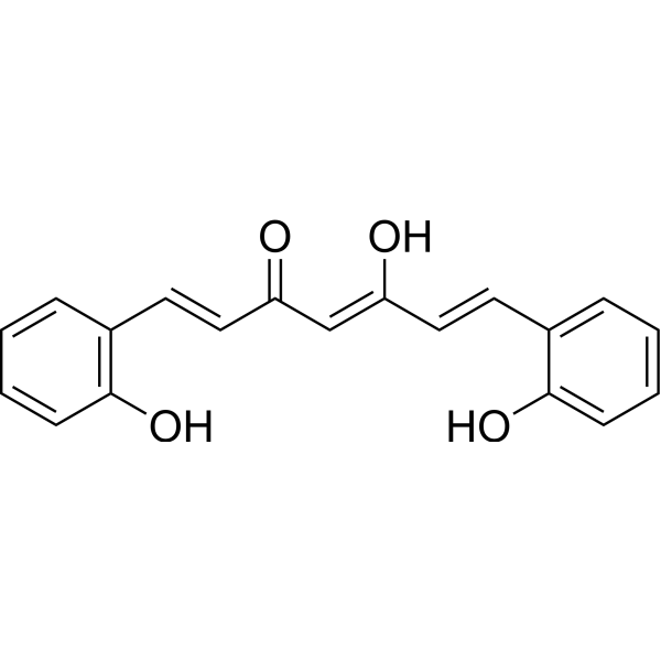
Salicylcurcumin is a synthetic curcumin analogue with antioxidant activity. Salicylcurcumin may affect lipid peroxidation in fish, favoring increased survival, disease resistance, and ultimately growth rate.
For research use only. We do not sell to patients.
- CAS No.: 1236545-54-2
- Formula: C19H16O4
- Molecular Weight:308.33
-
Storage:
Please store the product under the recommended conditions in the Certificate of Analysis.

To place orders, for customer services and technical support, please contact: MedChemExpress USA
Tel: 609-228-6898 E-mail: [email protected] [email protected]


-
 Biological Activity
-
Chemical Information
- Protocols
- Purity & Documentation
- References
-
 Help & FAQs




Biological Activity
Description
Cellular Effect
|
Cell Line

|
Type | Value | Description | References |
|---|---|---|---|---|
| SW-620 | GI50 |
35 μM
Compound: 5
|
Cytotoxicity against human SW620 cells after 48 hrs by WST1 assay
Cytotoxicity against human SW620 cells after 48 hrs by WST1 assay
|
[PMID: 21183341] |
In Vitro
Salicylcurcumin (0.1-10 mM) can inhibit the formation of lipid peroxidation products and the content of thiobarbituric acid reactive substances (TBARS) in vitro[1]. br/
MedChemExpress (MCE) has not independently confirmed the accuracy of these methods. They are for reference only. Further protocols information, click here.
In Vivo
MedChemExpress (MCE) has not independently confirmed the accuracy of these methods. They are for reference only.


Chemical Information
-
CAS No. 1236545-54-2
-
Molecular Weight 308.33
-
Formula C19H16O4
-
SMILES
OC1=C(C=CC=C1)/C=C/C(/C=C(/C=C/C2=CC=CC=C2O)O)=O
-
Structure Classification
-
Initial Source
Anabas testudineus
-
Shipping
Room temperature in continental US; may vary elsewhere.
-
Storage
Please store the product under the recommended conditions in the Certificate of Analysis.

Protocols
-
Protocol for Fluorescence In Situ Hybridization (FISH)
Fluorescence in situ hybridization detects specific DNA or RNA sequences inside fixed cells or tissue sections by hybridizing fluorescently labeled nucleic-acid probes to complementary target sequences, allowing the target’s copy number, chromosomal position, spatial distribution, or transcript abundance to be visualized microscopically. DNA-FISH detects genomic loci, chromosomal gains/losses, amplifications, deletions, and rearrangements, while RNA-FISH detects RNA molecules or transcript localization; in cancer cells, mouse tumors, neurons, organoids, macrophages, or drug-screening samples, the readout is fluorescent puncta, fusion/split signals, or localized RNA signal interpreted relative to validated controls.
-
Ferroptosis Solutions
Ferroptosis is an iron-dependent, non-apoptotic form of regulated cell death characterized by lethal lipid peroxidation and sensitivity to suppression by iron chelators or lipophilic radical-trapping antioxidants. The core pathway links cystine uptake through system Xc−, glutathione availability, GPX4-dependent detoxification of phospholipid hydroperoxides, iron-dependent oxidative reactions, and polyunsaturated-phospholipid metabolism into a cell-death program that is biochemically and morphologically distinct from apoptosis, necrosis, and autophagy. The ferroptosis pathway is experimentally linked to phenotype through chemical and genetic perturbation. Erastin induces ferroptosis by inhibiting cystine uptake through system Xc− and weakening antioxidant defenses, while GPX4 inhibition or depletion causes lipid peroxide accumulation and ferroptotic cancer-cell death. ACSL4 and oxidizable arachidonoyl- or adrenoyl-containing phosphatidylethanolamines shape ferroptosis sensitivity by con


Purity & Documentation
References

Calculators
Concentration (start) × Volume (start) = Concentration (final) × Volume (final)


Salicylcurcumin Related Classifications
HY-116117 Related Classifications
 Powered by Bioz