

325 Results for "

microscopy

" in MedChemExpress (MCE) Product Catalog:
Products (325)

325 Results for "microscopy" in MCE Product Catalog:

1
1 Cited Publications
Cat. No.: HY-P2031
CAS No.: 26645-35-2
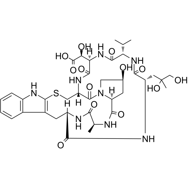
Phallacidin is a natural bicyclic heptapeptide derived from the poisonous mushroom Amanita phalloides. Phallacidin binds to filamentous actin specifically with high affinity, with a Kd of 20 nM. After binding to F-actin, Phallacidin strongly inhibits its depolymerization, stabilizes microfilament structures, and prevents their disruption by drugs such as cytochalasins. When conjugated with a fluorophore, Phallacidin serves as a specific fluorescent probe for F-actin, which is used to clearly visualize the distribution of actin in the cytoskeleton (e.g., stress fibers, cortical peripheral bands) under fluorescence microscopy .
loading...
    loading...
1
1 Cited Publications
Cat. No.: HY-D1668
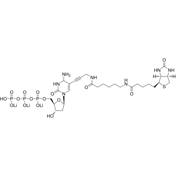
Biotin-11-dCTP is a biotinylated deoxynucleoside triphosphate and an important DNA labeling reagent. In random primer DNA labeling reactions, Biotin-11-dCTP incorporates into newly synthesized DNA strands to generate labeled DNA probes suitable for hybridization applications. In addition, Biotin-11-dCTP can serve as a substrate for terminal deoxynucleotidyl transferase to end-label oligonucleotides for telomere sequence detection, or to label the cut ends of linearized DNA molecules, thereby supporting streptavidin-based electron microscopy analysis. For example, Biotin-11-dCTP can label the cut ends of linearized DNA molecules under the action of dGTP and avian myeloblastosis virus reverse transcriptase .
loading...
    loading...
1
1 Cited Publications
Cat. No.: HY-118667
CAS No.: 516-85-8
Purity:  ≥99.0%
Target:  

Fluorescent Dye

Research Areas:  

Others

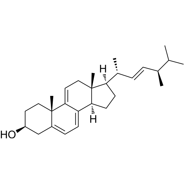
Dehydroergosterol is a fluorescent cholesterol analog (Ex/Em = 325/375 nm) used in cholesterol transport, membrane structure research, and live-cell imaging. Unlike cholesterol, Dehydroergosterol contains three additional double bonds, an extra methyl group, and an ergosterol side chain, forming a conjugated system that produces intrinsic fluorescence. In its monomeric form, Dehydroergosterol exhibits excitation peaks at 310-311, 324, and 338-340 nm, as well as emission peaks at 354, 371-375, 390-394, and 414 nm; in its microcrystalline form, Dehydroergosterol shows enhanced excimer emission around 404 and 426 nm. Dehydroergosterol mimics the properties of cholesterol, integrates into membranes and lipoproteins, and localizes to organelles .
loading...
    loading...
Cat. No.: HY-162543
CAS No.: 384833-00-5
Synonyms: 18:1 Lissamine rhodamine PE
Target:  

Fluorescent Dye

Research Areas:  

Others

18:1 Liss Rhod (18:1 Lissamine rhodamine) PE is a fluorescent phospholipid and fluorescent probe.18:1 Liss Rhod PE admixes into phospholipid inks for large-scale monitoring of dip-pen nanolithography-generated lithographic structures via fluorescence microscopy.18:1 Liss Rhod PE undergoes phase separation or self-quenching under certain conditions in thin lipid membrane stacks .
loading...
    loading...
Cat. No.: HY-D2001
CAS No.: 863772-22-9
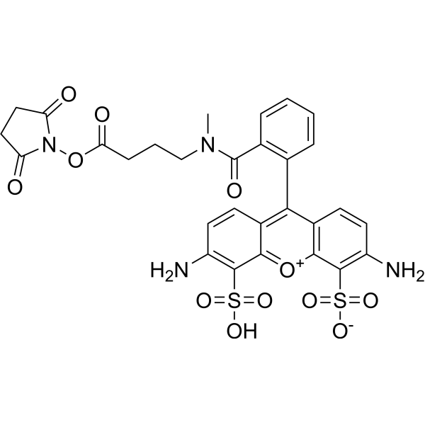
ATTO 488 NHS ester is a new fluorescent marker based on the Rhodamine structure. It has strong absorption, high fluorescence quantum yield, high thermal stability and photochemical stability, and is suitable for single molecule detection and high-resolution microscopy. ATTO 488 NHS ester is an NHS ester derivative of ATTO 488 that can be used to label proteins or antibodies.
loading...
    loading...
Cat. No.: HY-DY1004
CAS No.: 121207-31-6
BODIPY 493/503 (solution) is a BODIPY dye. BODIPY dye is a small molecule dye with strong ultraviolet absorption ability, its fluorescence peak is relatively sharp, and the quantum yield is high. They are relatively insensitive to the polarity and pH of the environment and are relatively stable under different physiological conditions. Due to its structural asymmetry, BODIPY derives a variety of structural products. BODIPY lipid droplet dyes can well pass through the cell membrane into the cell, and localize the neutral lipids in the cell to specifically stain the lipid droplets, which can be used for labeling of live cells and fixed cells . Maximum excitation/emission wavelength: 493/503 nm.
Solvent and concentration: DMSO: 5 mM.
loading...
    loading...
Cat. No.: HY-P10932A
Research Areas:  

Neurological Disease

pGk13a TFA is an azide (azide group can be combined with fluorophore)-containing amphiphilic membrane labeling probe. pGk13a TFA enables high-resolution imaging of cell membranes in the ultrastructural membrane expansion microscopy (umExM) technique, facilitating the observation of membrane-associated structures and proteins. pGk13a TFA can be used for neuronal structural studies .
loading...
    loading...
Cat. No.: HY-D0721
CAS No.: 3348-03-6
Synonyms: 6-Carboxyfluorescein diacetate
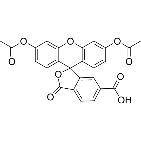
6-CFDA is a common aliphatic luciferin-line organism. CFDA conducts free diffusion into cells, and then it is hydrolyzed into carboxyl fluorescein (CF) by intracellular non-specific lipase. CF containing portion contains an additional negative charge so that it is better retained in cells, compared to fluorescein dyes .
loading...
    loading...
Cat. No.: HY-D0953
CAS No.: 1330-38-7
Synonyms: Solvent Blue 38
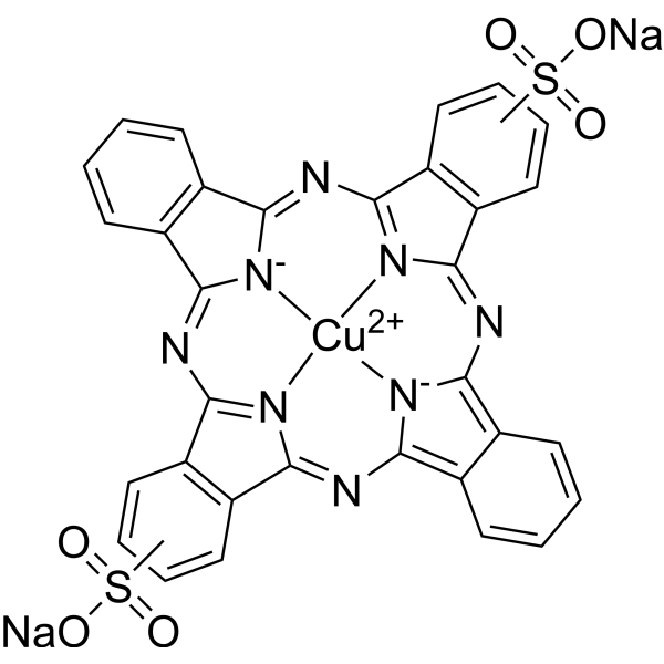
Direct Blue 86 (Solvent Blue 38) is a myelin-sheath stain, commonly utilized in microscopy to detect demyelination in the central nervous system. Direct Blue 86 also is a dye with various applications including as a commercial dye in the printing of cotton and mucilage glue fabrics .
loading...
    loading...
Cat. No.: HY-D2381
AF 488 maleimide is a thiol-reactive dye used to label SH groups of proteins, which can attach the AF 488 fluorophore to cysteine residue-containing proteins and peptides as well as other thiolated molecules. AF 488 maleimide enables real-time visualization of dynamic pilus extension and retraction in live bacterial cells via epifluorescence microscopy (Ex/Em = 470/520 nm) .
loading...
    loading...
Cat. No.: HY-D0950A
CAS No.: 7114-03-6
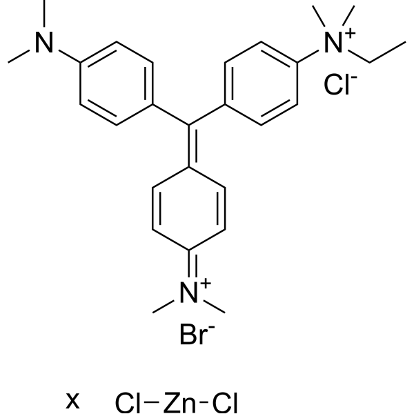
Methyl Green zinc chloride is a potent fluorescent dye. Methyl Green zinc chloride is a DNA stains of cells and electrophoretic gels. Methyl Green zinc chloride can be used as direct measuring of viability by both microscopy and flow cytometry, with peaks at 633 and 677 nm .
loading...
    loading...
Cat. No.: HY-NP163A
Synonyms: WGA-AF488
Wheat germ agglutinin-AF488 (WGA-AF488) is a cell membrane-specific staining agent prepared by conjugating wheat germ agglutinin with the Alexa Fluor 488 (HY-D1304) fluorescent dye, and it binds to cell surface glycoproteins with high affinity. Wheat germ agglutinin-AF488 is applied in fluorescence microscopy and confocal imaging techniques, and it can clearly label the membrane structures of various cells including breast cancer cells, enabling high-resolution visual observation. Wheat germ agglutinin-AF488 is used in studies of breast cancer and triple-negative breast cancer to observe cell morphology and membrane dynamic changes .
loading...
    loading...
Cat. No.: HY-DY3002
Glutaraldehyde Fixative (4%, For Electron Microscopy) is composed of glutaraldehyde, phosphate, deionized water, etc., with a pH range of 7.2-7.4. This fixative is effective in fixing fine structures of cell nuclei and cytoplasm and is commonly used for fixing electron microscopy specimens.
loading...
    loading...
Cat. No.: HY-D2001A
TTO 488 NHS ester TEA is a new fluorescent marker based on the Rhodamine structure. It has strong absorption, high fluorescence quantum yield, high thermal stability and photochemical stability, and is suitable for single molecule detection and high-resolution microscopy. TTO 488 NHS ester TEA is an NHS ester derivative of ATTO 488 that can be used to label proteins or antibodies.
loading...
    loading...
Cat. No.: HY-D0954
CAS No.: 62851-42-7
Jenner's Stain is a dye that is used in microscopy for staining blood smears. Jenner's Stain can be used for the chromosome stain by C-banding technique. Jenner's Stain can be used for the stain for routine blood examinations and malarial staining .
loading...
    loading...
Cat. No.: HY-D1950
CAS No.: 2982225-57-8
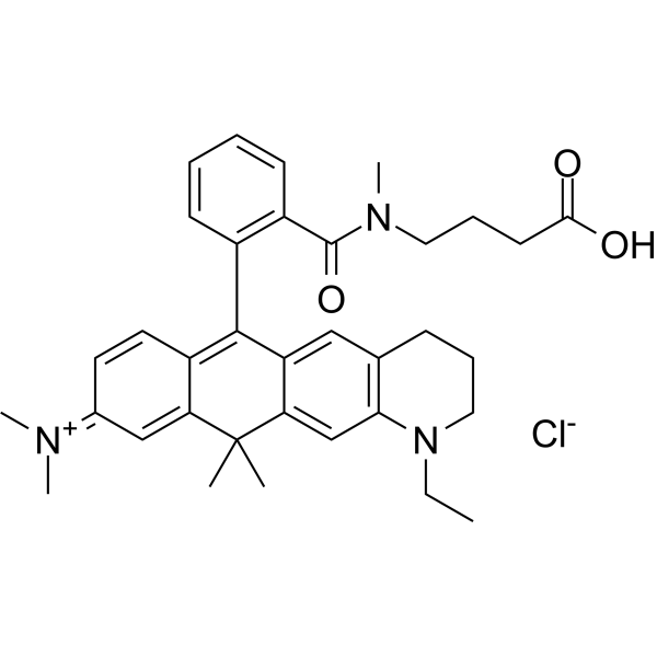
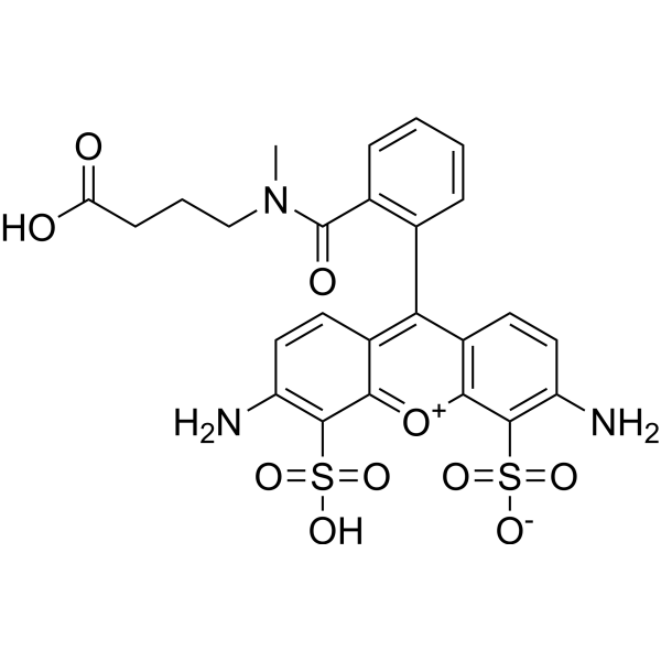
ATTO 633 is a fluorescent dye with an absorption peak at approximately 630 nm and a fluorescence emission peak at 651 nm. ATTO 633 can be used in nanomechanical photothermal microscopy studies .
loading...
    loading...
Cat. No.: HY-D1305
CAS No.: 1443553-08-9
Target:  

Fluorescent Dye

Research Areas:  

Others

ATTO 488 carboxylic acid is a bright green fluorescent dye with high photostability, which is commonly used in protein fluorescent labeling, single-molecule fluorescence imaging and super-resolution microscopy studies. The carboxylic acid form of ATTO 488 can be covalently coupled to primary amines of proteins via activation, or prepared into a maleimide derivative for coupling with thiol groups; it emits green fluorescence after labeling. Ex/Em = 488/524 nm .
loading...
    loading...
Cat. No.: HY-P10932
Research Areas:  

Neurological Disease

pGk13a is an amphipathic membrane-labeling probe containing an azide group, which can bind to fluorophores. pGk13a enables high-resolution imaging of cell membranes in the ultrastructure expansion microscopy (umExM) technique, facilitating the observation of membrane-associated structures and proteins. pGk13a is applicable to neuronal structure research .
loading...
    loading...
Cat. No.: HY-150743A
Purity:  93.84%
Research Areas:  

Inflammation/Immunology

Biotin-labeled ODN 2395 (sodium), a C class oligodeoxynucleotide, is a TLR9 agonist. Biotin-labeled ODN 2395 (sodium) can be used to evaluate CpG ODN cellular uptake and localization using a biotin detection system and light microscopy.
loading...
    loading...
Cat. No.: HY-146245A
Purity:  95.88%
Research Areas:  

Inflammation/Immunology

Biotin-labeled ODN 1826 (sodium), a class B CpG ODN (oligodeoxynucleotide), is a TLR9 agonist. Biotin-labeled ODN 1826 (sodium) can be used to evaluate CpG ODN cellular uptake and localization using a biotin detection system and light microscopy.
loading...
    loading...

