
396 Results for "

Fluorescence Probe

" in MedChemExpress (MCE) Product Catalog:
Products (396)

396 Results for "Fluorescence Probe" in MCE Product Catalog:

Cat. No.: HY-DY1097
CAS No.: 107091-89-4
Thiazole Orange (solution) is an asymmetric anthocyanin dye that can be coupled with oligonucleotides (ONs) to prepare fluorescent hybridization probes. Thiazole Orange has been widely used in biomolecular detection and staining of DNA/ RNA in gels and can be used for reticulocyte analysis. Thiazole orange generates a significant fluorescence enhancement and high quantum yield when it binds with nucleic acids, especially RNA. Thiazole orange can permeate living cell membranes. Thiazole orange can use UV light for detection, but can also be detected with blue light. The excitation and emission of Thiazole orange are λex = 510 nm (488 nm and 470 nm also show strong excitation) and λem = 527 nm, respectively .
Solvent and concentration: DMSO: 10 mM
The 1 mL volume is defined as the base specification. All larger sizes correspond to incremental volumes of this base.
loading...
    loading...
Cat. No.: HY-D3105
Target:  

Fluorescent Dye

Research Areas:  

Others

DCA is a Fluorescent probe for visualization of phase behavior in ER membranes. DCA is an ER-targeting, polarity-responsive NIR ratiometric probe, with its p-toluenesulfonamide group responsible for ER localization; its sensitivity to polarity relies on its donor-π-acceptor (D-π-A) structure, where aniline acts as the donor and dicyanomethylene acts as the acceptor, driving an intramolecular charge transfer (ICT) process upon excitation. In environments with low polarity, such as the closely packed, low water content ERₒ phase of ER membranes, DCA emits at a shorter wavelength, while in high polarity environments like the loosely packed, higher water content ERd phase, ICT leads to a red-shifted emission, allowing discrimination of the two phases via dual NIR emission colors and ratiometric imaging. Ex/Em = 488/570–620 nm and 488/665–735 nm; additional excitation/emission pairs include Ex/Em = 488/631 nm in low polarity 1,4-dioxane and Ex/Em = 488/677 nm in 1,4-dioxane with 30% water, the higher polarity condition. It shows a large Stokes shift of ~170 nm, and pH, viscosity, and biologically relevant species including Cys, GSH, H₂O₂, and metal ions do not exert marked interference on its fluorescence spectra[1].
loading...
    loading...
Cat. No.: HY-112636
CAS No.: 14696-39-0
Purity:  98.34%
Target:  

Amyloid-β

Research Areas:  

Others

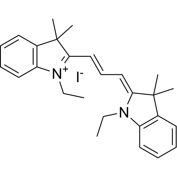
Astrophloxine is a fluorescent imaging probe capable of targeting antiparallel dimers. Astrophloxine can be used to detect aggregated Aβ in brain tissue and cerebrospinal fluid samples of Alzheimer's disease (AD) mice .
loading...
    loading...
Cat. No.: HY-181453
Synonyms: 2’-O-Benzyl-3’-MANT-GTP sodium
Research Areas:  

Others

2'-O-Benzyl-3'-O-(N'-methylanthraniloyl) guanosine-5'-O-triphosphate sodium (Mant-GTP R=CH3, B=Gua) (2'-O-Benzyl-3'-MANT-GTP sodium) is a methylanthraniloyl derivative of GTP (HY-113225). Methylanthraniloyl-GTP exhibits fluorescent properties (λmax: 442 nm in water; 432 nm in ethanol) .
loading...
    loading...
Cat. No.: HY-D3075
CAS No.: 2490504-23-7
Target:  

Fluorescent Dye

Research Areas:  

Others

TPE-2E N-oxide is a fluorescent probe used for hypoxia detection and tumor hypoxia imaging. Due to its active intramolecular motion, TPE-2E N-oxide shows no fluorescence in aqueous solution. In hypoxic environments, its N-oxide group undergoes two-electron reduction by CYP450 reductase and other heme protein reductases expressed under hypoxia, forming hydrophobic aggregates that trigger aggregation-induced emission through the restriction of intramolecular motion. TPE-2E N-oxide exhibits selective lipid droplet localization in cells. The absorption wavelength of TPE-2E N-oxide in dichloromethane is 313 nm; in a 99% hexane/dichloromethane mixed system, its excitation wavelength is 330 nm and emission wavelength is 460 nm. For in vitro cell imaging, excitation is performed at 405 nm, with an emission wavelength range of 430-560 nm .
loading...
    loading...
Cat. No.: HY-D3210
CAS No.: 2098632-04-1
Target:  

Fluorescent Dye

Research Areas:  

Inflammation/Immunology

DCM-KPV is a fluorescent probe targeting the human intestinal oligopeptide transporter PEPT1/SLC15A1 receptor (λex=480 nm, λem=620-670 nm). DCM-KPV specifically binds to PepT1 via its KPV domain and mediates receptor-targeted internalization, thus effectively accumulating in the cytoplasm and nucleus of cells overexpressing this receptor. DCM-KPV has the advantages of long emission wavelength, high emission efficiency, low photobleaching, and negligible cytotoxicity. DCM-KPV maintains stable fluorescence intensity under continuous illumination, exhibiting extremely high live cell compatibility. DCM-KPV can specifically accumulate at colonic inflammatory sites through the intestinal mucosa, enabling direct non-invasive visual differentiation between chronic and acute ulcerative colitis groups and the normal group .
loading...
    loading...
Cat. No.: HY-D3525
Target:  

Fluorescent Dye

Research Areas:  

Others

BDL-E5 is a fluorescent probe for detecting live induced pluripotent stem cells at the early reprogramming stage. Its detection mechanism involves specific binding to authentic reprogramming cells, which exhibit increased expression of pluripotency and epithelial genes and decreased expression of mesenchymal genes compared to non-reprogramming cells; it appears to co-localize more significantly with the Golgi complex than other organelle markers in reprogramming cells, and it does not require washing after staining to reduce background signals; fluorescence is generated upon binding to these early reprogramming cells, with positive cells appearing 7 days before iPS colonies are visible and stain positive for the conventional pluripotent marker TRA-1-60, and sorting BDL-E5-positive cells enriches populations that generate higher numbers and quality of iPS colonies. Its detection wavelength is Ex/Em = 578/599 nm .
loading...
    loading...
Cat. No.: HY-W115607
CAS No.: 24991-53-5
Synonyms: Poly(ethylene glycol)-bis-amine 4000
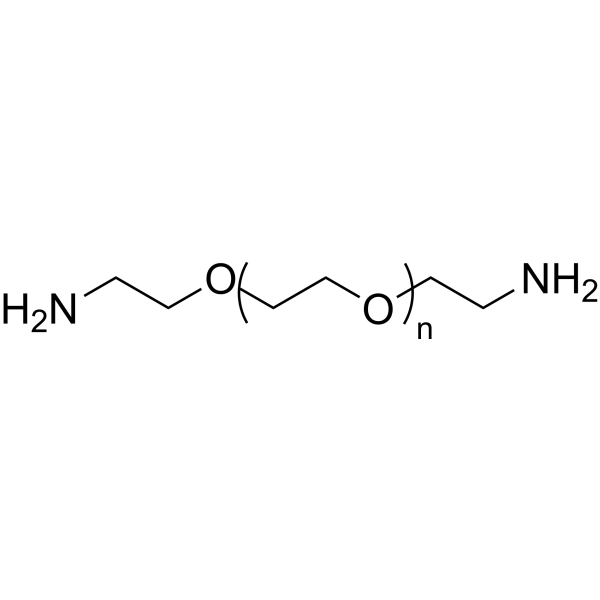
PEG4000-bis-amine (Poly(ethylene glycol)-bis-amine 4000) is a diamine-terminated polyethylene glycol that acts as a linker for conjugate synthesis. PEG4000-bis-amine facilitates the development of MUC1 aptamer-based near-infrared fluorescent probes, and also conjugates folic acid with the near-infrared dye ICG-Der-01 to form a tumor-targeting probe. PEG4000-bis-amine supports the synthesis of folic acid-PEG-DSPE conjugates for the preparation of folic acid-targeted biliposomes. PEG4000-bis-amine is widely used in studies related to breast cancer, non-small cell lung cancer, hepatocellular carcinoma, rheumatoid arthritis, and tumors with high folate receptor expression .
loading...
    loading...
Cat. No.: HY-D3003
CAS No.: 2522921-17-9
M-H2S is an endoplasmic reticulum-targeted ratio-type near-infrared fluorescent probe (Ex/Em : 480 nm/560 nm) specifically designed for the detection of hydrogen sulfide (H2S) in living cells and zebrafish. M-H2S exhibits detection limit for H2S of 39.1 nM, and the fluorescence intensity ratio (F₆₅₀/F₅₆₀) within the range of 0-40 μM shows a linear relationship with the H2S concentration. M-H2S responds best under pH = 7.2 (the normal physiological pH of the endoplasmic reticulum) conditions, making it suitable for the detection of the microenvironment of the endoplasmic reticulum. M-H2S can be used to study the role of H2S in endoplasmic reticulum function, stress response, and related diseases .
loading...
    loading...
Cat. No.: HY-D3072
Research Areas:  

Neurological Disease

P6-Aggrate is a fluorescent probe for aggregated proteome detection. P6-Aggrate specifically recognizes amorphous aggregated proteomes through non-covalent reversible binding, and its fluorescence enhances after heat-induced protein aggregation. P6-Aggrate reflects the polarity and compactness heterogeneity within aggregated proteomes via emission wavelength shift: short-wavelength emission (blue shift) corresponds to large aggregates with high compactness, while long-wavelength emission (red shift) corresponds to small spots with low compactness (Ex/Em = 488/520-580 nm). P6-Aggrate enables reversible monitoring of the dynamic processes of formation and clearance of stress-induced proteome aggregation such as that induced by MG132 (HY-13259) in living cells. P6-Aggrate can be used in studies related to protein homeostasis imbalance, neurodegenerative diseases and protein aggregation .
loading...
    loading...
Cat. No.: HY-D3074
Research Areas:  

Others

TCM-4COOH is an AIE-active fluorescent probe for calcium ions. TCM-4COOH relies on aggregation-induced emission activity, and its carboxyl chelating group can bind to Ca 2+ and Mg 2+ to form insoluble aggregates, triggering fluorescence enhancement via restricting intramolecular motion (Ex/Em = 477/603 nm). TCM-4COOH shows pH independence in physiological environments (pH 4-9), can be strongly activated at Ca 2+ concentrations ranging from low nM to mM, and has a stronger binding ability to Ca 2+ than to Mg 2+. TCM-4COOH, as its acetoxymethyl ester derivative TCM-AM, can penetrate cell membranes for intracellular detection. TCM-4COOH can be used for imaging of cellular calcium overload, detection of bone microcracks, and studies related to calcium-rich biological environments .
loading...
    loading...
Cat. No.: HY-D3401
CAS No.: 3121511-17-6
Research Areas:  

Others

LHA585 is a fluorescent probe targeting the lipid hydrogen abstraction process for the early detection of ferroptosis (Ferroptosis). Its detection mechanism relies on the reactivity of its own 4-phenyl-3-methylbut-2-enyl moiety with biologically relevant oxidative radicals, including peroxyl radicals (ROO·), alkoxyl radicals (RO·), hydroxyl radicals (HO·), and peroxynitrite anions (ONOO−); upon hydrogen abstraction from this structure, radical rearrangement and radical cleavage occur, releasing a xanthene radical which converts into a highly fluorescent rhodamine dye in biological environments, thereby achieving a fluorescence turn-on ratio of up to 360-fold. LHA585 is non-fluorescent in its native state, possesses excellent membrane permeability due to its non-polar property, and the rhodamine dye generated after oxidation diffuses into mitochondria. The excitation wavelength of LHA585 is 561 nm, and its emission wavelength ranges from 570 to 620 nm .
loading...
    loading...
Cat. No.: HY-160062
Target:  

Mucin

Research Areas:  

Cancer

S2.2 aptamer sodium is a nucleic acid-based MUC1-binding aptamer with high affinity and low toxicity. Upon binding to its target, S2.2 aptamer sodium undergoes a conformational switch and restores fluorescence signal, serving as a targeted imaging agent for MUC1-positive cancer cells. S2.2 aptamer sodium enables targeted delivery to breast cancer cells with overexpressed MUC1. When formulated as the S2.2-PEG-MZF molecular probe, S2.2 aptamer sodium possesses the functions of T2 signal inhibition, magnetic field-induced hyperthermia and targeted magnetic resonance molecular imaging. In the S2.2-PEG-MZF/DOX nanoliposome, S2.2 aptamer sodium supports targeted thermochemotherapy, effectively inhibiting cancer cell proliferation and invasion as well as inducing apoptosis, and is widely used in studies related to breast cancer .
loading...
    loading...
Cat. No.: HY-W327027
CAS No.: 314742-00-2
Target:  

Fluorescent Dye

Research Areas:  

Others

7-(2,4-Dinitrophenoxy)-4-methyl-2H-chromen-2-one (Compound 1) is a fluorescent probe for the detection of hydrogen sulfide (H2S). 7-(2,4-Dinitrophenoxy)-4-methyl-2H-chromen-2-one has a low detection limit (4×10 -6 mol/L), good selectivity and high sensitivity. 7-(2,4-Dinitrophenoxy)-4-methyl-2H-chromen-2-one shows almost no cytotoxicity at concentrations of 150 µg/mL. 7-(2,4-Dinitrophenoxy)-4-methyl-2H-chromen-2-one has the excitation peak of 331 nm, and the emission peak about 385 nm in DMSO solvent. Upon the addition of increasing amounts of HS -, the fluorescence intensity increases obviously at about 392 nm .
loading...
    loading...
Cat. No.: HY-W1061291
CAS No.: 3101697-53-1
Research Areas:  

Others

Quasar670 Phosphoramidite is a modified phosphoramidite monomer that can be used for oligonucleotide synthesis and fluorescent in situ hybridization probe synthesis. Oligonucleotide probes synthesized with Quasar670 Phosphoramidite can be used to detect eGFP mRNA in fixed cells; they can target the 26S rRNA D1/D2 region of Hanseniaspora uvarum to enable tracking and detection of this yeast strain .
loading...
    loading...
Cat. No.: HY-D2465
Target:  

Fluorescent Dye

Research Areas:  

Infection

CY5-Dextran (MW60-80k) is a fluorescent probe formed by covalent conjugation of the cyanine dye Cy5 to dextran. It is a pH-insensitive fluid-phase endocytosis tracer (the pH of endosomal lumen is precisely quantified by the fluorescence ratio of FITC/Cy5). CY5-Dextran (MW60-80k) is internalized by cells via a clathrin-independent fluid-phase pinocytosis pathway, and is sequentially evidenced by early endosomes, late endosomes are transported to lysosomes. CY5-D I cannot pass through the limited pore size formed by uncoating of human rhinovirus type 2 (HRV2), but can be released into the neutral cytoplasm upon adenovirus-mediated endosomal rupture, thus distinguishing between the two viral modes of "pore-forming escape" and "lysis escape". CY5-Dextran (MW60) -80k) has an excitation wavelength of 635 nm, and it can also be used for time-lapse imaging of endocytosis dynamics of stromal cells in tissues such as rat salivary glands under in vivo two-photon microscopy .
loading...
    loading...

