396 Results for "

Fluorescence Probe

" in MedChemExpress (MCE) Product Catalog:
Products (396)

396 Results for "Fluorescence Probe" in MCE Product Catalog:

Cat. No.: HY-D3091
CAS No.: 2757681-99-3
Target:  

Fluorescent Dye

Research Areas:  

Others

NRLyso is a lysosome-targeted solvatochromic fluorescent probe derived from Nile Red (HY-D0718). It can localize to lysosomes and is applicable for imaging lysosomal lipid order and polarity. The excitation wavelength of NRLyso is 520 nm, and its emission maximum varies with the environment. NRLyso partitions well into lipid membranes: it exhibits fluorescence enhancement and emission blue-shift in low-polarity, tightly packed lipid environments, while shows emission red-shift in more polar, disordered lipid environments. NRLyso maintains organelle specificity under oxidative and hyperosmotic stress conditions, and can detect changes in lysosomal lipid order and polarity in response to these stresses, cholesterol extraction, and cholesterol enrichment .
loading...
    loading...
Cat. No.: HY-D3109
CAS No.: 148043-22-5
Target:  

Fluorescent Dye

Research Areas:  

Others

pH-Ap-1 is a fluorescent probe used for ratiometric monitoring of pH fluctuations during autophagy in living cells, and it can detect intracellular pH changes in the autophagy process. For fluorescence detection of pH-Ap-1, its excitation wavelength is 475 nm, with an emission wavelength of 538 nm under alkaline conditions and 645 nm under acidic conditions. For intracellular imaging, excitation is performed at 488 nm: the emission light ranging from 500-550 nm is collected via the green channel corresponding to 538 nm, while the emission light ranging from 625-750 nm is collected via the red channel corresponding to 645 nm. pH-Ap-1 exhibits excellent photostability and chemical stability .
loading...
    loading...
Cat. No.: HY-D3123
Target:  

Fluorescent Dye

Research Areas:  

Cardiovascular Disease

Ac-MtFluNox is a mitochondria-targeted Fluorescent probe that can be used for the selective detection of labile Fe (II). Ac-MtFluNox can enter living cells, where intracellular esterases cleave its acetyl protecting group to generate activated MtFluNox. Ac-MtFluNox functions through an Fe (II)-mediated deoxygenation mechanism of its amine N-oxide group, which eliminates the fluorescence quenching effect and oxidizes Fe (II) to Fe (III) to deplete the catalytically active Fe (II) pool. A FITC filter set (excitation 465-500 nm, emission 516-556 nm) is used for live-cell imaging. Ac-MtFluNox can be applied to studies related to myocardial ferroptosis .
loading...
    loading...
Cat. No.: HY-D3192
CAS No.: 2028316-07-4
CDy11 is a fluorescent probe and amyloid-binding dye (λex=590 nm; λem=612 nm), with a Ka of 29 μM for Pseudomonas aeruginosa Fap. CDy11 specifically recognizes amyloid fibrils in bacterial biofilms and exhibits significantly enhanced fluorescence upon binding to the target. CDy11 shows no staining effect on amyloid-deficient mutant strains, planktonic cells or protein monomers. CDy11 supports in vivo imaging of Pseudomonas aeruginosa biofilms in mouse implant and corneal infection models. CDy11 is widely used in studies of Staphylococcus aureus biofilm infections, dental caries, and Pseudomonas aeruginosa-associated implant and corneal infections .
loading...
    loading...
Cat. No.: HY-D3443
Target:  

Fluorescent Dye

Research Areas:  

Cancer

NR-HOCl-TFMU is a HOCl-gated phosphonite fluorescent probe based on the Nebraska Red scaffold. NR-HOCl-TFMU undergoes HOCl-triggered oxidative ring-opening of the spirocyclic thioether and phosphonite hydrolysis to synchronously release the near-infrared reporter group NR666-MSA (Ex 666 nm/Em 698 nm) and the cargo TFMU (Ex 405 nm/Em 510 nm) . NR-HOCl-TFMU enables HOCl-selective fluorescence imaging and cargo release monitoring in vitro, in live cells and in vivo, and exerts gated selective cytotoxicity in HOCl-positive cells. NR-HOCl-TFMU can be used for studies related to acute myeloid leukemia .
loading...
    loading...
Cat. No.: HY-DY1111
CAS No.: 1539318-40-5
Target:  

DNA Stain

Research Areas:  

Neurological Disease

DFHBI-2T solution is a membrane-permeable, RNA aptamer-activated fluorescent probe (Ex/Em=500 nm/523 nm), with a Kd value of 1300 nM for the RNA aptamer Spinach2. DFHBI-2T solution binds to Spinach2, converting the fluorophore to a fluorescent state, stabilizing its planar structure to facilitate the radiative fluorescence decay pathway, and forming a complex with red-shifted excitation and emission peaks. DFHBI-2T solution can be used for RNA imaging in live cells. It is also applicable to research related to fragile X-associated tremor/ataxia syndrome .
Solvent and concentration: DMSO: 5 mM
loading...
    loading...
Cat. No.: HY-P992459

Research Areas:  

Cancer

SGM-101 Antibody is a chimeric monoclonal antibody targeting carcinoembryonic antigen (CEA). SGM-101 Antibody can serve as a tumor-specific fluorescent imaging probe after being covalently conjugated with the near-infrared fluorescent dye BM104 . SGM-101 Antibody accumulates in CEA-positive tumor tissues via antigen-antibody specific binding, and emits near-infrared fluorescence to enable imaging of tumor lesions. SGM-101 Antibody can be used in research related to gastric cancer, colorectal cancer, pancreatic cancer, non-small cell lung cancer, breast cancer, peritoneal carcinomatosis, and liver metastases .
loading...
    loading...
Cat. No.: HY-D3187
CAS No.: 2484831-02-7
HMRef-αMan is a substrate-based green fluorescent probe (Ex/Em=465 nm/515 nm) targeting MAN2C1 (α-mannosidase). HMRef-αMan can be specifically cleaved by MAN2C1 to generate a highly fluorescent product, which thus gets activated to produce green fluorescence in malignant breast tissues, benign lesions and living cancer cells. The signal intensity of HMRef-αMan is directly correlated with MAN2C1 activity, and it can effectively detect tiny breast cancer lesions with a diameter of less than 1 mm. When used in combination with the red-emitting γ-glutamyl transpeptidase (GGT) probe gGlu-2OMe SiR600 (HY-D3188), HMRef-αMan enables precise optical differentiation of breast tissue types via a dual-color imaging strategy. HMRef-αMan has been widely used in the research of breast diseases such as breast cancer, fibroadenoma, phyllodes tumor and various types of papilloma .
loading...
    loading...
Cat. No.: HY-D3094
CAS No.: 2687198-42-9
Target:  

Fluorescent Dye

Research Areas:  

Others

TPE-SQ5 is a ratiometric Fluorescent probe for hypochlorite (ClO -) detection with a limit of detection of 5.6 nM. TPE-SQ5 exhibits high selectivity and high sensitivity towards ClO -. TPE-SQ5 can be oxidized by ClO - to form an epoxide, which quenches its deep red fluorescence; subsequently, the epoxide decomposes into a hemicyanine and a sulfonic acid-substituted oxindole, producing blue-green fluorescence to achieve a ratiometric response. TPE-SQ5 can form nanoparticles with Pluronic F-127 via the nanoprecipitation method, and these nanoparticles possess cell permeability. TPE-SQ5 has unique Ex/Em spectra in different solvents and nanoparticle forms, with emission peaks at 643 nm (380 nm excitation, 95% H2O/THF), 631 nm (564 nm excitation, DMSO), 652 nm (536 nm excitation, THF) and 630 nm (524 nm excitation, nanoparticles); the Ex/Em settings used for cell imaging are 488/550-650 nm (red channel) and 405/450-550 nm (green channel) .
loading...
    loading...
Cat. No.: HY-D1725
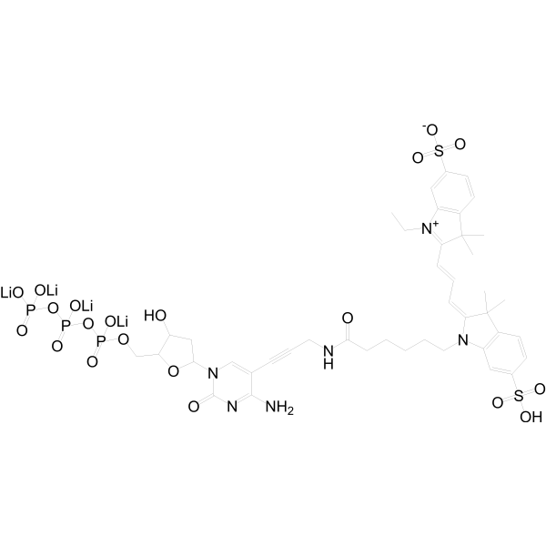
Cy3-dCTP is a directly fluorescently labeled deoxyribonucleotide, in which Cy3 is a cyanine fluorescent dye. Cy3-dCTP is used for direct enzymatic labeling of DNA and cDNA: with the aid of DNA polymerases, this modified nucleotide is incorporated into the extending DNA strand during processes such as reverse transcription, PCR, nick translation or random primer labeling .
loading...
    loading...
Cat. No.: HY-D3117
CAS No.: 2146114-18-1
Target:  

Fluorescent Dye

Research Areas:  

Others

MBCB is a two-photon Fluorescent probe for dual-detection of mitochondrial SO₂ derivatives and viscosity. For SO₂ derivatives detection, the probe utilizes a Michael addition mechanism: nucleophilic addition of SO₂ derivatives to the C=C bond between the carbazole skeleton and 3-methylbenzothiazolium moiety destroys the strong intramolecular charge transfer (ICT) system between these groups, while enhancing the weak ICT system between the benzothiazole group and carbazole framework; this causes the red emission at 600 nm to decrease and the blue emission at 434 nm to increase, creating a ratiometric response based on the I₄₃₄ₙₘ/I₆₀₀ₙₘ intensity ratio. For viscosity detection, in low-viscosity environments, steric hindrance creates a twisted ICT (TICT) system with weak fluorescence, while in high-viscosity environments, intramolecular rotation is blocked, the TICT state is disrupted, and the strong ICT system is recovered, leading to a strong red emission at 567 nm with negligible change to the short-wavelength emission at 415 nm, creating a ratiometric response based on the I₅₆₇ₙₘ/I₄₁₅ₙₘ intensity ratio that has a logarithmic linear relationship with viscosity. The probe has excitation/emission wavelengths of Ex/Em = 351/434, 600 nm for SO₂ derivatives detection and Ex/Em = 351/567 nm for viscosity detection, with two-photon excitation at 740 nm for bioimaging; it also exhibits good mitochondrial targeting ability with a Pearson's colocalization coefficient of 0.93 when paired with Mito-Tracker Green. The probe shows high sensitivity and selectivity for SO₂ derivatives, has low cell cytotoxicity, and can be applied to detect exogenous/endogenous HSO₃⁻ in living cells and in vivo, as well as visualize mitochondrial viscosity changes induced by nystatin[1].
loading...
    loading...
Cat. No.: HY-D0074
CAS No.: 70504-01-7
Target:  

Fluorescent Dye

Research Areas:  

Others

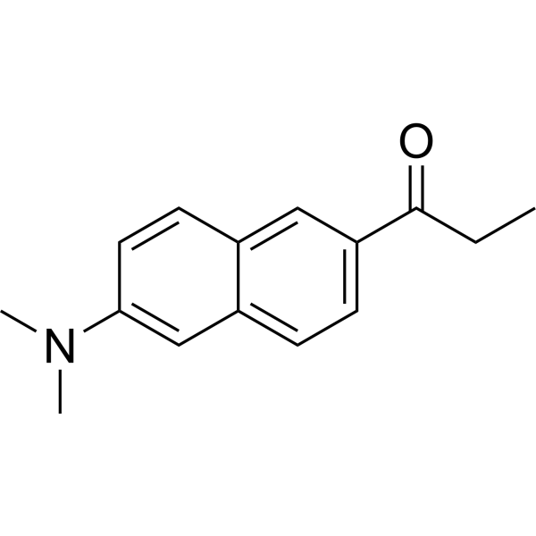
Prodan is an environment-sensitive fluorescent probe used to investigate the polarity, fluidity, and structure of lipid membranes. Prodan exhibits environment-dependent excitation/emission wavelengths, with its wavelength range covering Ex = 360-410 nm and Em = 430-530 nm when in solvents, bound to DNA, located in lipid bilayers, or associated with proteins. Prodan can interact with the major groove of DNA, regions of lipid bilayers, and hydrophobic pockets of proteins. During phase transitions, Prodan localizes to different regions of the bilayer: the emission minimum is 430-440 nm in the gel phase, while it shifts to 480-500 nm in the liquid-crystalline phase or interdigitated gel phase. When bound to rigid hydrophobic sites of proteins, the emission peak of Prodan undergoes a blue shift and its fluorescence intensity increases .
loading...
    loading...
Cat. No.: HY-D1991
CAS No.: 906664-68-4
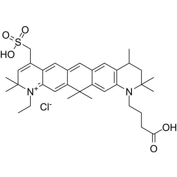
ATTO 647 is a carborhodamine fluorophore and imaging tracer with photostable properties. ATTO 647 serves as a fluorescent probe to investigate cell membrane structure and diffusion characteristics. When conjugated with wheat germ agglutinin, ATTO 647 specifically binds to N-acetyl-β-D-glucosamine and sialic acid residues on membrane glycoproteins, enabling single-molecule tracing of glycoprotein diffusion. ATTO 647 exhibits highly stable fluorescence properties with significantly reduced blinking in mounting media such as ROXS (AA/MV) and ROXS (TX/TQ), whereas its brightness properties vary in Ibidi-MM and Vectashield. ATTO 647 can also be used to label histone H2B-GFP in fixed cells for confocal microscopy photobleaching experiments .
loading...
    loading...
Cat. No.: HY-D3191
L&M-D-MR is a highly specific fluorescent "AND" logic probe with response moieties for leucine aminopeptidase (LAP) and monoamine oxidase (MAO). The coexistence of both LAP and MAO is required for L&M-D-MR to trigger intramolecular cyclization, release fluorophores and activate fluorescence. In the presence of only a single enzyme, L&M-D-MR generates only an extremely weak signal. L&M-D-MR enables bioimaging in living cells and mouse models, and can effectively distinguish different subtypes of liver diseases via blood samples or test strips. L&M-D-MR is widely used in studies related to liver cirrhosis, hepatitis B and drug-induced liver injury .
loading...
    loading...
Cat. No.: HY-D3370
CAS No.: 3114128-41-2
Target:  

Fluorescent Dye iGluR

Research Areas:  

Others

CL-NC is a chemiluminescent probe used for in vivo imaging of N-methyl-D-aspartate receptors (NMDARs), and its detection mechanism relies on bioorthogonal activation achieved via a tetrazine-triggered click-to-release reaction. The isonitrile group of CL-NC undergoes a click reaction with tetrazine, followed by hydrolysis and β-elimination to release a phenoxide anion, which induces the departure of adamantane and generates chemiluminescence. CL-NC has a maximum absorption wavelength of approximately 425 nm, a maximum fluorescence emission wavelength of approximately 670 nm, and a maximum chemiluminescence emission wavelength of 710 nm. When used in combination with Tz-IFDL (HY-D3369), an NMDAR ligand conjugated with tetrazine, CL-NC enables targeted imaging of NMDARs in living cells, as well as in the brain and spinal cord of living mice .
loading...
    loading...
Cat. No.: HY-P5520
CAS No.: 2413262-74-3
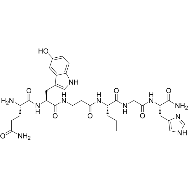
GB-6 is a short linear peptide that targets the gastrin releasing peptide receptor (GRPR). GRPR is overexpressed in pancreatic cancer. Based on the tumor selectivity and tumor-specific accumulation properties of GB-6, GB-6 labeled with near infrared (NIR) fluorescent dyes or radionuclide netium-99m (99mTc) can be used as a high-contrast imaging probe. GB-6 has excellent in vivo stability, with tumor to pancreatic and intestinal fluorescence signal ratios of 5.2 and 6.3, respectively, in SW199 0 subcutaneous xenograft models. GB-6 can rapidly target tumors and accurately delineate tumor boundaries, which has broad application prospects .
loading...
    loading...
Cat. No.: HY-D3073
CAS No.: 2477607-64-8
Target:  

Fluorescent Dye

Research Areas:  

Others

DXB-NIR is a push-pull dioxaboron fluorescent probe with solvatochromic properties, featuring high fluorescence brightness, photostability, and a large two-photon absorption cross-section. DXB-NIR exhibits Ex/Em of 550/620–780 nm (with two-photon excitation at 930 nm). DXB-NIR quantifies local polarity via the intensity ratio I (>640)/I (<640) between the far-red channel (<640 nm) and the near-infrared channel (>640 nm), and it can simultaneously label the plasma membrane, endoplasmic reticulum, and lipid droplets in live cells. DXB-NIR monitors the increase in local polarity of various cell compartments under conditions of cholesterol depletion, starvation, and oxidative stress. DXB-NIR can be used for polarity imaging of biological membranes and lipid droplets, as well as studies related to cellular stress .
loading...
    loading...
Cat. No.: HY-D3085
CAS No.: 2882924-23-2
SSR-LDs is a fluorescent probe for lipid droplet polarity detection and lipid droplet imaging. SSR-LDs exhibits polarity-dependent fluorescence: it produces intense short-wavelength emission in low-polarity environments, while generating weak long-wavelength emission in high-polarity environments. SSR-LDs enters cells via free diffusion, specifically targets lipid droplets, and is unaffected by intracellular viscosity or pH values ranging from 4.5 to 9.0. SSR-LDs shows variable Ex/Em wavelengths in different solvents, including 590/663 nm in THF, 600/660 nm in mouse tissue and in vivo imaging, and an excitation wavelength of 580 nm with an emission wavelength range of 610-750 nm in cell imaging. SSR-LDs can be used for research related to fatty liver, liver injury and hepatitis .
loading...
    loading...
Cat. No.: HY-D3086
CAS No.: 1668565-72-7
Target:  

Fluorescent Dye

Research Areas:  

Cancer

Mito-polarity is a mitochondria-targeted polarity probe. Mito-polarity can be used to detect mitochondrial polarity. The detection mechanism of Mito-polarity is based on an intramolecular charge transfer (ICT) system designed with a donor-π-bridge-acceptor (D-π-A) structure. Among them, the 467 nm green emission generated by the Coumarin (HY-N0709) structure is extremely sensitive to polarity changes, while the 642 nm red emission produced by the extended π-conjugation and ICT effect between the Coumarin and benzothiazine structures shows only a weak response, thus forming a ratiometric fluorescence response that is linearly correlated with solvent polarity. When Mito-polarity is excited at 405 nm in methanol, its excitation/emission wavelengths are Ex/Em = 405/467 nm and Ex/Em = 405/642 nm, with maximum absorption wavelengths of 426 nm and 561 nm .
loading...
    loading...
Cat. No.: HY-D3391
RMR-Tre is a fluorescent probe targeting the mycobacterial acyltransferase Ag85. Under the catalysis of Ag85, RMR-Tre undergoes 6-position mycoloylation and anchors to the mycobacterial membrane, while achieving fluorescence activation by inhibiting the intramolecular twisted charge transfer state transition. RMR-Tre can distinguish live mycobacteria from dead ones through metabolism-driven labeling, enabling rapid, wash-free, low-background detection of viable bacteria. RMR-Tre reports the drug resistance of Mycobacterium tuberculosis via the trehalose catalytic shift activity readout associated with TreS. In addition, RMR-Tre can be combined with flow cytometry or high-content imaging techniques to visualize and quantitatively analyze the metabolic heterogeneity of Mycobacterium tuberculosis related to persistence and drug resistance. RMR-Tre is widely used in tuberculosis-related research .
loading...
    loading...

