



Prodan

Based on 1 Customer Validation
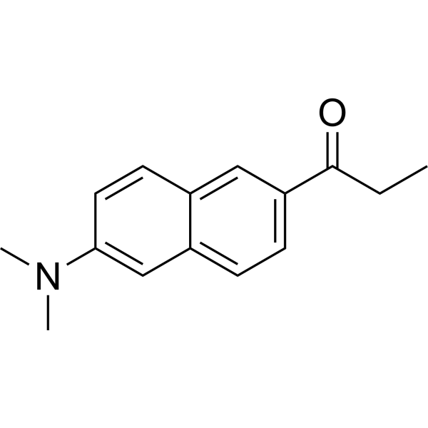
Prodan is an environment-sensitive fluorescent probe used to investigate the polarity, fluidity, and structure of lipid membranes. Prodan exhibits environment-dependent excitation/emission wavelengths, with its wavelength range covering Ex = 360-410 nm and Em = 430-530 nm when in solvents, bound to DNA, located in lipid bilayers, or associated with proteins. Prodan can interact with the major groove of DNA, regions of lipid bilayers, and hydrophobic pockets of proteins. During phase transitions, Prodan localizes to different regions of the bilayer: the emission minimum is 430-440 nm in the gel phase, while it shifts to 480-500 nm in the liquid-crystalline phase or interdigitated gel phase. When bound to rigid hydrophobic sites of proteins, the emission peak of Prodan undergoes a blue shift and its fluorescence intensity increases.
For research use only. We do not sell to patients.
- Purity : 99.71%
- CAS No.: 70504-01-7
- Formula: C15H17NO
- Molecular Weight:227.31
-
Storage:
4°C, protect from light
* In solvent : -80°C, 6 months; -20°C, 1 month (protect from light)

To place orders, for customer services and technical support, please contact: MedChemExpress USA
Tel: 609-228-6898 E-mail: [email protected] [email protected]


-
 Biological Activity
-
Chemical Information
-
Solvent & Solubility
- Purity & Documentation
- References
-
 Help & FAQs


-
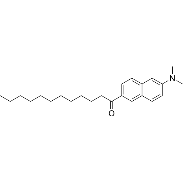
HY-D0080
LaurdanFluorescent Probe -
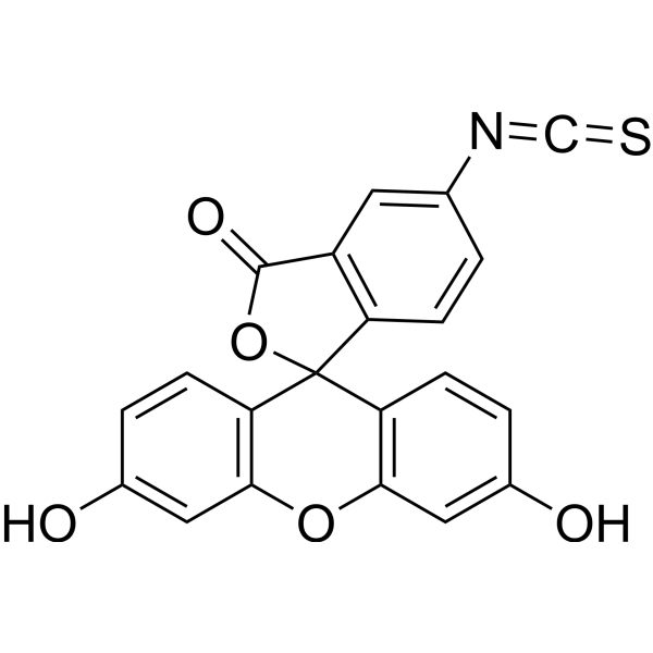
HY-66019
FITCFluorochrome -
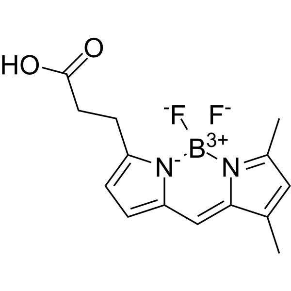
HY-43520
BODIPY-FLFluorescent Dye -

HY-158779
Sulfane sulfur probe 4Fluorescent Indicator -

HY-DY1007
FITC (solution)Fluorochrome -
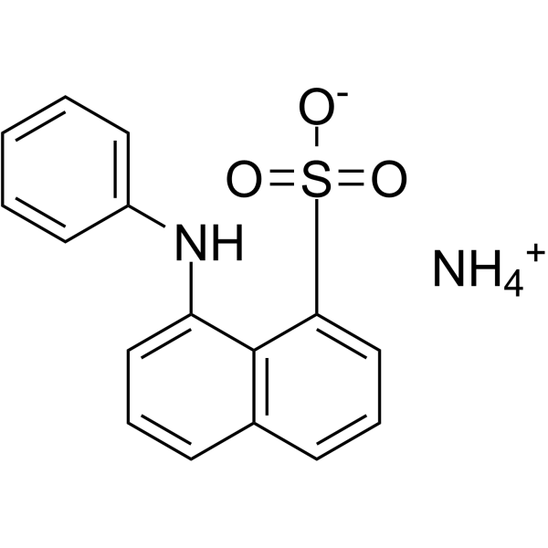
HY-D0896A
ANS ammoniumAntibacterial Agent -
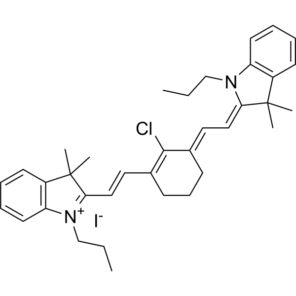
HY-D1063
IR-780Fluorescent Probe -

HY-D0896
ANSAntibacterial Agent -
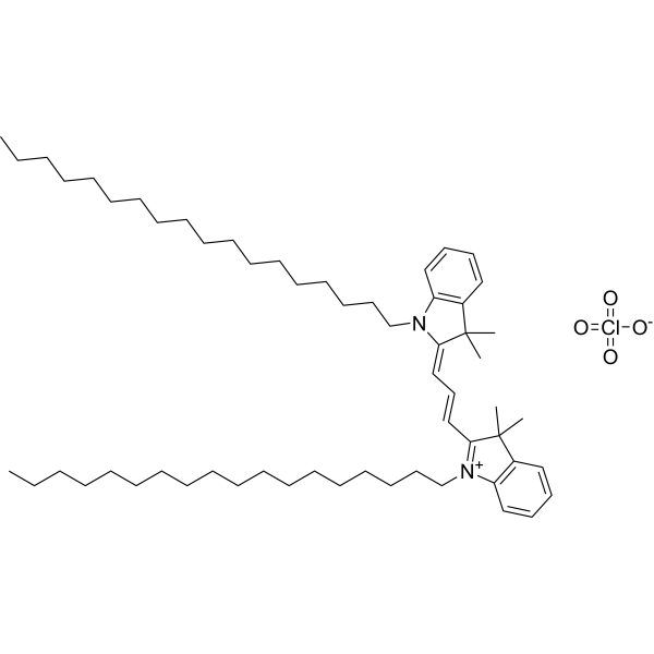
HY-D0083
DiIFluorescent Dye -
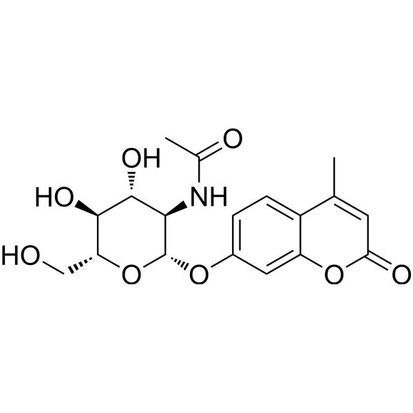
HY-137853
4-Methylumbelliferyl-2-acetamido-2-deoxy-β-D-GlucopyranosideFluorogenic Substrate -
HY-126831
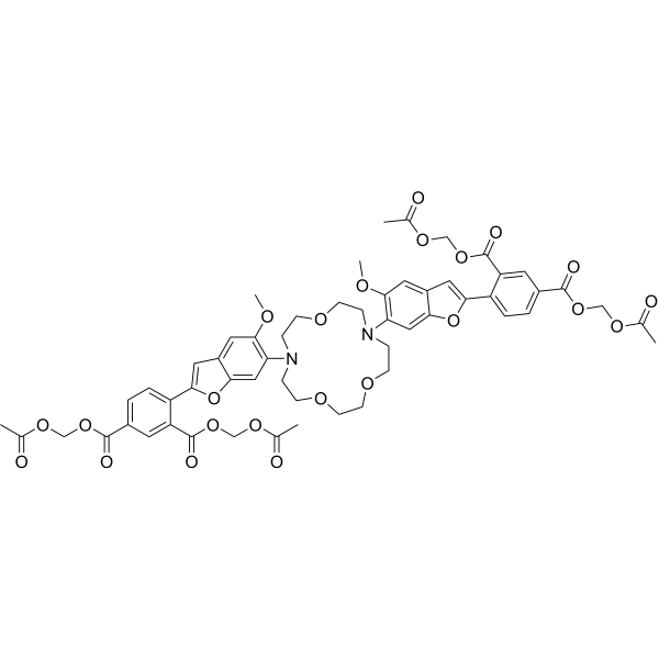
SBFI-AMFluorescent Indicator -
HY-145519
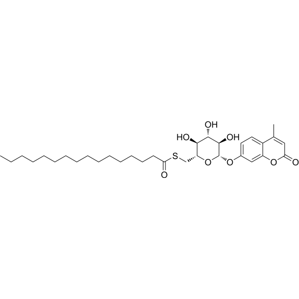
Mu-6S-Palm-β-GlcFluorescent Dye


Biological Activity
Description
In Vitro
1. Stock Solution Preparation
1.1 Solvents: Acetonitrile[2], ethanol[3], or DMF[4] are optional.
2. Working Solution Preparation
2.1 Diluents: Optional options include 50 mM sodium phosphate buffer containing 0.1 M NaCl (pH 7.0, universal type), 25 mM sodium phosphate buffer containing 50 mM NaCl (pH 7.0, for SNP genotyping)[1], water[2], or buffer containing 5 mM phosphate, 20 mM KCl, 1 mM EDTA, pH 8.0 supplemented with 0.2 mM DTT[4].
2.2 Working concentration: 0-5 μM.
2.3 Notes: Adjust the working solution concentration as needed; prepare fresh before use.
3. Staining Procedures
3.1 Sample Type Description
3.1.1 Oligodeoxynucleotide (ODN) samples (single-stranded or double-stranded ODNs)[1]:
3.1.1.1 Incubation conditions: Incubate 2.5 μM single-stranded ODN containing PDNX with 2.5 μM complementary ODN at room temperature to form double strands; perform fluorescence detection at 25 °C.
3.1.2 Large unilamellar vesicles (LUVs) composed of phospholipids (lecithin, DOPC, or their mixtures) with optional cholesterol addition[2]:
3.1.2.1 Incubation conditions: Hydrate dried lipid membranes with 2 μM Prodan aqueous solution at 25°C for approximately 5 min; for vesicles derived from electrospun membranes, dissolve 10 mg of membrane in 5 mL water containing Prodan, and stir to induce self-assembly.
3.1.3 Multilamellar vesicle (MLV) dispersions of symmetric saturated diacylphosphatidylcholines (CnPCs) with acyl chain lengths n=12-15 and 19-22[3]:
3.1.3.1 Incubation conditions: Mix Prodan stock solution with CnPC stock solution, remove the solvent under vacuum, add water, then vortex/sonicate at a temperature slightly above the main transition temperature of each CnPC bilayer to prepare MLV dispersions.
3.1.4 Purified dimeric spectrin solution[4]:
3.1.4.1 Incubation conditions: Incubate with 0.5 μM to 1 μM Prodan working solution at 25°C.
4. Control Setup
4.1 ODN samples[1]:
4.1.1 Set single-stranded ODN containing PDNX as a control to distinguish double-stranded samples.
4.2 Lipid vesicle samples[2]: Prepare control samples with the same phospholipid concentration but without Prodan for subtracting background absorption and fluorescence.
4.3 Spectrin samples[4]: Use aqueous buffer containing only Prodan but no spectrin as a blank control to detect the fluorescence of unbound Prodan.
5. Detection and Analysis
5.1 Instrument types: Fluorescence spectrophotometer, fluorescence microplate reader (for SNP genotyping), etc.
5.2 Excitation/Emission Wavelengths
5.2.1 ODNs containing PDNX[1]:
5.2.1.1 PDNU: 380 nm/524 nm (free nucleoside), 389 nm/520 nm (single-stranded ODN), 404 nm/522 nm (matched double strand); base-selective detection uses 450 nm excitation/520 nm emission; SNP genotyping uses 440±10 nm excitation filter/510±10 nm emission filter.
5.2.2 Lipid LUV samples[2]: Excitation at 350 nm or 370 nm; monitor emission at 434 nm, 445 nm, 465 nm, 490 nm, and 525 nm.
5.2.3 Lipid MLV samples[3]: Excitation at 361 nm; monitor emission in the range of 400 nm to 600 nm.
5.2.4 Spectrin samples[4]: Excitation at 360 nm; measure emission at 430 nm and 520 nm.
5.3 Result Analysis
5.3.1 ODN samples[1]:
5.3.1.1 Under 450 nm excitation, the fluorescence intensity of fully matched Watson-Crick double strands is significantly higher than that of single-stranded ODNs and mismatched double strands.
5.3.1.2 The Prodan fluorophore anchors to the major groove surface of the DNA double strand.
5.3.1.3 Fully matched double strands have a smaller Stokes shift value (lower wavenumber) compared with single-stranded ODNs and mismatched double strands.
5.3.2 Lipid LUV samples[2]:
5.3.2.1 The emission band blue-shifts from 530 nm in aqueous solution to approximately 498 nm, and an emission band at 434 nm appears as the vesicle concentration increases.
5.3.2.2 The emission band at 434 nm corresponds to PRODAN localized in the deep nonpolar region of the lipid bilayer.
5.3.2.3 PRODAN exhibits triexponential decay in lecithin LUVs (corresponding to the water-bilayer interface, bilayer nonpolar region, and deep nonpolar core of the bilayer, respectively); it exhibits biexponential decay in pure DOPC LUVs (corresponding to the polar interface and bilayer nonpolar region, respectively).
5.3.2.4 Prodan in LUVs derived from electrospun membranes shows a red edge excitation shift (REES) of up to 8 nm when the excitation wavelength changes from 350 nm to 400 nm, indicating that it is in a motion-restricted microenvironment.
5.3.3 Lipid MLV samples[3]:
5.3.3.1 Prodan localizes in regions of the bilayer corresponding to phase states, and the shift of the emission minimum correlates with changes in bilayer polarity.
5.3.3.2 Fluorescence second-derivative spectra show characteristic minima specific to bilayer phases: 430-440 nm for the bilayer gel phase (Lβ' or Pβ'), 480-490 nm for the liquid crystalline phase (Lα), and approximately 500 nm for the interdigitated gel phase (LβI).
5.3.3.3 Stack the second-derivative spectra obtained at every 1°C to construct a three-dimensional (3D) spectral imaging map for visualizing the change in bilayer phase state with temperature.
5.3.4 Spectrin samples[4]:
5.3.4.2 Compared with unbound Prodan in aqueous buffer, the emission intensity of Prodan bound to spectrin increases by 2-fold; the fluorescence polarization degree rises from 0.05 in the unbound state to 0.27 in the bound state.
5.3.4.3 Prodan binds to the hydrophobic sites of spectrin.
5.3.4.4 The emission maximum blue-shifts by 87 nm (from 520 nm for unbound Prodan to 433 nm for Prodan bound to spectrin).
MedChemExpress (MCE) has not independently confirmed the accuracy of these methods. They are for reference only.


Chemical Information
-
CAS No. 70504-01-7
-
Appearance Solid
-
Molecular Weight 227.31
-
Formula C15H17NO
-
Color Light yellow to yellow
-
SMILES
CCC(C1=CC=C2C=C(N(C)C)C=CC2=C1)=O
-
Shipping
Room temperature in continental US; may vary elsewhere.
-
Storage
4°C, protect from light
* In solvent : -80°C, 6 months; -20°C, 1 month (protect from light)


Solvent & Solubility
In Vitro:
DMSO : 50 mg/mL (219.96 mM; Need ultrasonic; Hygroscopic DMSO has a significant impact on the solubility of product, please use newly opened DMSO)
Please refer to the solubility information to select the appropriate solvent. Once prepared, please aliquot and store the solution to prevent product inactivation from repeated freeze-thaw cycles.
Storage method and period of stock solution: -80°C, 6 months; -20°C, 1 month (protect from light). When stored at -80°C, please use it within 6 months. When stored at -20°C, please use it within 1 month.
Please refer to the solubility information to select the appropriate solvent. Once prepared, please aliquot and store the solution to prevent product inactivation from repeated freeze-thaw cycles.
Storage method and period of stock solution: -80°C, 6 months; -20°C, 1 month (protect from light). When stored at -80°C, please use it within 6 months. When stored at -20°C, please use it within 1 month.
Concentration (start) × Volume (start) = Concentration (final) × Volume (final)


Purity & Documentation
-
Data Sheet (286 KB)
-
SDS (393 KB)
- English - EN (393 KB)
- Français - FR (393 KB)
- Deutsch - DE (393 KB)
- Norwegian - NO (393 KB)
- Español - ES (393 KB)
- Swedish - SV (393 KB)
- Italian - IT (393 KB)
- Korean - KR (393 KB)
- Portuguese - PT (393 KB)
-
Handling Instructions (2659 KB)
References
[1]. Tainaka K, et al. PRODAN-conjugated DNA: synthesis and photochemical properties. Journal of the American Chemical Society. 2007 Apr 18;129(15):4776-84. [Content Brief]
[2]. Luna MA, et al. PRODAN Photophysics as a Tool to Determine the Bilayer Properties of Different Unilamellar Vesicles Composed of Phospholipids. Langmuir : the ACS journal of surfaces and colloids. 2024 Jan 09;40(1):657-667. [Content Brief]
[3]. Tamai N, et al. Phase Imaging of Phosphatidylcholine Bilayer Membranes by Prodan Fluorescence. Membranes. 2022 Dec 02;12(12):1219. [Content Brief]
[4]. Chakrabarti A, et al. Fluorescence of spectrin-bound prodan. Biochemical and biophysical research communications. 1996 Sep 13;226(2):495-7. [Content Brief]


Complete Stock Solution Preparation Table
Please refer to the solubility information to select the appropriate solvent. Once prepared, please aliquot and store the solution to prevent product inactivation from repeated freeze-thaw cycles.
Storage method and period of stock solution: -80°C, 6 months; -20°C, 1 month (protect from light). When stored at -80°C, please use it within 6 months. When stored at -20°C, please use it within 1 month.
| Optional Solvent | Concentration Solvent Mass | 1 mg | 5 mg | 10 mg | 25 mg |
|---|---|---|---|---|---|
| DMSO | 1 mM | 4.3993 mL | 21.9964 mL | 43.9928 mL | 109.9820 mL |
| 5 mM | 0.8799 mL | 4.3993 mL | 8.7986 mL | 21.9964 mL | |
| 10 mM | 0.4399 mL | 2.1996 mL | 4.3993 mL | 10.9982 mL | |
| 15 mM | 0.2933 mL | 1.4664 mL | 2.9329 mL | 7.3321 mL | |
| 20 mM | 0.2200 mL | 1.0998 mL | 2.1996 mL | 5.4991 mL | |
| 25 mM | 0.1760 mL | 0.8799 mL | 1.7597 mL | 4.3993 mL | |
| 30 mM | 0.1466 mL | 0.7332 mL | 1.4664 mL | 3.6661 mL | |
| 40 mM | 0.1100 mL | 0.5499 mL | 1.0998 mL | 2.7495 mL | |
| 50 mM | 0.0880 mL | 0.4399 mL | 0.8799 mL | 2.1996 mL | |
| 60 mM | 0.0733 mL | 0.3666 mL | 0.7332 mL | 1.8330 mL | |
| 80 mM | 0.0550 mL | 0.2750 mL | 0.5499 mL | 1.3748 mL | |
| 100 mM | 0.0440 mL | 0.2200 mL | 0.4399 mL | 1.0998 mL |


Prodan Related Classifications
HY-D0074 Related Classifications
 Powered by Bioz
Keywords
- Prodan
- 70504-01-7
- Fluorescent Dye
- lipid bilayer characterization
- multilamellar vesicle dispersions
- single-nucleotide polymorphism typing
- large unilamellar vesicles
- microenvironment sensing
- oligodeoxyribonucleotides
- DNA major grooves
- phosphatidylcholine bilayer phase state detection
- protein hydrophobic binding site investigation
- spectrin protein
- Inhibitor
- inhibitor
- inhibit