4660 Results for "

specificity

" in MedChemExpress (MCE) Product Catalog:
Products (4660)

4660 Results for "specificity" in MCE Product Catalog:

Cat. No.: HY-145491
CAS No.: 578008-43-2
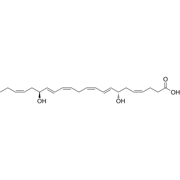
Resolvin D5 is an anti-inflammatory and analgesic agent produced in M2 macrophages. Resolvin D5 alleviates Paclitaxel (HY-B0015)-induced mechanical allodynia and inflammatory pain by activating the GPR32 receptor, with gender specificity (effective only in male mice) and independence from TRPV1 or TRPA1 channels. Resolvin D5 attenuates LPS-induced ERK phosphorylation and NF-κB nuclear translocation, downregulates proinflammatory mediators such as IL-6 and CCL5, inhibits Th17 cell differentiation and osteoclastogenesis, promotes regulatory T cell differentiation, and shows no cytotoxicity to human monocytes. The level of Resolvin D5 is elevated in arthritic SKG mice, but Resolvin D5 has no effect on dendritic cell differentiation or M1 macrophage polarization, nor does it prevent ZyA-induced arthritis progression. Resolvin D5 is suitable for research related to chemotherapy-induced peripheral neuropathy, inflammatory pain and rheumatoid arthritis .
loading...
    loading...
Cat. No.: HY-164429
CAS No.: 2418533-90-9
Target:  

Integrin Elastase

Research Areas:  

Cancer

VIP236 is a small-molecule drug conjugate targeting αvβ3 integrin. VIP236 achieves tumor homing via specific binding to αvβ3 integrin and delivers its payload to the tumor microenvironment. The linker of VIP236 is cleavable by neutrophil elastase, which is highly expressed in the tumor microenvironment, to release the payload 7-ethylcamptothecin. This payload induces DNA damage by inhibiting topoisomerase 1, thereby exerting anti-tumor effects. VIP236 exhibits excellent plasma stability and tumor targeting property, with a tumor/plasma payload ratio 10-fold higher than that of the single administration. It effectively induces tumor regression, reduces metastasis formation, and shows good tolerance in mouse models. VIP236 has been used in studies related to non-small cell lung cancer, clear cell renal cell carcinoma, colon cancer, triple-negative breast cancer, small cell lung cancer, and metastatic solid tumors .
loading...
    loading...
Cat. No.: HY-169798
CAS No.: 23481-50-7
Target:  

Fluorescent Dye VSV

Research Areas:  

Infection

1,9-Dimethylmethylene blue is a photosensitizer, virus inactivator and hemoglobin oxidant derived from methylene blue. When activated, 1,9-Dimethylmethylene blue generates reactive oxygen species including singlet oxygen, and acts as a metachromatic dye. When activated in monomeric or dimeric form, 1,9-Dimethylmethylene blue induces photoinactivation of R17 phage and vesicular stomatitis virus and oxidizes hemoglobin via non-singlet oxygen reactive oxygen species or singlet oxygen-mediated pathways, respectively. The monomeric form, with higher nucleic acid affinity, achieves virus inactivation under specific conditions without forming methemoglobin. 1,9-Dimethylmethylene blue binds to substances such as glycosaminoglycans to produce color changes. Although it is susceptible to interference from non-glycosaminoglycan components in urine, it is still applicable to spectrophotometric analysis for glycosaminoglycan quantification. With these unique photochemical and binding properties, 1,9-Dimethylmethylene blue is widely used in studies of viral infections and related biochemical analyses .
loading...
    loading...
Cat. No.: HY-188056
CAS No.: 121714-70-3
Biotin-11-UTP is a biotinylated uridine triphosphate that can be incorporated into short RNA strands via co-transcription with T7 RNA polymerase, and is used for synthesizing labeling reagents and detection markers. After binding to streptavidin, Biotin-11-UTP generates characteristic downward ionic current peaks during nanopore translocation, which allows the differentiation between modified and unmodified RNA. Biotin-11-UTP enables site-specific labeling of short RNA, and is suitable for single-molecule solid-state nanopore sensing detection. Biotin-11-UTP can serve as a raw material for synthesizing biotinylated single-stranded RNA probes, which is applied to rotavirus nucleic acid detection; it can also label single-stranded RNA probes targeting and immobilizing 28S ribosomal RNA in leukemia cells. Biotin-11-UTP can alter the electrophoretic mobility of transcribed RNA in agarose gels, and can be used to monitor the biotinylation degree of RNA probes .
loading...
    loading...
Cat. No.: HY-19872
CAS No.: 942436-93-3
Target:  

GABA Receptor

Research Areas:  

Neurological Disease

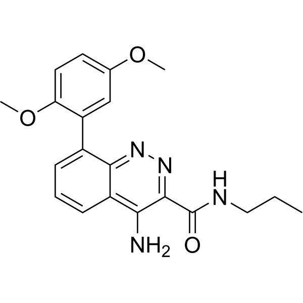
AZD6280 is an orally active, blood-brain barrier-permeable GABAA receptor modulator, with Ki values of 21 nM, 31 nM and 1680 nM against human receptors containing α2, α3 and α5 subtypes, respectively, and subtype-specific Ki values of < 30 nM for α2 and α3 subtypes. AZD6280 selectively potentiates GABAA receptors containing α2/α3 subtypes with higher potency than α1/α5 subtypes, exerts no modulatory effect on receptors containing α1 subtypes, and regulates the tuberoinfundibular dopaminergic pathway. AZD6280 decreases peak saccadic velocity, increases serum and plasma prolactin levels, rescues dendritic abnormalities of cortical neurons induced by DISC1 knockdown. AZD6280 can be used in studies related to anxiety disorders .
loading...
    loading...
Cat. No.: HY-B0426AR
CAS No.: 140462-76-6
Synonyms: ALO4943A (Standard); KW4679 (Standard)
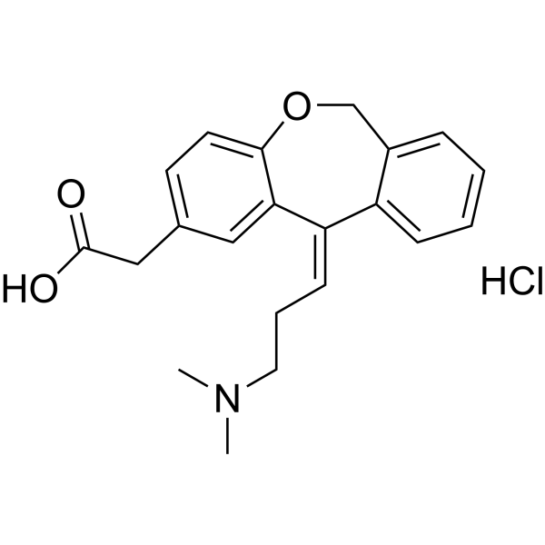
Olopatadine (hydrochloride) (Standard) is the analytical standard of Olopatadine (hydrochloride). This product is intended for research and analytical applications. Olopatadine hydrochloride (ALO4943A; KW4679) is an orally active histamine H1 receptor antagonist and mast cell stabilizer. Olopatadine hydrochloride exerts antiallergic effects by blocking histamine H1 receptor-mediated activities. Olopatadine hydrochloride inhibits exocytosis, chemokine release, F-actin polymerization, CXCL10-induced calcium influx, and T cell chemotactic activity. Olopatadine hydrochloride also reduces the expression levels of CXCR3 on the surface of CD4 + and CD8 + T cells. Olopatadine hydrochloride inhibits scratching behavior, improves dermatitis scores, and suppresses intraepidermal neurite outgrowth. Olopatadine hydrochloride simultaneously decreases the levels of inflammatory markers, growth factors, histamine, and specific IgE, while increasing the expression of ErbB3A/HER3A. Olopatadine hydrochloride can be used in research related to seasonal pollinosis, chronic rhinitis, urticaria, allergic conjunctivitis, alopecia areata, and atopic dermatitis .
loading...
    loading...
Cat. No.: HY-B0426AS
CAS No.: 1331635-21-2
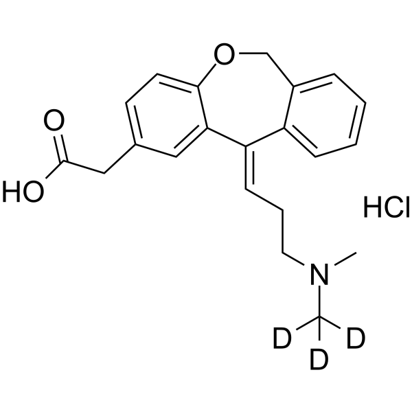
Olopatadine-d3 hydrochloride (ALO4943A-d3) is the deuterium labeled Olopatadine hydrochloride. Olopatadine hydrochloride (ALO4943A; KW4679) is an orally active histamine H1 receptor antagonist and mast cell stabilizer. Olopatadine hydrochloride exerts antiallergic effects by blocking histamine H1 receptor-mediated activities. Olopatadine hydrochloride inhibits exocytosis, chemokine release, F-actin polymerization, CXCL10-induced calcium influx, and T cell chemotactic activity. Olopatadine hydrochloride also reduces the expression levels of CXCR3 on the surface of CD4 + and CD8 + T cells. Olopatadine hydrochloride inhibits scratching behavior, improves dermatitis scores, and suppresses intraepidermal neurite outgrowth. Olopatadine hydrochloride simultaneously decreases the levels of inflammatory markers, growth factors, histamine, and specific IgE, while increasing the expression of ErbB3A/HER3A. Olopatadine hydrochloride can be used in research related to seasonal pollinosis, chronic rhinitis, urticaria, allergic conjunctivitis, alopecia areata, and atopic dermatitis .
loading...
    loading...
Cat. No.: HY-D3120
CAS No.: 2246946-53-0
Target:  

Fluorescent Dye

Research Areas:  

Others

LysoAIE2 is a Fluorescent probe for lysosomal viscosity detection and live-cell imaging. Its detection mechanism relies on the aggregation-induced emission effect: it exhibits only weak fluorescence in non-viscous media; in viscous environments, restricted intramolecular motion inhibits non-radiative energy dissipation pathways, thereby significantly enhancing fluorescence intensity. It achieves specific targeting of lysosomes through the proton acceptor property of its indole ring structure, while its hydroxyl group endows it with excellent water solubility. This probe is basically unaffected by microenvironmental polarity and pH within the range of pH 4.0 to pH 8.0, which avoids interference from these factors in viscosity measurement. LysoAIE2 has an emission wavelength of 570 nm. It can be used to monitor lysosomal viscosity changes during processes such as Dexamethasone (HY-14648)-induced lysosomal migration and starvation-induced mitophagy in live cells; at concentrations up to 40 μM, cell viability remains above 80%, demonstrating excellent biocompatibility .
loading...
    loading...
Cat. No.: HY-D3416
Synonyms: TAMRA-2,4-dinitroaniline
Target:  

Fluorescent Dye

Research Areas:  

Others

TMR-DN (TAMRA-2,4-dinitroaniline) is a fluorescent probe for RNA imaging that consists of a covalently coupled fluorophore (5-carboxy tetramethylrhodamine (TMR)) and quencher (dinitroaniline (DN)). The Kd value of TMR-DN with SRB-2 is 35 nM, and its Kd value with RhoBAST is approximately 30 nM. TMR-DN has a contact quenching function and forms a non-fluorescent intramolecular ground-state dimer in the free state; the excitation/emission wavelengths of the free probe are Ex/Em = 557/579 nm, those of the complex formed by binding to SRB-2 are Ex/Em = 561/587 nm, and those of the probe bound to the DNB aptamer are Ex/Em = 555/582 nm. TMR-DN reduces non-specific binding to genomic nucleic acids via negatively charged carboxyl-functionalized aromatic rings. Fluorescence dequenching occurs when TMR-DN specifically binds to SRB-2, RhoBAST or the DNB aptamer, among which the binding of RhoBAST requires Mg 2+ for proper folding .
loading...
    loading...
Cat. No.: HY-N0394R
CAS No.: 56-89-3
L-Cystine (Standard) is the analytical standard of L-Cystine. This product is intended for research and analytical applications. L-Cystine, the extracellular form of L-Cysteine (HY-Y0337), is a nutritionally dispensable semiessential sulfur-containing amino acid, occurring in proteins of plants and animals. L-Cystine induces Nrf2 protein elevation in a Keap1 (HY-P75897)-dependent manner and activates Nrf2 transcription factor. L-cystine can elicit cytoprotection by reducing ROS generation and protecting against oxidant- or doxorubicin-induced apoptosis. The reduced reabsorption of L-Cystine in renal tubules and its poor solubility in urine are the important causes of cystine precipitation and cystine crystal formation eventually leading to kidney stones. L-Cystine combined with L-theanine (HY-15121) enhances the production of antigen-specific IgG by increasing glutathione (GSH) levels and T helper 2 (Th2) mediated responses in mice. L-Cystine is promising for research of cystinuria and cystinosis
loading...
    loading...
Cat. No.: HY-N2600R
CAS No.: 76472-87-2
Kuwanon H (Standard) is the analytical standard of Kuwanon H. This product is intended for research and analytical applications. Kuwanon H is a selective non-peptide bombesin receptor antagonist and platelet activation inhibitor. Kuwanon H also acts as a specific antagonist of GRP-preferring receptors, with a Ki value of 290 nM for mouse GRP-R and 6500 nM for rat NMB-R. Kuwanon H inhibits the phosphorylation of AKT, mTOR, ERK, cPLA2 and p38, and upregulates TRIB3. It induces endoplasmic reticulum stress, apoptosis and autophagosome formation. Kuwanon H blocks calcium mobilization, mitogenic signaling, DNA synthesis, dense granule secretion, thromboxane A2 generation and integrin αIIb/β3 activity. It inhibits collagen-induced platelet aggregation and fibronectin adhesion, and delays clot retraction. Kuwanon H inhibits melanoma cell growth in vitro and in vivo, and enhances the sensitivity of melanoma cells to CDDP. It can be used in research related to melanoma, small cell lung cancer and thrombosis .
loading...
    loading...
Cat. No.: HY-P5107
CAS No.: 121052-30-0
Synonyms: LMWP; TDSP5
Target:  

VEGFR

Research Areas:  

Cancer

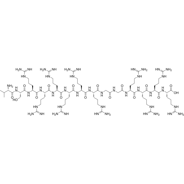
Low molecular weight protamine (LMWP;TDSP5) is a truncated arginine-rich protamine peptide, as well as a heparin/low-molecular-weight heparin antidote and a cell-penetrating delivery carrier. Low molecular weight protamine neutralizes heparin-induced anticoagulant activities, including aPTT, anti-Xa and anti-IIa activities, and also neutralizes anti-Xa activity of commercially available low-molecular-weight heparin preparations. Low molecular weight protamine translocates across mammalian cell membranes, delivers conjugated impermeable molecules across tumor tissues, enhances skin permeability of conjugated epidermal growth factor, and accelerates wound healing when conjugated with epidermal growth factor. Low molecular weight protamine retains the in vitro cell proliferation activity of conjugated EGF, and also enables site-specific conjugation with peptides or proteins via genetic recombination. Low molecular weight protamine can be used in studies related to colon adenocarcinoma, skin wounds and diabetic skin wounds .
loading...
    loading...
Cat. No.: HY-P992062

Target:  

PD-1/PD-L1

Research Areas:  

Inflammation/Immunology

Anti-Mouse CD80 Antibody (TKMG48) is an antibody that targets mouse CD80. By specifically binding to and disrupting the CD80:PD-L1 complex to release PD-L1, Anti-Mouse CD80 Antibody (TKMG48) functions as an indirect PD-1 agonist without blocking CD28 co-stimulation or CD80-CTLA4 binding. Anti-Mouse CD80 Antibody (TKMG48) inhibits T cell activation, reduces T cell effector functions and antigen-specific CD8 + T cell populations, and does not interfere with the differentiation, migration, antigen presentation or surface marker expression of dendritic cells. Anti-Mouse CD80 Antibody (TKMG48) significantly attenuates disease severity in mouse models of arthritis, spondyloarthritis, multiple sclerosis and Sjögren's syndrome, and its activity depends on the expression of PD-1 and PD-L1 .
loading...
    loading...
Cat. No.: HY-W720629
CAS No.: 21420-37-1
Cyanamide- 15N2 is the 15N-labeled Cyanamide (HY-Y0070). Cyanamide is a cell division and plant growth inhibitor, as well as an allelochemical derived from Vicia villosa. Cyanamide inhibits root growth and biomass accumulation in a dose-dependent manner by disrupting the formation of mitotic spindles and phragmoplast complexes, reducing the number of mitotic cells and blocking the cell cycle. The effects of Cyanamide are partially reversible after removal from low-concentration environments. Cyanamide is also a specific inhibitor of aldehyde dehydrogenase (ALDH). Although Cyanamide has no direct effect on tumor growth, it can significantly enhance the anti-tumor efficacy of Cyclophosphamide (HY-17420) at non-toxic doses by inhibiting the inactivation of Cyclophosphamide. Cyanamide enables Cyclophosphamide to exert equivalent therapeutic effects at lower doses, effectively inhibiting the growth of primary and metastatic tumors and prolonging the lifespan of tumor-bearing mice. Cyanamide is commonly used in studies related to ha-1 hepatoma and rls lymphosarcoma .
loading...
    loading...
Cat. No.: HY-W740027
CAS No.: 1160707-78-7
Synonyms: 5-Methyldeoxycytidine-d3
5-Methyl-2'-deoxycytidine-d3 (5-Methyldeoxycytidine-d3) is the deuterium labeled Methyl-2'-deoxycytidine (HY-W012078). 5-Methyl-2'-deoxycytidine (5mdC) is an endogenous substrate of DNA methyltransferases (such as mammalian 5-C-MTase) and binds to DNA dependent on the formation of DNA stem-loop structures. 5-Methyl-2'-deoxycytidine guides de novo DNA methylation by acting as a methylation mark and activates the methylation of adjacent CpG sites in single-stranded DNA through cis action. 5-Methyl-2'-deoxycytidine regulates DNA methylation patterns by recruiting methyltransferases to specific chromatin regions, affecting chromatin condensation and gene expression. Its distribution in plant cells is related to cell proliferation and differentiation stages. The methylation level of 5-Methyl-2'-deoxycytidine is low in proliferating cells and high in differentiated cells .
loading...
    loading...
Cat. No.: HY-L237
338 compounds

Pattern Recognition Receptors (PRRs) are a crucial class of protein molecules expressed in cells of the innate immune system. The core function of Pattern Recognition Receptors is to recognize Pathogen-Associated Molecular Patterns (PAMPs) and Damage-Associated Molecular Patterns (DAMPs). Upon recognizing and binding to PAMPs or DAMPs, PRRs rapidly initiate intracellular signaling pathways (such as the NF-κB, IRF, and inflammasome pathways). This triggers the production of inflammatory factors, chemokines, and type I interferons, thereby initiating inflammatory responses to eliminate pathogens or repair damage. PRRs represent the body's first line of defense against infection, and the rapidity and broad specificity of their response are crucial for host survival. However, aberrant activation of PRR signaling is also a cause of many chronic inflammatory diseases, autoimmune disorders, and neurodegenerative diseases. Therefore, precisely regulating PRR activity has become a key therapeutic strategy for these conditions.

MCE has cataloged 338 inhibitors targeting key PRRs, such as NLRs, TLRs, C-type Lectin Receptors (CLRs), and cGAS, to support drug discovery efforts for chronic inflammatory diseases.

Cat. No.: HY-L221
626 compounds

Cosmetics are complex mixtures formulated through the rational blending and processing of various natural, synthetic, or extracted raw materials. The ingredients used in cosmetics are diverse in type and functionality. Based on their properties and applications, cosmetic ingredients can generally be classified into two main categories: base materials and auxiliary ingredients. The former constitutes the primary component of cosmetics, accounting for a significant proportion in formulations and serving as the key functional substances. Auxiliary ingredients, on the other hand, play roles in shaping the product, ensuring stability, or imparting color, fragrance, and other specific characteristics.Establishing a systematic ingredient database is of great significance for cosmetic research and development. By analyzing the physicochemical properties (such as oil-water partition coefficients and molecular weight distribution), biological activity mechanisms (such as antioxidant pathways and cellular signaling regulation), and compatibility of ingredients, the database can provide precise guidance for formulation design. This approach helps shorten the development cycle of cosmetic/skincare products and reduces trial-and-error costs.

MCE offers 626 types of cosmetic ingredient compounds, including antioxidants, humectants, emulsifiers, film-forming agents, and more.

Cat. No.: HY-L008
738 compounds

The Janus kinase (JAK)/signal transducer and activator of transcription (STAT) pathway is central to signaling by cytokine receptors, a superfamily of more than 30 transmembrane proteins that recognize specific cytokines, and is critical in blood formation and immune response. Canonical JAK/STAT signaling begins with the association of cytokines and their corresponding transmembrane receptors. Activated JAKs then phosphorylate latent STAT monomers, leading to dimerization, nuclear translocation, and DNA binding. In mammals, there are four JAKs (JAK1, JAK2, JAK3, TYK2) and seven STATs (STAT1, STAT2, STAT3, STAT4, STAT5a, STAT5b, STAT6). Since the JAK/STAT pathway plays a major role in many fundamental processes, such as apoptosis and inflammation, dysfunctional proteins in the pathway may lead to a number of diseases. For example, alterations in JAK/STAT signalling can result in cancer and diseases affecting the immune system, such as severe combined immunodeficiency disorder (SCID).

MCE provides 738 compounds that can be used in the study of the JAK/STAT signaling pathway and related diseases.

Cat. No.: HY-L027
1,237 compounds

Viruses are much simpler organisms than bacteria, and they are made from protein substances and nucleic acid. Despite the fact that the exact mechanism of infection is extremely specific to each type of virus, the general scheme of infection can be represented in the following manner: A virus is absorbed at the surface of a host cell and then permeates through the membrane, where it releases nucleic acid from its protein protection. Then the viral nucleic acid begins to replicate, and transcription of the viral genome takes place either in the cytoplasm, or in the nucleus of the host cell. As a result of these events, a large amount of viral nucleic acid and protein are made to make new generations of virions. Therefore, one mechanism of action of antiviral drugs is to interfere with the ability of a virus to get into a target cell. A second mechanism of action is to target the processes that synthesize virus components after a virus invades a cell, such as nucleotide or nucleoside analogs.

MCE designs a unique collection of 1,237 anti-virus compounds that target several viruses, including SARS-CoV, HBV, HCV, HIV, HSV and Influenza Virus. It’s an effective tool for anti-virus drug discovery.

Cat. No.: HY-101488R
CAS No.: 1010100-07-8
CC-885 (Standard) is the analytical standard of CC-885 (HY-101488). This product is intended for research and analytical applications. CC-885, a cereblon (CRBN) modulator, acts as a molecular glue degrader targeting GSPT1 with a Kd of 350 nM. CC-885 binds to CRBN to alter the substrate specificity of the CRL4 E3 ligase, promoting the ubiquitination and proteasomal degradation of GSPT1, BNIP3L, PLK1, CDK4, IKZF1/3, GSPT2, WIZ, CHD7, GOLM1, CK1α and ETS1. CC-885 induces apoptosis, cell cycle arrest, transcriptional downregulation and antiproliferation in cancer cells. CC-885 is applicable to research related to relapsed/refractory acute myeloid leukemia, MYC-driven tumors, metastatic castration-resistant prostate cancer, multiple myeloma, hepatocellular carcinoma, glioblastoma, VHL-deficient clear cell renal cell carcinoma and non-small cell lung cancer .
loading...
    loading...

