
192 Results for "

visualize

" in MedChemExpress (MCE) Product Catalog:
Products (192)

192 Results for "visualize" in MCE Product Catalog:

Cat. No.: HY-165470
CAS No.: 945400-24-8
AZD-2184 is a β-amyloid (Aβ(1-40)) fibril binder with blood-brain barrier permeability, exhibiting a Kd of 8.4 nM for hAβ(1-40). AZD-2184 binds to the same site as CPIB, shows low levels of non-specific binding to brain tissue, rapidly enters and clears from the brain tissue of normal rats, and binds reversibly to plaques in vivo. The radiolabeled form of AZD-2184 can serve as a PET ligand for the visualization of deposits. AZD-2184 is applicable to research related to Alzheimer's disease .
loading...
    loading...
Cat. No.: HY-D3068
CAS No.: 3077446-86-4
Target:  

Fluorescent Dye

Domaines de recherche:  

Metabolic Disease

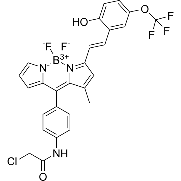
LDs-BCA is a BF2-chelated AIE fluorescent probe with a positive solvatochromic effect, which can specifically target intracellular lipid droplets (Ex/Em = 565/620-730 nm). LDs-BCA monitors the dynamic changes in the morphology, size and quantity of intracellular lipid droplets under oleic acid induction and starvation stress. LDs-BCA, after intraperitoneal injection, accumulates in the gastric fat of living obese mice, allowing continuous monitoring via its fluorescent signal. LDs-BCA is applicable to studies related to intracellular lipid droplet polarity imaging and in vivo gastric fat visualization .
loading...
    loading...
Cat. No.: HY-D3366
Target:  

Fluorescent Dye

Domaines de recherche:  

Others

Th-TMR-S-Halo is a fluorescent probe for HaloTag labeling and live-cell imaging, including super-resolution imaging and real-time visualization of endoplasmic reticulum (ER) vortex formation under ER stress conditions. Th-TMR-S-Halo shows weak fluorescence in the free state, and upon covalent binding to HaloTag, fluorescence activation is achieved by restricting the rotation of the thiophene ring. Th-TMR-S-Halo can be used in studies related to intracellular protein localization, cytoskeleton structure, and ER vortex formation. Live-cell NSPARC super-resolution detection is performed using Ex/Em = 561/570-616 nm .
loading...
    loading...
Cat. No.: HY-D3582
Target:  

Fluorescent Dye

Domaines de recherche:  

Others

AF633 C5 Maleimide is a fluorescent dye used for surface protein labeling of extracellular vesicles and peptide labeling. AF633 C5 Maleimide covalently binds to protein thiol residues under neutral pH conditions (6.5-7.5), enabling visualization and quantitative analysis of extracellular vesicle uptake by target cells. AF633 C5 Maleimide undergoes maleimide-cysteine conjugation with cysteine residues on peptides in acetonitrile-water buffer solution, supporting detection of peptide-functionalized gold nanorods via confocal laser scanning microscopy, as well as tracking their cellular uptake and localization .
loading...
    loading...
Cat. No.: HY-K0606

MCE Methenamine Silver Stain Kit For Basement Membrane (PASM) is developed based on the classical staining principle. Through an optimized staining system and standardized reagent combination, it enables clear visualization of basement membranes and related reticular structures in tissue sections. This method is particularly widely used in renal pathology research, where it is commonly applied to examine morphological alterations of the glomerular capillary basement membrane, such as thickening, rupture, folding, double-contour (tram-track) appearance, or abnormal proliferation caused by inflammatory injury. In addition, this method can also be applied to the histological investigation and morphological observation of glomerular diseases, diabetic nephropathy, and other basement membrane–associated pathological changes.

loading...
    loading...
Cat. No.: HY-DY2037
Target:  

Fluorescent Dye

Domaines de recherche:  

Others

Lignin Staining Solution (Safranin Method) is a histochemical staining solution for lignin with Safranin O as the active component. Lignin Staining Solution (Safranin Method) enables lignified cell walls to appear red to deep red (pinkish in regions with low lignification) via the high affinity of the dye for lignin. Lignin Staining Solution (Safranin Method) can be used to visualize lignin distribution in ex vivo thick sections of annual shoots from woody plants and vibratome sections of stems from herbaceous plants (such as Arabidopsis and flax), as well as for in situ quantification of cell wall substructural regions (cell corners, intercellular layers, secondary walls) based on Safranin O ratiometric fluorescence .
loading...
    loading...
Cat. No.: HY-152154
CAS No.: 3035008-40-0
Pureté:  99.41%
Synonyms: Nampt degrader-2
Target:  

PROTACs NAMPT

Domaines de recherche:  

Cancer

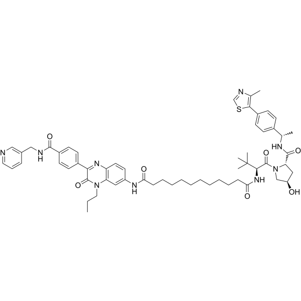
PROTAC NAMPT Degrader-30 is a PROTAC degrader targeting nicotinamide phosphoribosyltransferase (NAMPT), with a DC50 of 8.4 nM in A2780 cells. PROTAC NAMPT Degrader-30 binds to the VHL E3 ligase and forms a ternary complex with NAMPT, which induces ubiquitination and subsequent proteasomal degradation of NAMPT, thereby inhibiting the enzymatic activity of NAMPT. PROTAC NAMPT Degrader-30 reduces intracellular NAD + levels and suppresses tumor cell proliferation. PROTAC NAMPT Degrader-30 exhibits a fluorescence turn-on response to the NAMPT protein, enabling real-time visualization of the NAMPT degradation process in living cells. PROTAC NAMPT Degrader-30 is applicable for research on ovarian cancer .
loading...
    loading...
Cat. No.: HY-183665
CAS No.: 76778-24-0
Target:  

Dopamine Receptor

Domaines de recherche:  

Neurological Disease

GBR-13119 is a blood-brain barrier-permeable inhibitor of the presynaptic dopamine uptake system. GBR-13119 binds to dopamine uptake sites with high selectivity, without being affected by other neurotransmitter reuptake inhibitors. GBR-13119 exhibits a lipophilic systemic distribution profile with extremely low defluorination in rats, while it shows a differential characteristic of slow striatal washout in the brain of primates. GBR-13119 can serve as a PET tracer to achieve visualized imaging of the striatum in primates, and can reflect MPTP-induced dopaminergic neuron degeneration through reduced uptake. GBR-13119 can be widely applied to studies related to Parkinson's disease and dopaminergic neuron degeneration .
loading...
    loading...
Cat. No.: HY-D1422
CAS No.: 1445108-32-6
Synonyms: BDNCA-346
Target:  

Fluorescent Dye

Domaines de recherche:  

Others

ER Tracker Yellow (BDNCA-346) is a Fluorescent probe for targeted visualization of temperature at the endoplasmic reticulum. Its detection mechanism depends on temperature, with fluorescence intensity decreasing at higher temperatures due to preferential non-radiative processes; this intensity reversibly returns to baseline when cooled, provided the probe does not precipitate or degrade. It localizes specifically to the endoplasmic reticulum, where it may covalently attach to biomacromolecules via a thiol-reactive moiety, and its fluorescence remains stable after cell fixation; its fluorescence intensity is unaffected by physiological pH ranges of 5.0–8.0 and free Ca²⁺ concentrations of 0–800 μM. Its excitation/emission wavelength is Ex/Em = 559/581 nm[1].
loading...
    loading...
Cat. No.: HY-D3311
M1219 is a GSH/ATP dual near-infrared activated fluorescent probe that enables independent real-time monitoring of dynamic changes in intracellular GSH and ATP without spectral crosstalk (GSH: Ex=640 nm, Em=740~800 nm; ATP: Ex=594 nm/610 nm, Em=650~700 nm). M1219 not only visualizes the metabolic regulatory mechanism of TNBC under single/dual-target inhibition of SLC7A11/GLUT1 and accurately evaluates its in vivo efficacy, but also achieves precise localization of the TNBC tumor invasion boundary. M1219 can be used for the research of triple-negative breast cancer .
loading...
    loading...
Cat. No.: HY-DY1089
CAS No.: 797785-10-5
Di-4-ANEPPDHQ (solution) is a polarity-sensitive membrane probe with a doubly positive charge that can be used to visualize membrane microdomains in living Arabidopsis cells. Di-4-ANEPPDHQ produces a fluorescence spectrum shift by changing the polarity of the lipid environment after embedding into the cell membrane, thereby quantifying the degree of lipid accumulation. The excitation wavelength of Di-4-ANEPPDHQ is 488 nm, and the emission wavelengths are 560 nm and 650 nm in the liquid ordered and disordered phases, respectively. Di-4-ANEPPDHQ is often used for fluorescence imaging studies of membrane microstructure, lipid order, and membrane tension response in living cells[1][2][3].
Solvent and concentration: DMSO: 5 mM
loading...
    loading...
Cat. No.: HY-153843
Domaines de recherche:  

Others

RNA Aptamer Corn (sodium) is a 28-nt-long aptamer that is substantially shorter than Spinach and Spinach2 and exhibits bright red fluorescence upon binding DFHO (a soluble analog of the intrinsic fluorophore of red fluorescent protein), RNA Aptamer Corn (sodium) can be used to visualize RNA expression or localization in live cells which have been soaked with chromophores. The Corn-DFHO does not become appreciably cytotoxic when illuminated. And most importantly, Corn-DFHO exhibits markedly increased photostability compared to other aptamer-chromophore complexes both in vitro and in vivo. (36 nt Corn construct: 5'-GGCGCGAGGAAGGAGGUCUGAGGAGGUCACUGCGCC-3'; A 36-nt RNA construct, comprised of the 28-nt minimal Corn sequence extended proximally with a 4 base-pair stem.)
loading...
    loading...
Cat. No.: HY-D3189
CAS No.: 2370825-44-6
5GluAF-2MeTG is an activatable fluorescent probe targeting the glutamate carboxypeptidase (CP) activity of PSMA (Ex/Em=490/500-600 nm). After being hydrolyzed by PSMA, 5GluAF-2MeTG releases a cell membrane-permeable fluorescent product, and achieves fluorescence activation by disrupting donor-excited photoinduced electron transfer (d-PeT). 5GluAF-2MeTG enables fluorescence imaging of live PSMA-expressing prostate cancer cells in vitro and visualizes the carboxypeptidase activity of PSMA. 5GluAF-2MeTG can be used to detect prostate cancer regions in preclinical excised tissue specimens .
loading...
    loading...
Cat. No.: HY-D3197
CDg16 is a selective fluorescent dye targeting SLC18B1 (λabsem=458/544 nm) that is actively transported into lysosomal vesicles of activated macrophages independent of the endocytic pathway. CDg16 enables highly specific vesicle localization in live cells. CDg16 exhibits no cytotoxicity and accurately distinguishes activated M1 and M2 subsets from different origins. CDg16 shows low background staining in non-activated cells and normal organs, making it suitable for time-lapse imaging. In preclinical animal models of inflammatory sites, atherosclerotic plaques and liver inflammation, CDg16 allows visualization of activated macrophages. CDg16 can be used to study inflammation-related diseases and atherosclerosis .
loading...
    loading...
Cat. No.: HY-D3389
sCy5DL-amide is an amidated fluorescent D-amino acid conjugated to Sulfo-Cyanine 5, and a peptidoglycan labeling agent. sCy5DL-amide incorporates into bacterial peptidoglycan cell wall via transpeptidase-mediated reactions, replacing the fourth or fifth D-amino acid of the peptidoglycan stem peptide for visualization of nascent peptidoglycan biosynthesis. sCy5DL-amide produces clear cell outline, septum labeling, and high localization density in Bacillus subtilis, including a 'V-shape' pattern at cell-cell contact areas. sCy5DL-amide exhibits robust incorporation into Gram-positive bacteria and mycobacteria, with reduced incorporation into Gram-negative bacteria (Ex/Em = 646/666 nm) .
loading...
    loading...
Cat. No.: HY-W800702
CAS No.: 2770422-53-0
BP Fluor 555 Azide is a water-soluble, bright orange-fluorescent dye with excitation ideally suited for the 532 nm or 555 nm laser lines and visualized with TRITC (tetramethylrhodamine) filter sets. BP Fluor 555 conjugates of antibodies, peptides, and proteins are pH insensitive from pH 4 to pH 10. AF 555 conjugates ideally suited for detection of low abundance targets. BP Fluor 555 Azide can be reacted with terminal alkynes via a copper-catalyzed click reaction (CuAAC). It also reacts with strained cyclooctyne via a copper-free “click chemistry” reaction to form a stable triazole and does not require Cu-catalyst or elevated temperatures. The brightness and photostability of this dye are best suited to direct imaging of low-abundance targets.
loading...
    loading...
Cat. No.: HY-D1320
CAS No.: 1267539-32-1
Target:  

Fluorescent Dye

Domaines de recherche:  

Others

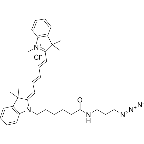
Cyanine5 azide chloride is a Fluorescent probe for cell-selective bacterial protein labeling, as well as a fluorescent dye for single-molecule imaging and dynamic tracking of intracellular organelles. Cyanine5 azide chloride can conjugate with the alkynyl side chain of propargylglycine incorporated into bacterial proteins by the engineered methionyl-tRNA synthetase variant PraRS via copper (I)-catalyzed azide-alkyne [3+2] cycloaddition reaction, thereby enabling in-gel fluorescent visualization of labeled proteins. Cyanine5 azide chloride can diffuse across cell membranes and undergo copper-free click reaction with DBCO-functionalized scAVD pre-bound to DBCO-functionalized biotin, forming fluorescent nanoprobes with enhanced brightness, photostability and localization accuracy, and exhibiting discrete photobleaching steps. The excitation wavelength of Cyanine5 azide chloride is Ex = 633 nm .
loading...
    loading...
Cat. No.: HY-D1556
CAS No.: 384832-91-1
Synonyms: 1,2-dioleoyl-sn-glycero-3-phosphoethanolamine-N-(carboxyfluorescein) ammonium
Target:  

Fluorescent Dye

Domaines de recherche:  

Cancer

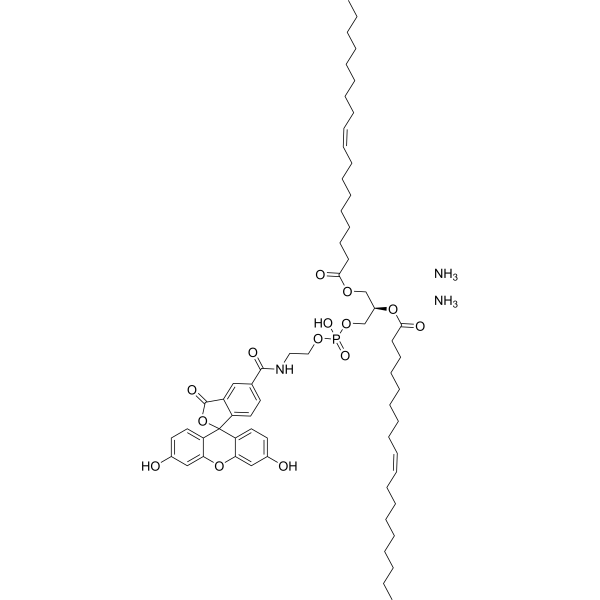
18:1 PE CF (1,2-dioleoyl-sn-glycero-3-phosphoethanolamine-N-(carboxyfluorescein) (ammonium)) is a pH-sensitive fluorescent indicator. 18:1 PE CF exhibits changes in fluorescence intensity in response to pH variations and reflects the electrostatic states of cationic liposomes and lipoplexes. 18:1 PE CF supports real-time visualization of pH distribution and membrane deformation in giant unilamellar vesicles and is suitable for steady-state fluorescence testing in large unilamellar vesicle membranes. 18:1 PE CF is capable of monitoring the surface electrical potential of cationic liposomes and lipoplexes and can effectively label lipid bilayers and nanomicelles. 18:1 PE CF can be used for the research of the investigation of lung cancer (Ex = 495 nm; Em = 520 nm) .
loading...
    loading...
Cat. No.: HY-D3186
CAS No.: 2163840-29-5
HMRef-βGal is a fluorescent probe and a substrate responsive to β-galactosidase (β-galactosidase) (Ex/Em=498 nm/505-600 nm). After being cleaved by β-galactosidase, HMRef-βGal triggers significant fluorescence enhancement via intramolecular spirocyclic function regulation. HMRef-βGal generates bright fluorescence in cancer cells with elevated β-galactosidase activity, enabling visualization of tiny peritoneal metastases in mouse models. HMRef-βGal exhibits low in vitro cytotoxicity and low acute in vivo toxicity in mice. HMRef-βGal can be used for preclinical fluorescence-guided diagnosis and cytoreductive surgery of peritoneal metastases, including compatibility with real-time naked-eye detection and endoscopic imaging, as well as for studies related to peritoneal metastases of ovarian cancer .
loading...
    loading...
Cat. No.: HY-D3391
RMR-Tre is a fluorescent probe targeting the mycobacterial acyltransferase Ag85. Under the catalysis of Ag85, RMR-Tre undergoes 6-position mycoloylation and anchors to the mycobacterial membrane, while achieving fluorescence activation by inhibiting the intramolecular twisted charge transfer state transition. RMR-Tre can distinguish live mycobacteria from dead ones through metabolism-driven labeling, enabling rapid, wash-free, low-background detection of viable bacteria. RMR-Tre reports the drug resistance of Mycobacterium tuberculosis via the trehalose catalytic shift activity readout associated with TreS. In addition, RMR-Tre can be combined with flow cytometry or high-content imaging techniques to visualize and quantitatively analyze the metabolic heterogeneity of Mycobacterium tuberculosis related to persistence and drug resistance. RMR-Tre is widely used in tuberculosis-related research .
loading...
    loading...

