6 Results for "

spectral analysis

" in MedChemExpress (MCE) Product Catalog:
Products (6)

6 Results for "spectral analysis" in MCE Product Catalog:

Cat. No.: HY-114907
CAS No.: 596-03-2
Purity:  ≥95.0%
Synonyms: Eosinic acid; Solvent Red 72
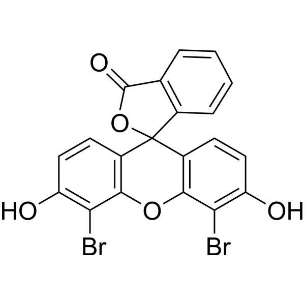
4',5'-Dibromofluorescein (Eosinic acid) is a fluorescein derivative that can used as a ligand for studying of protein properties by spectral analysis .
loading...
    loading...
Cat. No.: HY-120963
CAS No.: 52514-04-2
Purity:  98%
Target:  

Endogenous Metabolite

Research Areas:  

Metabolic Disease

Several different arachidonoyl amino acids, including N-arachidonoyl dopamine and N-arachidonoyl serine, have been isolated and characterized from bovine brain.1 During mass spectral lipidomics analysis of rat brain, a series of fatty acyl amides of a third amino acid, taurine, were discovered.2 This novel class of compounds is present in kidney and activates members of the transient receptor potential (TRP) family of calcium channels.3 N-Oleoyl taurine is an amino-acyl endocannabinoid isolated from rat brain that may activate TRPV1 and TRPV4.
loading...
    loading...
Cat. No.: HY-120965
CAS No.: 83982-06-3
Purity:  99.00%
Target:  

Endogenous Metabolite

Research Areas:  

Metabolic Disease

Several different arachidonoyl amino acids, including N-arachidonoyl dopamine (NADA) and N-arachidonoyl serine (ARA-S), have been isolated and characterized from bovine brain.1 During mass spectral lipidomic analysis of rat brain, a series of fatty acyl amides of a third amino acid, taurine, is discovered. N-Palmitoyl taurine is a prominent amino-acyl endocannabinoid isolated from rat brain during lipidomics profiling. Its function is currently under investigation.
loading...
    loading...
Cat. No.: HY-120964
CAS No.: 63155-80-6
Purity:  99.00%
Target:  

Endogenous Metabolite

Research Areas:  

Metabolic Disease

Several different arachidonoyl amino acids, including N-arachidonoyl dopamine (NADA) and N-arachidonoyl serine (ARA-S), have been isolated and characterized from bovine brain.1 During mass spectral lipidomic analysis of rat brain, a series of fatty acyl amides of a third amino acid, taurine, is discovered.2 This novel class of compounds is present in kidney and activates members of the transient receptor potential (TRP) family of calcium channels.3 N-Stearoyl taurine is a prominent amino-acyl endocannabinoid isolated from rat brain during lipidomics profiling.
loading...
    loading...
Cat. No.: HY-D0293
CAS No.: 116-95-0
Synonyms: Xylene blue free acid; C.I. Acid blue 1 free acid
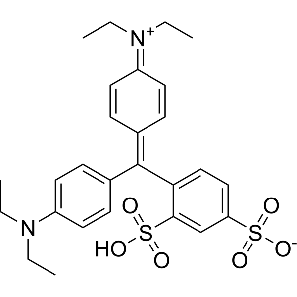
Acid blue 1 free acid (Patent Blue V) is a novel biological dye that can be used as an intraocular dye for retinectomy. Retinectomy refers to the removal of the translucent inner limiting membrane (ILM). The application of appropriate dyes in vitreoretinal surgery can achieve the purpose of complete removal. Acid blue 1 free acid can be used to stain retinal premembranous structures. Spectral analysis shows that Acid blue 1 free acid has strong absorption below 450 nm and above 600 nm, showing a blue-green color. Acid blue 1 free acid is also used as a marker in lymphangiography for resection of neoplastic lymph nodes .
loading...
    loading...
Cat. No.: HY-D3101
Target:  

Fluorescent Dye

Research Areas:  

Others

Nile-TPA is a fluorescent probe for polarity detection, which can be used for lipid droplet targeting, cell polarity analysis, in vivo polarity imaging, and blood polarity monitoring in circulating tumor mouse models. The detection mechanism of Nile-TPA relies on the dual intramolecular charge transfer (ICT) effect generated by the electron-donating triphenylamine and Nile Red moieties and the electron-withdrawing carbonyl group; this probe emits strong red fluorescence in a low-polarity environment, and as polarity increases, the fluorescence intensity decreases and a red shift occurs due to changes in the excited-state dipole moment. In spectral tests, its excitation wavelength is 540 nm, and the emission wavelength varies in the range of 550-800 nm depending on solvent polarity; for cell, zebrafish and mouse imaging, Ex/Em = 561/570-620 nm, and for in vivo mouse imaging, Ex/Em = 560/620 nm .
loading...
    loading...

