



Based on 1 Customer Validation
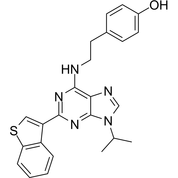
Stemregenin 1 (SR1) (GMP) is a GMP-grade Stemregenin 1 (HY-15001). GMP-grade small molecules can be used as adjuvants in cell therapy. Stemregenin 1 is a potent aryl hydrocarbon receptor (AhR) antagonist with an IC50 of 127 nM. Stemregenin 1 (GMP) can competitively bind to AhR and inhibit its nuclear translocation, inhibiting osteoclastogenesis and promoting hematopoietic progenitor cell expansion by blocking the AhR-c-src-NF-κB/p-ERK MAPK-NFATc1 signaling pathway. Stemregenin 1 (GMP) can be used for the study of osteoporosis, in vitro expansion of hematopoietic stem cells, and the study of the mechanism of bone metabolic diseases.
For research use only. We do not sell to patients.
- CAS No.: 1227633-49-9
- Formula: C24H23N5OS
- Molecular Weight:429.54
-
Storage:
Please store the product under the recommended conditions in the Certificate of Analysis.


To place orders, for customer services and technical support, please contact: MedChemExpress USA
Tel: 609-228-6898 E-mail: [email protected] [email protected]


-
 Biological Activity
-
Chemical Information
- Protocols
- Purity & Documentation
- References
-
 Help & FAQs


-
HY-15001
Stemregenin 1AhR Antagonist -
HY-12684
CH-223191AhR Antagonist -
HY-104026
L-KynurenineAryl Hydrocarbon Receptor Agonist -
HY-19317
ITEAryl Hydrocarbon Receptor Agonist -
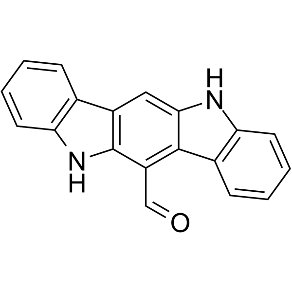
HY-12451
FICZAhR Agonist -
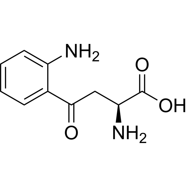
HY-104026R
L-Kynurenine (Standard)Aryl Hydrocarbon Receptor Agonist -
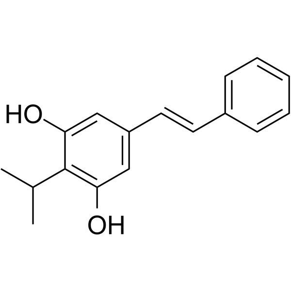
HY-109044
TapinarofAhR Agonist -
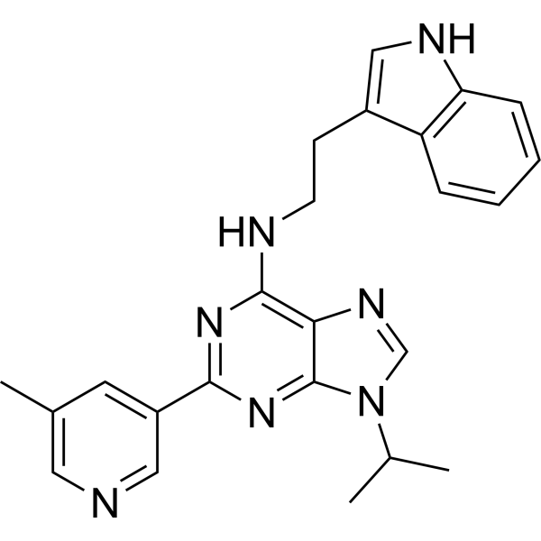
HY-102023
GNF351AHR Antagonist -
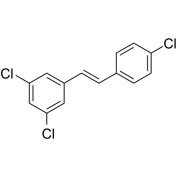
HY-112629
PDM2Aryl Hydrocarbon Receptor Antagonist -
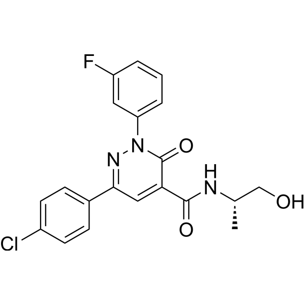
HY-111449
BAY-218AHR Antagonist -

HY-15001R
Stemregenin 1 (Standard)AhR Antagonist -
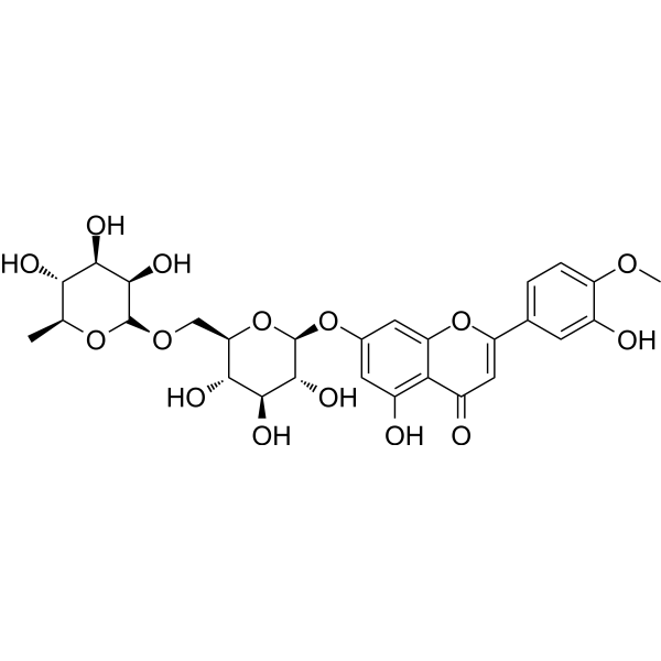
HY-N0178
DiosminAryl Hydrocarbon Receptor Agonist


Biological Activity
Description
IC50 & Target
IC50: 127 nM (AhR)[4]
In Vitro
Stemregenin 1 (GMP) (1, 5, 10 μM; 7 days) promotes CD34+ cell expansion and enhance colony formation ability in human embryonic stem cell-derived hematopoietic progenitor cell experiments in a concentration-dependent manner, but inhibits cell proliferation at 10 μM[1].
Stemregenin 1 (GMP) (0.75 μM; 7 days) combined with DMOG increases the proportion of primitive hematopoietic subsets and improves the number of long-term colony-forming units in CD34+ umbilical cord blood cell experiments[2].
Stemregenin 1 (GMP) (3, 6, 8, 10 μM; 4-7 days) inhibits RANKL-induced osteoclast differentiation in bone marrow macrophage experiments, reduces the expression of genes such as NFATc1 and ACP5, and F-actin ring formation[3].
MedChemExpress (MCE) has not independently confirmed the accuracy of these methods. They are for reference only. Further protocols information, click here.
-
Cell Line:BMMs, hESC-derived hematopoietic progenitors, CD34+ cord blood cells
-
Concentration:0-10 μM
-
Incubation Time:48-96 h for BMMs; 7 days for others
-
Result:Hed no cytotoxicity in BMMs at ≤10 μM, and maintained CD34+ cells viability without significant loss at 0.75 μM.
In Vivo
MedChemExpress (MCE) has not independently confirmed the accuracy of these methods. They are for reference only.
-
Animal Model:C57BL/6J mice (female, 8 weeks old, 20-25 g) with ovariectomized (OVX) osteoporosis model[3]
-
Dosage:2.5 mg/kg
-
Administration:Intraperitoneal injection, once every 3 days, for 42 days
-
Result:Reversed OVX-induced bone loss, as shown by micro-CT analysis demonstrating increased trabecular bone volume/total volume (BV/TV).
Improved bone mineralization.
Serum markers of bone resorption were reduced, and bone formation markers were enhanced.


Chemical Information
-
CAS No. 1227633-49-9
-
Appearance Solid
-
Molecular Weight 429.54
-
Formula C24H23N5OS
-
Color Off-white to yellow
-
SMILES
CC(C)N1C2=NC(C3=CSC4=C3C=CC=C4)=NC(NCCC5=CC=C(O)C=C5)=C2N=C1
-
Synonyms
SR1 (GMP)
-
Shipping
Room temperature in continental US; may vary elsewhere.
-
Storage
Please store the product under the recommended conditions in the Certificate of Analysis.

Protocols
-
Cytoplasmic-Nuclear Fractionated Protein Extraction
Cytoplasmic-nuclear fractionated protein extraction separates soluble cytoplasmic proteins from nuclear-enriched proteins by mild plasma-membrane permeabilization, differential centrifugation, washing of nuclei, and extraction of nuclear proteins for downstream immunoblotting or related molecular analysis. The readout is the relative abundance of a protein in cytoplasmic and nuclear fractions, commonly assessed by western blotting together with compartment markers such as tubulin or pyruvate kinase for cytoplasm and lamin, nucleoporin, hnRNP, H2AX, or Lamin B for nuclear fractions.
-
Cell Cytotoxicity Assay
Cytotoxicity assays are usually based on the assessment of cell membrane damage, which can also be indirectly detected by measuring cell viability. Detection methods include MTT assay, CKK-8 assay, LDH assay and ATP assay, etc.
-
Research Protocol for Inflammation-related Diseases
The NLRP3 inflammasome is a cytosolic innate immune signaling platform that integrates priming signals and danger-signal activation to promote caspase-1 activation, maturation of IL-1β and IL-18, and gasdermin D-mediated pyroptotic cell death. The core experimental logic is to determine whether inflammatory disease phenotypes are driven by increased NLRP3 expression, ASC-containing inflammasome assembly, caspase-1 cleavage, GSDMD cleavage, and extracellular release of IL-1β/IL-18 rather than by nonspecific cell injury alone. The pathway is strongly linked to inflammation-related disease phenotypes because monosodium urate crystals activate NALP3/NLRP3 inflammasome signaling in gout-like crystal inflammation, cholesterol crystals activate NLRP3 inflammasomes in atherogenesis models, and DSS-induced intestinal inflammation has been reported to involve NLRP3 inflammasome activity. However, experimental colitis studies also show context-dependent protective effects of NLRP3 inflammasome co
-
Directly Induced Neuron Culture
Directly induced neuron culture converts somatic cells, most commonly fibroblasts, into induced neurons without passing through a pluripotent or neural progenitor stage; classic evidence shows that mouse fibroblasts can be converted by Ascl1, Brn2/Pou3f2, and Myt1l, human fibroblasts can be converted by defined neuronal transcription factors, and human fibroblasts can also be converted by miR-9/9-124 with neurogenic or subtype-specifying transcription factors. The readout is acquisition of neuronal identity and function, assessed by neuronal morphology, neuronal markers such as Tuj1/βIII-tubulin, MAP2, synapsin, and subtype markers when relevant, together with functional assays such as action-potential firing, synaptic activity, and electrophysiology.
-
Osteoclast differentiation from monocyte/macrophage precursors
Osteoclast differentiation is an in vitro induction assay in which monocyte/macrophage-lineage precursors are exposed to macrophage colony-stimulating factor (M-CSF) and receptor activator of NF-κB ligand (RANKL), generating multinucleated osteoclasts that are commonly identified by tartrate-resistant acid phosphatase (TRAP) staining and functionally confirmed by resorption pits on dentin, bone, or mineralized substrates. M-CSF supports survival and expansion of osteoclast precursors, while RANKL binding to RANK drives osteoclast commitment, fusion, maturation, and resorptive function; osteoprotegerin inhibits this pathway by binding RANKL and preventing RANK activation. The main readouts are the number of TRAP-positive multinucleated cells, formation of F-actin rings, and resorbed surface area; TRAP-positive multinucleated cells indicate osteoclast differentiation, whereas pit formation on dentin, bone, or mineralized coating indicates functional bone-resorbing activity.


Purity & Documentation
-
Data Sheet (278 KB)
-
SDS (393 KB)
- English - EN (393 KB)
- Français - FR (393 KB)
- Deutsch - DE (393 KB)
- Norwegian - NO (393 KB)
- Español - ES (393 KB)
- Swedish - SV (393 KB)
- Italian - IT (393 KB)
- Korean - KR (393 KB)
- Portuguese - PT (393 KB)
-
Handling Instructions (2659 KB)
References
[1]. Tao L, et al. StemRegenin 1 selectively promotes expansion of Multipotent Hematopoietic Progenitors derived from Human Embryonic Stem Cells. J Stem Cells Regen Med. 2017 Dec 18;13(2):75-79. [Content Brief]
[2]. Jackson CS, et al. Targeting the aryl hydrocarbon receptor nuclear translocator complex with DMOG and Stemregenin 1 improves primitive hematopoietic stem cell expansion. Stem Cell Res. 2017 May;21:124-131. [Content Brief]
[3]. Zhou S, et al. StemRegenin 1 attenuates the RANKL-induced osteoclastogenesis via inhibiting AhR-c-src-NF-κB/p-ERK MAPK-NFATc1 signaling pathway. iScience. 2024 Apr 6;27(5):109682. [Content Brief]
[4]. Boitano AE, et al. Aryl Hydrocarbon Receptor Antagonists Promote the Expansion of Human Hematopoietic Stem Cells. Science. 2010 Sep 10;329(5997):1345-8. [Content Brief]

Calculators
Concentration (start) × Volume (start) = Concentration (final) × Volume (final)


Stemregenin 1 (GMP) Related Classifications
HY-15001G Related Classifications
 Powered by Bioz