



Tanaproget
Based on 1 publication(s) in Google Scholar
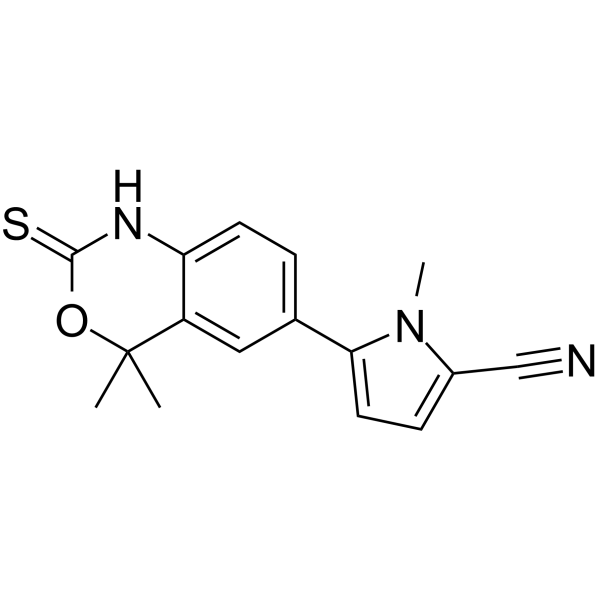
Tanaproget is an orally effective, selective nonsteroidal progesterone receptor (PR) agonist targeting human PR with an IC50 of 1.7 nM. Tanaproget promotes the interaction between PR receptors and coactivators (such as SRC-1), thereby inhibiting the secretion of matrix metalloproteinases (MMP-3/MMP-7) and reducing endometrial tissue invasion and angiogenesis. Tanaproget can be used for contraception and endometriosis inhibition research.
For research use only. We do not sell to patients.
- Purity : 99.73%
- CAS No.: 304853-42-7
- Formula: C16H15N3OS
- Molecular Weight:297.37
-
Storage:Powder -20°C, 3 years , 4°C, 2 years ; In solvent -80°C, 2 years , -20°C, 1 year

To place orders, for customer services and technical support, please contact: MedChemExpress USA
Tel: 609-228-6898 E-mail: [email protected] [email protected]


-
 Biological Activity
-
Chemical Information
-
Solvent & Solubility
- Protocols
- Purity & Documentation
- References
-
 Help & FAQs


-
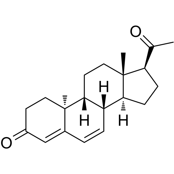
HY-B0257A
DydrogesteroneProgestogen -
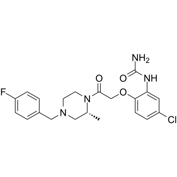
HY-12080
BX471CCR1 Antagonist -
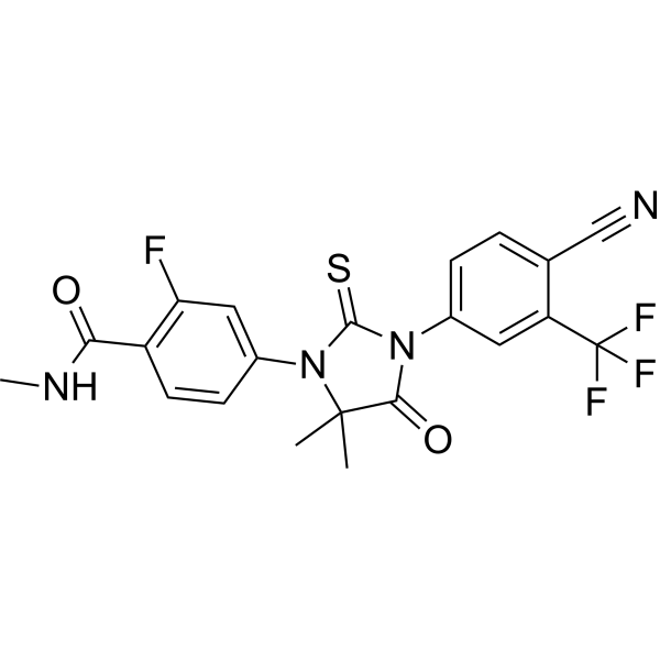
HY-70002
EnzalutamideAndrogen Receptor Antagonist -
HY-12518
OF-1pan-BRD Inhibitor -
HY-13071
NestoronProgesterone Receptor Agonist -
HY-13702
NilutamideAndrogen Receptor Antagonist -
HY-70002A
N-Desmethyl enzalutamideActive Metabolite of Enzalutamide -
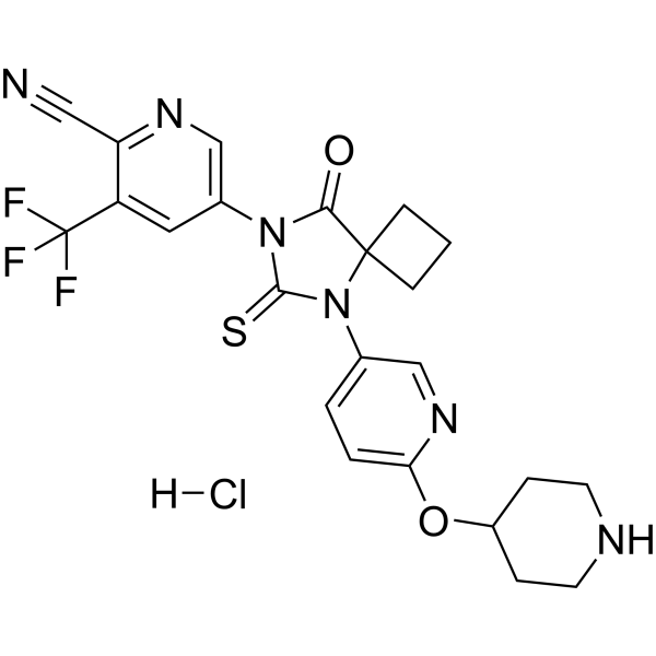
HY-115282A
JNJ-63576253AR Antagonist -
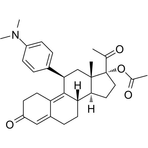
HY-16508
Ulipristal acetateSelective Progesterone Receptor Modulator -
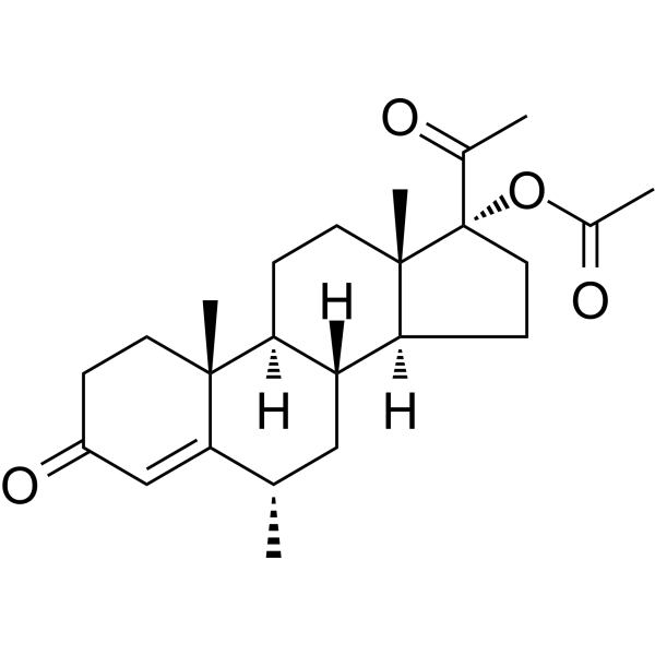
HY-B0469
Medroxyprogesterone acetateProgesterone Receptor Agonist -
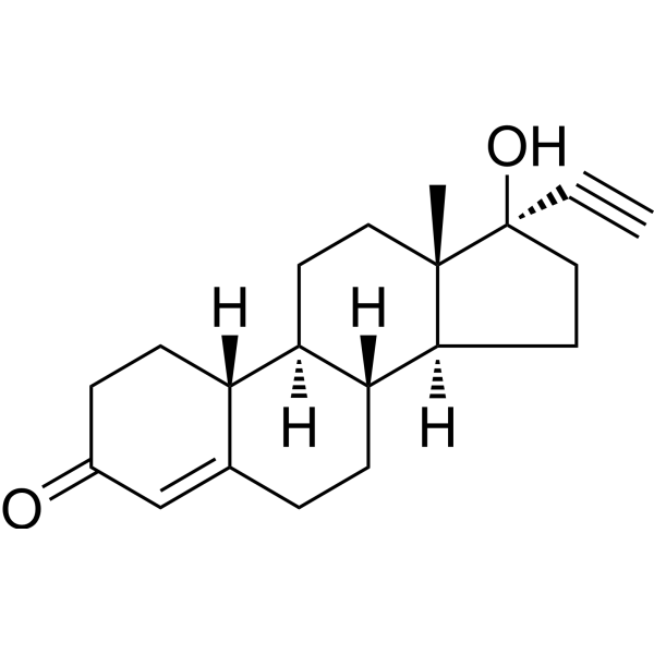
HY-B0554
NorethindroneProgestin -
HY-12516
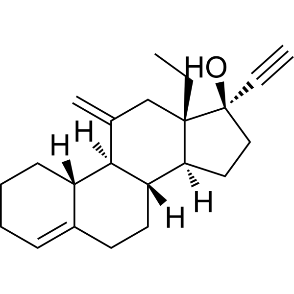
DesogestrelProgesterone Receptor Agonist
Publications Citing Use of MedChemExpress (MCE) Tanaproget
More

Biological Activity
Description
Cellular Effect
|
Cell Line

|
Type | Value | Description | References |
|---|---|---|---|---|
| T47D | EC50 |
0.15 nM
Compound: 13
|
Effective concentration against alkaline phosphatase activity in human T47D breast carcinoma cell line
Effective concentration against alkaline phosphatase activity in human T47D breast carcinoma cell line
|
[PMID: 16078826] |
| T47D | EC50 |
0.15 nM
Compound: 8
|
Agonist activity at human PR expressed in human T47D cells assessed as stimulation of alkaline phosphatase
Agonist activity at human PR expressed in human T47D cells assessed as stimulation of alkaline phosphatase
|
[PMID: 18318463] |
| T47D | IC50 |
0.5 nM
Compound: 8
|
Displacement of [3H]R5020 from human PR in human T47D cells by whole cell assay
Displacement of [3H]R5020 from human PR in human T47D cells by whole cell assay
|
[PMID: 18318463] |
In Vitro
Tanaproget (1 nM; 3-5 days) significantly down-regulated pro-MMP-3 expression in endometrial stromal cells isolated from normal women, even under IL-1α stimulation[1].
Tanaproget (1-100 pM; 48-72 h) inhibited the secretion of pro-MMP-3 and pro-MMP-7 in endometrial organ cultures from exogenous endometriosis patients[1].
Tanaproget (0.1 nM; 24 h) induced alkaline phosphatase activity in T47D cells with an EC50 of 0.15 nM (human), which is comparable to steroidal progestins[2].
Tanaproget against PR of different species with IC50s of 1.7 nM (human), 0.3 nM (monkey), and 0.5 nM (rat, rabbit)[2].
MedChemExpress (MCE) has not independently confirmed the accuracy of these methods. They are for reference only. Further protocols information, click here.
-
Cell Line:Human endometrial stromal cells (normal and endometriosis patients)
-
Concentration:1-500 nM (natural progesterone), 1-100 pM (synthetic progestins including Tanaproget)
-
Incubation Time:3-5 d
-
Result:Suppressed pro-MMP-3 expression in normal stromal cells, even after IL-1α challenge.
Reduced pro-MMP-3 and pro-MMP-7 secretion compared to placebo.
-
Cell Line:T47D breast carcinoma cells
-
Concentration:0.001–100 nM
-
Incubation Time:24 h
-
Result:Induced alkaline phosphatase activity with an EC50 of 0.15 nM, demonstrating full agonist activity comparable to medroxyprogesterone acetate (MPA) and trimegestone (TMG).
In Vivo
Tanaproget (0.03 mg/kg; oral; once daily; 4 days) completely inhibits ovulation in the Sprague-Dawley rat ovulation inhibition model and is 30 times more potent than steroidal progestins[2].
MedChemExpress (MCE) has not independently confirmed the accuracy of these methods. They are for reference only.
-
Animal Model:Human endometriosis xenograft model in ncr/nude athymic mice (female, 5-week-old, ovariectomized)[1]
-
Dosage:300μg/kg (2% Tween 80/0.5% methylcellulose)
-
Administration:Gavage or slow-release capsule, daily for 14–16 days
-
Result:Reduced the number and size of human endometrial lesions derived from endometriosis patients, with complete regression in 33% of treated mice.
-
Animal Model:Sprague-Dawley rats (female, 200 g, estrus-synchronized)[2]
-
Dosage:0.03 mg/kg (corn oil)
-
Administration:Oral gavage, daily for 4 days
-
Result:Completely inhibited ovulation in all treated rats at 0.03 mg/kg, showing 30-fold higher efficacy than medroxyprogesterone acetate (MPA).


Chemical Information
-
CAS No. 304853-42-7
-
Appearance Solid
-
Molecular Weight 297.37
-
Formula C16H15N3OS
-
Color Light yellow to yellow
-
SMILES
N#CC1=CC=C(C2=CC=C(NC(OC3(C)C)=S)C3=C2)N1C
-
Synonyms
NSP-989
-
Shipping
Room temperature in continental US; may vary elsewhere.
-
Storage
Powder -20°C 3 years 4°C 2 years In solvent -80°C 2 years -20°C 1 year


Publications (1)
-
Journal Impact Factor
-
Most Recent
-
Nat Cancer
Discovering the anti-cancer potential of non-oncology drugs by systematic viability profiling. [Abstract]2020 Feb;1(2):235-248. PMID: 32613204


Solvent & Solubility
In Vitro:
DMSO : ≥ 50 mg/mL (168.14 mM; Hygroscopic DMSO has a significant impact on the solubility of product, please use newly opened DMSO)
* "≥" means soluble, but saturation unknown.
Please refer to the solubility information to select the appropriate solvent. Once prepared, please aliquot and store the solution to prevent product inactivation from repeated freeze-thaw cycles.
Storage method and period of stock solution: -80°C, 2 years; -20°C, 1 year. When stored at -80°C, please use it within 2 years. When stored at -20°C, please use it within 1 year.
Please refer to the solubility information to select the appropriate solvent. Once prepared, please aliquot and store the solution to prevent product inactivation from repeated freeze-thaw cycles.
Storage method and period of stock solution: -80°C, 2 years; -20°C, 1 year. When stored at -80°C, please use it within 2 years. When stored at -20°C, please use it within 1 year.
Concentration (start) × Volume (start) = Concentration (final) × Volume (final)
In Vivo:
Select the appropriate dissolution method based on your experimental animal and administration route.
- For the following dissolution methods, please ensure to first prepare a clear stock solution using an In Vitro approach and then sequentially add co-solvents:
- To ensure reliable experimental results, the clarified stock solution can be appropriately stored based on storage conditions. As for the working solution for In Vivo experiments, it is recommended to prepare freshly and use it on the same day.
- The percentages shown for the solvents indicate their volumetric ratio in the final prepared solution. If precipitation or phase separation occurs during preparation, heat and/or sonication can be used to aid dissolution.
In Vivo Dissolution Calculator
Please enter the basic information of animal experiments:
-
-
-
-
Recommended: Prepare an additional quantity of animals to account for potential losses during experiments.
Please enter your animal formula composition:
-
%DMSO +
Recommended: Keep the proportion of DMSO in working solution below 2% if your animal is weak.
-
%+
-
+%Tween-80 + +
-
%Saline +
The co-solvents required include: DMSO, . All of co-solvents are available by MedChemExpress (MCE). , Tween 80. All of co-solvents are available by MedChemExpress (MCE).
Working solution concentration: 0.22 mg/mL
Method for preparing stock solution: mg drug dissolved in μL DMSO. Stock solution concentration: mg/mL.
1. Take μL DMSO stock solution;
2. Add μL .
μL , mix evenly;
3. Then add μL Tween 80, mix evenly;
4. Then add μL
Please ensure that the stock solution in the first step is dissolved to a clear state, and add co-solvents in sequence. You can use ultrasonic heating (ultrasonic cleaner, recommended frequency 20-40 kHz), vortexing, etc. to assist dissolution.

Protocols
-
Mitochondrial membrane-potential fluorescent assay
Mitochondrial membrane potential fluorescent assays estimate ΔΨm in living cells using lipophilic cationic dyes such as TMRM, TMRE, rhodamine 123, and JC-1, which accumulate in mitochondria according to membrane polarization; loss of signal after FCCP or CCCP treatment is interpreted as mitochondrial depolarization. TMRM/TMRE and rhodamine 123 are commonly used for semi-quantitative live-cell microscopy or flow cytometry, while JC-1 can report a shift from red aggregate fluorescence to green monomer fluorescence during depolarization; interpretation requires controls because dye concentration, quenching mode, cell type, dye efflux, and mitochondrial mass can affect fluorescence independently of ΔΨm.
-
Human pluripotent stem cell endothelial-cell differentiation
Human pluripotent stem cell endothelial differentiation is based on stepwise developmental patterning: early activation of WNT/GSK3β inhibition promotes mesodermal or vascular progenitor entry, followed by endothelial specification using VEGF-related signaling, BMP4, FGF2, Notch modulation, or cAMP depending on the published protocol. Endothelial differentiation is read out by acquisition of CD31, CD34, VE-cadherin/CD144, KDR/VEGFR2, vWF, Tie2, NOS3, acetylated LDL uptake, tube/network formation, barrier function, and in vivo vessel-forming capacity where tested.
-
Cell Cytotoxicity Assay
Cytotoxicity assays are usually based on the assessment of cell membrane damage, which can also be indirectly detected by measuring cell viability. Detection methods include MTT assay, CKK-8 assay, LDH assay and ATP assay, etc.
-
Vascular/Branching Fractal Analysis
Vascular/branching fractal analysis quantifies the geometric complexity of vessel trees or vascular networks from segmented 2D images, commonly by converting vessels into binary and/or skeletonized maps and estimating fractal dimension using box-counting or related approaches. Fractal dimension is interpreted as an image-derived readout of vascular branching complexity, space filling, or density, and has been applied to retinal photographs, fluorescein angiography, OCT angiography, capillary perfusion maps, and in vitro Matrigel angiogenesis networks. The assay readout is generated from vessel-positive pixels after image preprocessing, vessel segmentation, binarization, and optional skeletonization; reported outputs include fractal dimension, vessel density, branchpoint density, endpoint density, vessel length density, tortuosity, and generation-based branching metrics when VESGEN-style analysis is used. The biological interpretation is limited to quantitative vascular patterning and s
-
Mitochondrial membrane-potential and mitochondrial mass staining
Mitochondrial membrane potential staining measures the electrochemical polarization across the mitochondrial inner membrane in live cells using lipophilic cationic fluorescent probes; early rhodamine-based work showed that selective mitochondrial dye accumulation is lost when the mitochondrial transmembrane potential is dissipated. JC-1 reports mitochondrial polarization by shifting from green monomer fluorescence to red J-aggregate fluorescence as dye concentration increases within energized mitochondria; therefore, the red/green fluorescence ratio is used as a relative readout of mitochondrial membrane potential. TMRE or TMRM staining provides a single-channel relative readout because these cationic rhodamine esters accumulate in polarized mitochondria, and lower fluorescence indicates reduced mitochondrial polarization when acquisition and dye-loading conditions are controlled. Mitochondrial mass staining is commonly performed with MitoTracker Green FM or related MitoTracker dyes as
-
Endothelial Tube Formation Assay
Endothelial tube formation assay evaluates the ability of endothelial cells to attach, migrate, align, and organize into capillary-like networks when cultured on gelled basement membrane extract or Matrigel; the readout is the morphology and quantity of tube-like networks, which reflects an in vitro endothelial morphogenesis step related to angiogenesis. Basement membrane extract/Matrigel provides laminin-rich extracellular matrix cues that support endothelial differentiation into capillary-like structures, but it can contain biologically active growth factors, so growth-factor-reduced matrix is preferred when testing defined angiogenic stimulators or inhibitors.
-
Cell invasion
Cell invasion is the ability of cells to migrate from one area to another via the extracellular matrix. Cell invasion is the response of normal and cancer cells to chemical and mechanical stimuli. Before migrating to a new region, the extracellular matrix is degraded by proteases within the cell. Cell invasion often occurs during wound repair, vascularization and inflammation, abnormal tissue invasion, and tumor cell metastasis.
-
Endothelial Cell Migration/Angiogenic Sprouting Assay
Endothelial cell migration and angiogenic sprouting assays are in vitro (and partially ex vivo-adapted) functional models that quantify the ability of endothelial cells to undergo coordinated migration, extracellular matrix invasion, and multicellular organization into capillary-like sprouts in response to pro-angiogenic stimuli such as VEGF, bFGF, or conditioned microenvironments. These assays are used to model early angiogenic events including tip-cell formation, directional migration, and lumen-like sprout extension, which collectively reflect angiogenic activation and vascular morphogenesis processes observed in vivo.


Purity & Documentation
-
Data Sheet (278 KB)
-
SDS (419 KB)
- English - EN (419 KB)
- Français - FR (419 KB)
- Deutsch - DE (419 KB)
- Norwegian - NO (419 KB)
- Español - ES (419 KB)
- Swedish - SV (419 KB)
- Italian - IT (419 KB)
- Korean - KR (419 KB)
- Portuguese - PT (419 KB)
-
Handling Instructions (2659 KB)
References
[1]. Bruner-Tran KL, et al. Down-regulation of endometrial matrix metalloproteinase-3 and -7 expression in vitro and therapeutic regression of experimental endometriosis in vivo by a novel nonsteroidal progesterone receptor agonist, tanaproget. J Clin Endocrin. [Content Brief]
[2]. Zhang Z, et al. Molecular and pharmacological properties of a potent and selective novel nonsteroidal progesterone receptor agonist tanaproget. J Biol Chem. 2005 Aug 5;280(31):28468-75. [Content Brief]
[3]. Bapst JL, et al. Pharmacokinetics and safety of tanaproget, a nonsteroidal progesterone receptor agonist, in healthy women. Contraception. 2006 Nov;74(5):414-8. [Content Brief]


Complete Stock Solution Preparation Table
Please refer to the solubility information to select the appropriate solvent. Once prepared, please aliquot and store the solution to prevent product inactivation from repeated freeze-thaw cycles.
Storage method and period of stock solution: -80°C, 2 years; -20°C, 1 year. When stored at -80°C, please use it within 2 years. When stored at -20°C, please use it within 1 year.
| Optional Solvent | Concentration Solvent Mass | 1 mg | 5 mg | 10 mg | 25 mg |
|---|---|---|---|---|---|
| DMSO | 1 mM | 3.3628 mL | 16.8141 mL | 33.6281 mL | 84.0704 mL |
| 5 mM | 0.6726 mL | 3.3628 mL | 6.7256 mL | 16.8141 mL | |
| 10 mM | 0.3363 mL | 1.6814 mL | 3.3628 mL | 8.4070 mL | |
| 15 mM | 0.2242 mL | 1.1209 mL | 2.2419 mL | 5.6047 mL | |
| 20 mM | 0.1681 mL | 0.8407 mL | 1.6814 mL | 4.2035 mL | |
| 25 mM | 0.1345 mL | 0.6726 mL | 1.3451 mL | 3.3628 mL | |
| 30 mM | 0.1121 mL | 0.5605 mL | 1.1209 mL | 2.8023 mL | |
| 40 mM | 0.0841 mL | 0.4204 mL | 0.8407 mL | 2.1018 mL | |
| 50 mM | 0.0673 mL | 0.3363 mL | 0.6726 mL | 1.6814 mL | |
| 60 mM | 0.0560 mL | 0.2802 mL | 0.5605 mL | 1.4012 mL | |
| 80 mM | 0.0420 mL | 0.2102 mL | 0.4204 mL | 1.0509 mL | |
| 100 mM | 0.0336 mL | 0.1681 mL | 0.3363 mL | 0.8407 mL |


Tanaproget Related Classifications
HY-15606 Related Classifications
 Powered by Bioz